DMI 181 MIDTERM: LOWER EXTREMITY ( FOOT, LEGS, PELVIC GIRDLE)
 Call Kai
 Learn
Learn Practice Test
 Spaced Repetition
 Match
Match Flashcards
 Knowt Play
1/107

There's no tags or description
Looks like no tags are added yet.
Name | Mastery | Learn | Test | Matching | Spaced | Call with Kai | Chat |
|---|
No analytics yet
Send a link to your students to track their progress
108 Terms
Phalanges
14 on each foot just like the hand
1st toe is known as the “great toe” / Hallux

Distal Interphalangeal (DIP) joint
between the distal and middle phalanges

Proximal interphalangeal ( PIP) joint
between the proximal and middle phalanges

MetaTARSALS
TARSALS TAKES YOU TO THE TOES.
Sesamoids: Under the 1st metatarsal, two small round bones embedded in tendons
5 each foot, 10 total

Metatarsophalangeal (MTP)
between the heads of the metatarsals and bases of the proximal phalanges.

Calcaneus
Also known as “Os calcis,” Most posterior and largest; it forms the heel of the foot. Superior surface is a shelf called Sustentaculum Tali

Talus
Sits on top of the calcaneus; ONLY tarsal bone that participates in forming the ankle bone; smooth upper surface= trochlea. Articulates Tibia, and where achilles attaches

Navicular
Located anterior to the talus, posterior to the cuneiform on the medial

Cuneiforms
3 small bones located anterior to the navicular.
*First ( medial)- articulates with the 1st metatarsal
Second ( intermediate)- articulates with the 3rd metatarsal
Third ( lateral)- articulates with the 3rd metatarsal

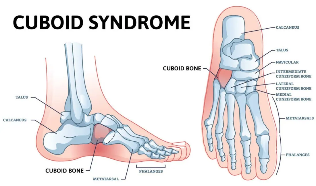
Cuboid
Located anterior to the calcaneus and posterior to the 4th and 5thmetatarsal on the lateral side of the foot.
Arches
stable bases and absorbers

Medial longitudinal
runs along the medial side of the foot ; calcaneus, navicular, cuneiforms, and metatarsals

Lateral longitudunal
runs along the lateral side; calcaneus, cuboid, and 4th and 5th metatarsals

Transverse
Cuboid, cuneiforms, and all the metatarsals

Pes planus
flat foot

Tarsometatarsal
between the proximal ends of the metatarsals, cuneiforms, and cuboid, with the bases of the metatarsals.

Intertarsal
between the tarsals

Subtalar
Inferior surface of the talus with the superior surface of the calcaneus. 3 facets: anterior, middle, posterior

Calcaneus tuberosity

Posterior surface of calcaneus

Tuberosity of 5th metatarsal
commonly breaks

Anterior surface Calcaneus

Middle surface Calcaneus

When articulated with the Talus, the Talus will sit superior to the Calcaneus

“ Shelf” of calcaneus
Horizontal shelf that arises from the anteromedial portion of the calcaneus

Talus Articulates with 4 bones
Inferiorly with Tib + Fib
Superiorly with Calcaneus
Anteriorly the Navicular
Hallux Valgus ( bunion)
Bony projection on the joint at the base of the big toe. It grows gradually and your toe turns inward over time.

Hallux
Also known as “ The great toe”, contains distal and proximal phalanges.

Tibia
Medial side
Parallel to the fibula; bears weight
Extends from the knee joint to the ankle joint

Medial Condyle ( Tibia) ( POSTERIOR)
Medial half of the proximal end

Lateral Condyle ( Tibia) ( POSTERIOR)
Lateral half of the proximal end.

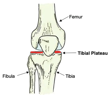
Tibial plateau
Flat superior surface of the condyles.
Intercondylar eminences (tubercles, spine)
Two small projections on top of the tibial plateau

Tibial tuberosity
Located on the anterior surface, just inferior to the condyles.
Tendons are attached to it.

Tibial spine

Anterior/ crest
sharp ridge along the anterior surfcae

Medial malleolus
Process that extends inferiorly from the medial side.

Fibular notch
depression on the lateral border from articulation with the fibula.

Tibial plafond
Fracture at the most distal portion of the tibia

Distal Tibiofibular joint
fibular notch of tibia with lateral malleolus of fibula

Proximal tibiofibular joint
lateral tibia condyle with the head of the fibula ( near the knee)

Fibula
Lateral to the tibia, parallel to the tibia, under the lateral condyle
Part of the ankle joint, but does not reach the knee joint

Head of Fibula
upper expanded part on the proximal end

Lateral malleolus
most distal portion

Styloid process ( apex)
process that sits on top of the head

Ankle mortise ( ankle joint, mortise joint)
Medial malleolus of the tibia
Lateral malleolus of the fibula
Superior surface of the talus

Femur
Extends from the hip to the knee. The longest bone in the body.

Femoral head
Round structure on the proximal end
Articulation with the hip
Sits in the pelvis

Femoral neck
Lateral to head

Greater Trochanter
Large prominence superiorly and lateral to the femoral shaft
Palpable landmark
MORE PROMINENT POSTERIOR

Lesser trochanter
Smaller process that projects medially and posteriorly from the junction of the neck and shaft
MORE PROMINENT POSTERIOR

Tibia and Femur articulate. What about the Fibula?
Fibula does NOT articulate with femur!

Intertrochanteric crest ( FEMUR)
Line/ ridge of bone between the greater and lesser trochanters on the posterior side.
POSTERIOR

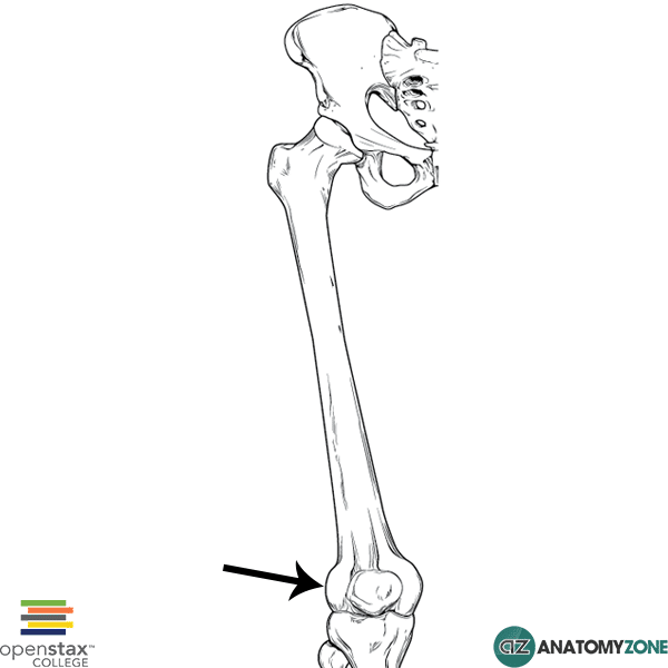
Medial Condyle
Rounded process that forms the medial half of the lower femur.

Lateral Condyle
Rounded process on the lateral side of the FEMUR

Intercondylar fossa ( notch)
Deep depression between the condyles
POSTERIOR

Patellar surface
Smooth area between the condyles anteriorly
Slight depression where the patella sits
ANTERIOR
Also referred to as the “trochlear groove surface”

Medial epicondyle
Small rounded prominence above the medial condyle

Lateral epicondyle
Small rounded process that sits above the lateral condyle
Popliteal surface
POSTERIOR
ABOVE the intercondylar fossa

Patella ( kneecap)
Flat triangular, 2inch diameter
Anterior surface convex and rough
Inner, POSTERIOR surface smooth and oval-shaped

Sesamoid
attaches to tibial tuberosity by patellar ligament

Apex of Patella
pointed INFERIOR part

Base
Pointed SUPERIOR part

Femorotibial joint
between 2 condlyes of femur and corresponding condyles of tibia

Patellofemoral joint
patella articulates with anterior surface of distal femur
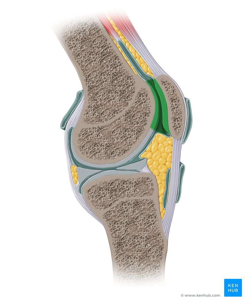
Condyle of distal femur articulates…
tibia plateau

Fat pads
located several areas of body.
protection
Burst/ calcify when trauma

Articular Capsule
holds joint together
Bursa cushions and reduces friction

Fovea Capitis
Opening in the femoral head where the blood vessels would go in (before fused) to supply blood to the femoral head/ neck

Instep
raised middle of the top of the foot

Ribs
12 pairs, 24 total
Protect heart
Attach to costal cartilage—> to sternum

True ribs
1-7, direct attachment through their pwn costal cartilage

False Ribs
8-10
Indirect attachment through the cartilage of the rib abovr

Floating ribs
11-12
No anterior attachment to sternum

Tarsals
7 each foot, 14 total

Osgood Schlatter
Condition that causes the tuberosity to separate from the tibia

Sinus Tarsi
Small, tunnel- like shape located between the talus and calcaneus
Lateral view

Pelvic Girdle
Consists of four bones: 2 hip bones ( Os Coxae, Innominate bone), 1 sacrum, 1 coccyx

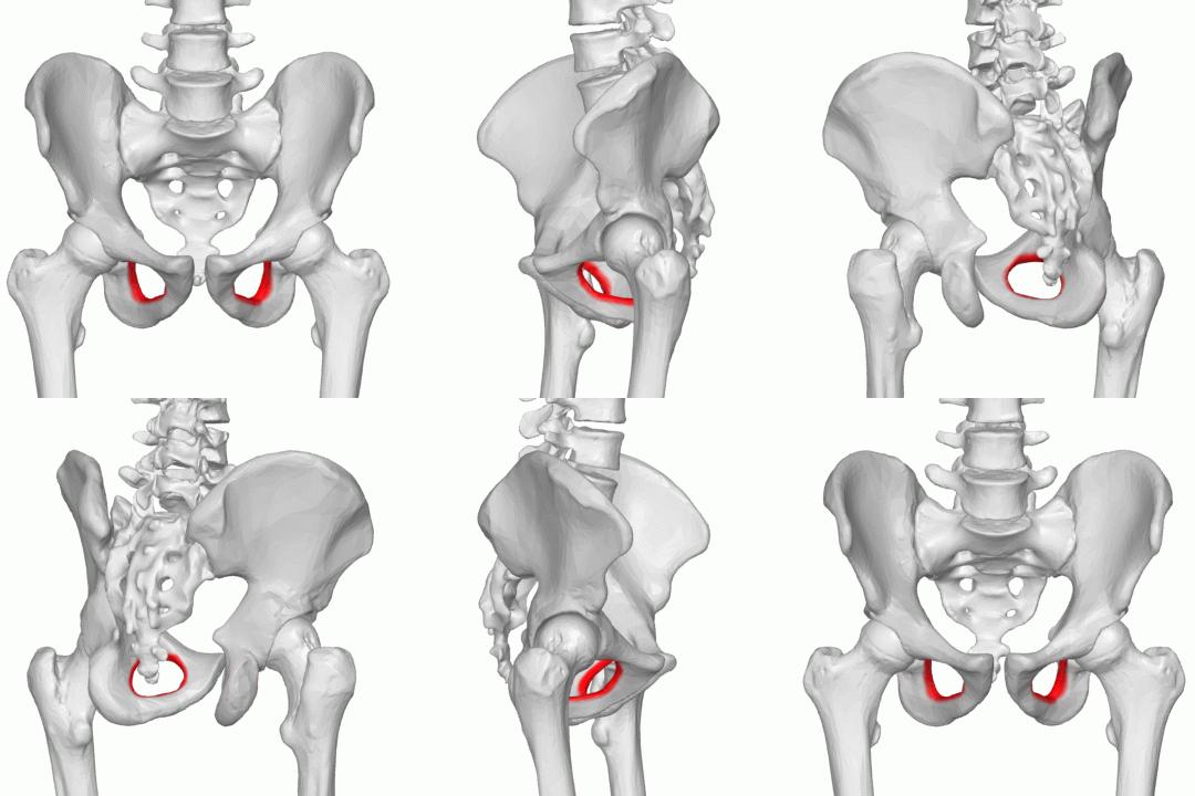
Acetabulum
Deep, cup-shaped cavity that accepts the head of the femur to form the hip joint.
Consists of 3 bones : illium, Pubis, Ischium
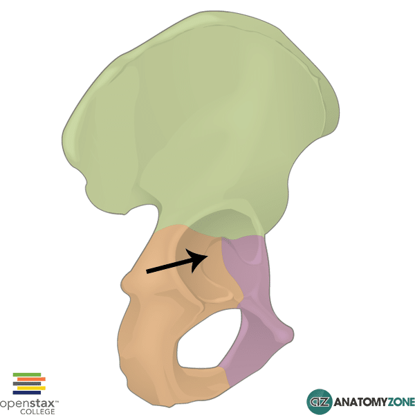
illium
Largest of three, superior to the acetabulum

Wing ( ala)
Thin, flared-shaped superior part of illium

Body of illium
Inferior portion near the acetabulum

illiac crest
upper curved border and most peak of the crest

ASIS
located at the anterior end of the crest.
palpable landmark

AIIS (Anterior inferior illiac spine)
Small process below the AIIS

PSIS (Posterior superior illiac spine)
Located on the POSTERIOR end of the crest

PIIS ( Posterior inferior illiac spine)
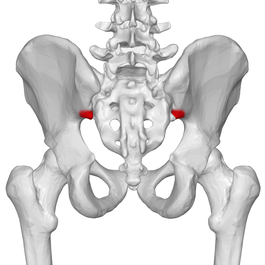
Ischium
Lies inferior AND posterior to the acetabulum

Body of Ischium

Ramus of Ischium
Projecting anteriorly from the ischial tuberosity

Ischial tuberosity
Large rounded process on the POSTERIOR surface on the superior ramus ( lowest part of the pelvis)
what we sit on

Ischial Spine
Bony projection posterior to the acetabulum. A small part of it is also visible on a frontal view of the pelvis

Greater sciatic notch
Directly SUPERIOR to the ischial spine is a deep notch
Posteriorly, between the PIIS and ischial spine

Lesser sciatic notch
INFERIOR to the ischial spine is a smaller notch
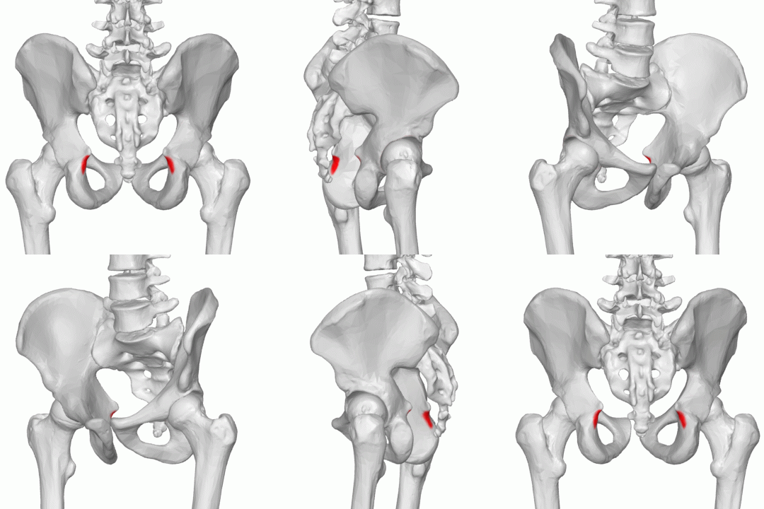
Pubis/ pubic bone
Anterior and inferior to the acetabulum and includes the anteroinferior one-fifth of the acetabulum

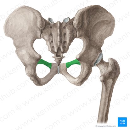
Superior ramus (pubis)
Extending anteriorly and medially from the body of each pubis
Symphysis pubis
midline a cartilage joint between the two superior rami

Inferior ramus ( pubis)
Passes down and posterior from the symphysis pubis to join the ramus of the respective ischium

Obturator foramen
A large opening formed by the ramus and body of each ischium and the pubis.
Largest foramen in the human skeletal system