Unit 4: Lower Limb Spaces, Lymph, Blood Supply, & Joints
 Call Kai
 Learn
 Practice Test
 Spaced Repetition
 Match
 Flashcards
 Knowt Play
1/58

There's no tags or description
Looks like no tags are added yet.
59 Terms
femoral triangle
- on anterior thigh
- base is inguinal lig.
- medial border is adductor longus
- lateral border is sartorius
- floor is adductor longus, pectineus, iliopsoas
- roof is fascia lata with saphenous opening for great saphenous vein

adductor / subsartorial canal
- runs deep to sartorius muscle from apex of femoral trainge to the adductor hiatus
- contains femoral artery and vein, saphenous nerve, nerve to vastus medialis

adductor hiatus
- opening in tendon of insertion for adductor magnus
- permits passage of femoral a/v, saphenous nerve, nerve to vastus medialis

popliteal fossa
- diamond shaped, fat-filled area posterior to knee
- superior borders: tendons of hamstring muscles
- inferior borders: gastrocnemius heads
- floor: popliteal surface of femur, joint capsule of knee
- roof: fascia lata proximally, crural fascia distally

subinguinal hiatus / retroinguinal space
- opening bordered by inguinal lig superiorly and superior pubic ramus inferiorly
- major passageway btwn abdominopelvic cavity and anterior femoral compartment

retroinguinal space contents
- iliopsoas muscle
- femoral sheath
- femoral nerve
- lateral femoral cutaneous nerve
- femoral branch of genitofemoral nerve
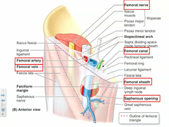
femoral sheath
- funnel-shaped fascial sheath (transversalis fascia) fusing distally with the CT of the femoral artery and vein
- divided by femoral septa into three compartments
- permits vessels to slidde in and out of hiatus during hip flexion and extension
- femoral nerve lies outside of sheath laterally
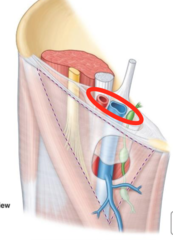
femoral canal
- medial compartment containing fat and lymph nodes
- femoral ring is the proximal entrance to the canal closed by extraperitoneal tissue and parietal peritoneum, bordered medially by lacunar lig.

femoral vein / artery
vein: - occupies the middle compartment of the sheath
- great saphenous vein enters sheath anteriorly to join the femoral vein
artery: occupies lateral compartment of the sheath

obturator artery
- emerges from obturator hiatus and supplies medial compartment muscles, hip joint, and head of femur through ligament of head of femur
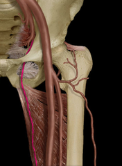
femoral artery
- passes through femoral triangle to enter adductor canal
- ends at adductor hiatus
- normally gives off superficial circumflex iliac artery, external pudendal artery, profunda femoris artery

deep femoral / profunda femoris artery
- arises from femoral a. in triangle, passes btwn pectineus and adductor longus (distally near their insertions)
- gives rise to medial/lateral circumflex femoral arteries
- runs deep on adductor magnus and is the major supply of blood to femoral region
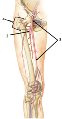
perforating arteries
- four arteries that pierce adductor magnus to supply posterior thigh compartment
- 1st perforating artery contributes to cruciate anastomosis
- 4th perforating artery is terminal branch of deep femoral artery
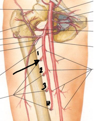
medial circumflex femoral artery
- runs around neck of femur, btwn pectineus and iliopsoas
- major supply to neck and head of femur

lateral circumflex femoral artery
- supplies neck and head of femur
- gives off descending branch which contributes to genicular anastomosis

cruciate anastomosis
- btwn inferior gluteal artery, medial/lateral circumflex femoral arteries, branch of 1st perforating artery

popliteal artery
- cont of femoral a distal to adductor hiatus
- runs through popliteal fossa and divides at inf border of popliteus muscle into ant and post tibial arteries
- gives rise ot 4 genicular arteries

genicular arteries
- superior lateral genicular
- superior medial genicular
- inferior lateral genicular
- inferior medial genicular

genicular anastomosis
- descending branch of lateral femoral circumflex and descending genicular branch of femoral artery
- superior medial & lateral, inferior medial & lateral genicular arteries from popliteal artery
- anterior tibial recurrent artery connects to both inferior genicular arteries

posterior tibial artery
- cont of popliteal artery running btwn superficial and deep muscle groups
- gives off fibular artery laterally which supplies posterior and lateral compartments
- passes deep to flexor retinaculum and divides into medial and lateral plantar arteries

medial / lateral plantar arteries
- form deep plantar arch (mainly from lateral) deep to digital flexor tendons and give rise to plantar metatarsal arteries
- each metarsal artery gives rise to two plantar digital arteries to adjacent digits
- deep plantar arch gives off perforating branches to dorsum of foot

anterior tibial artery
- smaller branch of popliteal artery which passes through a hiatus in the interosseus mem.
- runs on anterior surface of membrane with deep fibular nerve

dorsalis pedis artery
- continuation of anterior tibial artgery distal to talocrural joint
- continues as arcuate artery which gives rise to dorsal metatarsal arteries
- each dorsal metatarsal artery gives rise to two dorsal digital arteries to adjacent digits
- arcuate receives perforating arteries from deep plantar arch

deep veins
- travel with named arteries and have same name
- tibial and fibular veins are usually paired (venae comitantes)
superficial veins
- run in superficial fascia and eventually join deep veins
great saphenous vein
- longest vein in body
- drains medial aspect of foot, crural region, knee, and thigh
- passes through saphenous opening to join femoral vein in the femoral sheath

small saphenous vein
- drains lateral aspect of foot and dorsolateral crual region
- passes through roof of popliteal fossa to join popliteal vein

superficial lymph drainage
- vessels draining anteromedial aspect of lower limb follow course of great saphenous vein
- vessels draining posterolateral aspect of foot and leg follow course of small saphenous vein
deep lymph drainage
- follows the course of the arteries
popliteal nodes
- in popliteal fossa
- receives lymph from deep vessels distal to knee
- receives lymph from vessels along small saphenous vein
- drain into vessels traveling superiorly with femoral a/v

superficial inguinal nodes lower group
- at proximal end of great saphenous vein
- receive all superficial lymph drainage except for that which passes through popliteal nodes
- drain into external iliac nodes
superficial inguinal nodes upper group
- lies just inferior to inguinal lig
- receives lymph from gluteal and perineal regions, and ab wall below umbilicus
- drain into external iliac nodes
deep inguinal nodes
- in and just inferior to femoral canal
- receive all deep drainage and some drainage from superficial inguinal nodes
- drain into external iliac nodes, which drain into lateral aortic/lumbar nodes
pelvic organ lymph drainage
- most drain into internal iliac and latearl aortic nodes
- upper rectum drains into preaortic/visceral nodes along inferior mesenteric artery branches
- gonads drain into lateral aortic nodes along gonadal vessels
hip joint
- synovial, ball & socket joint
- capsule is strong, fibers spiral from ilium to femur and tighten in hip extension
- capsule attaches to neck of femur (intertrochanteric line and crest) and around acetabulum

acetabular labrum
- fibrocartilage which attaches to the edge of acetabulum

transverse acetabular ligament
- continuation of labrum across acetabular notch
- structures entering the acetabulum pass deep to the ligament

iliofemoral, pubofemoral, and ischiofemoral ligs.
- thickenings of capsule attaching respective pelvic bones to femur
- iliofemoral lig (y-shaped) is by far the thickest
- ligaments tighten on extension and limit this movement

ligament of head of femur
- also called round ligament of femur
- carries a branch of obturator artery to fovea capitis of the femur
- supplies blood to head of femur

knee joint
- modified hinge joint
- flexion/extension and slight rotation (lateral rotation at terminal extension)
- joint capsule incorporates patella and patellar lig., attached to menisci except anteriorly
medial collateral lig.
- broad, flat capsular lig which is attached to medial meniscus

lateral collateral lig.
- narrower, rounded band external to capsule, not attached to lateral meniscus
- attached to lateral epicondyle of femur and head of fibula

anterior and posterior cruciate ligaments
- attach to the intercondylar areas on tibia and walls of the intercondylar fossa of the femur
- named for location of the attachement on tibia

medial and lateral meniscus
- fibrocartilaginous structures (c-shaped)
- very thin centrally and taller peripherally
- deepen the concave articular surfaces of tibial plateau

horns of meniscus
- anterior and posterior horns attached to the intercondylar areas of the tibia
menisci and movement
- both attach to joint capsule (except anteriorly), but can move to stay out of the rolling condyle during knee flexion
- medial is less mobile
superior tibiofibular joint
- synovial plane joint btwn head of fibula and facet on lateral tibial condyle

inferior tibiofibular ligament
- syndesmosis class of fibrous joint

talocrural joint
- synovial hinge joint btwn distal tibia and both malleoli proximally and trochlea of talus distally
- permits plantar flexion/dorsiflexion only
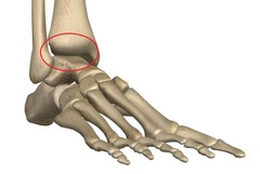
deltoid ligament
- fan-shaped capsular lig on medial aspect
calcaneofibular ligament
- narrow, rounded ligament on lateral aspect

anterior and posterior talofibular ligaments
- anterior is most commonly sprained in inversion injuries
- posterior attaches to fibula in the malleolar fossa

subtalar and transversel tarsal joints
- important in providing for inversion and eversion of foot about an anteroposterior axis
- subtalar : talocalcaneal and talonaviular
- transverse tarsal : calcaneocuboid and talonavicular
plantar calcaneonavicular ligament
- also called spring lig
- most important supporter of longitudinal arch of foot

tarsometarsal joints
- gliding joints between cuboid/cuneiforms and bases of metatarsals I-V
- contribute to transverse arch of the foot

metatarsophalangeal and interphalangeal joints
- similar to hand
- joint #1 has paired sesamoids embedded in flexor hallucis brevis tendon
short plantar ligament
- from anterior calcaneal tuberosity to cuboid

long plantar ligament
- deep fibers run from just anterior to the calcaneal tuberosity to the cuboid
- superficial fibers run to bases of metatarsals II-IV, sometimes V

cervical ligament
- from dorsum of calcaneus to neck of talus
- essentially inside sinus tarsi