15. Nervous System: Meninges, Venous Drainage, Subarachnoid Cisterns, Intercranial Bleeds
 Call Kai
 Learn
 Practice Test
 Spaced Repetition
 Match
 Flashcards
 Knowt Play
1/29

There's no tags or description
Looks like no tags are added yet.
30 Terms
meninges
fibrous membrane protecting brain + spinal cord
membrane layer b/w brain + skull → cushion + protection
has 3 layers (maters):
dura mater → double-layered
arachnoid mater
pia mater
surrounds + protects spinal cord
extends down through vertebral column
within vertebral column → epidural space = real space

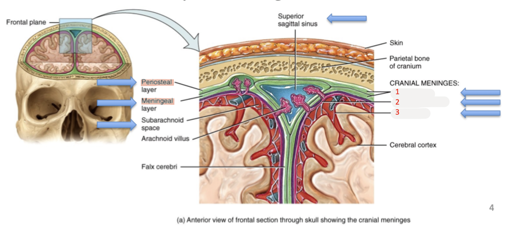
dura mater
(1) outermost layer of meninges
2 layers
periosteal layer → fused to inner surface of skull
meningeal layer
periosteal + meningeal layers fused together EXCEPT for 2 locations → separate
invaginations of meningeal layer deep into brain = fibrous partitions
falx cerebri
tentorium cerebelli

dural venous sinuses
separation areas b/w periosteal + meningeal layers of dura mater = space for drainage of:
venous blood
via cerebral veins accompanying cerebral arteries
CSF
via arachnoid granulations → delivers CSF from subarachnoid space

falx cerebri
(2) fibrous partition from meningeal invagination deep into brain
separates 2 cerebral hemispheres

tentorium cerebelli
(1) fibrous partition from meningeal invagination deep into brain
separates cerebral hemisphere + cerebellum

arachnoid mater
(2) middle layer of meninges
has weblike extensions = “spider”
attach to pia mater → b/w arachnoid mater + pia mater
ensures there is uniform distribution of CSF in subarachnoid space
pia mater
(3) innermost layer of meninges
clings to brain surface → fused to brain

layer spaces
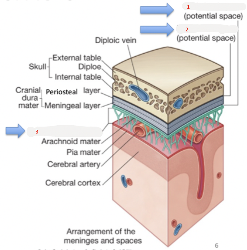
potential or real spaces b/w meningeal layers
potential
nothing within space
when bleeding occurs in this space → becomes real
real
has existing fluid (CSF) within space
3 spaces
epidural/extradural
subdural
subarachnoid

epidural space (extradural space)
(2) space b/w dura mater + skull
potential space
nothing within space
within vertebral column → real space
contains fat, blood vessels, nerves

subdural space
(1) space b/w dura mater + arachnoid mater
potential space
nothing within space

subarachnoid space
(3) space b/w arachnoid mater + pia mater
real space
contains CSF
epidural
anesthesia injection during childbirth or surgeries of pelvis + lower extremity
blocks nerve transmission through nearby spinal nerves
slip needle in b/w spinal processes → inject anesthesia into epidural space

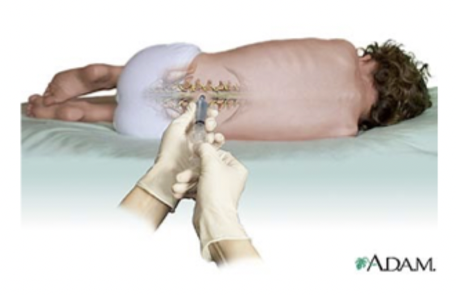
lumbar puncture (spinal tap)
needle insertion into subarachnoid space b/w L3/L4
withdrawal of CSF → test for infection or injury
NOTE: spinal cord ends at L2 but dura + arachnoid mater continues to S2
CSF circulation pathway
CSF produced by choroid plexus of each ventricle
CSF flows through ventricles → into subarachnoid space via median + lateral apertures
some CSF flows through central canal of spinal cord
CSF flows through subarachnoid space
CSF absorbed into dural venous sinuses via arachnoid villi (granulation)
into superior sagittal sinus

subarachnoid cisterns
larger cavities associated w/ certain regions of subarachnoid space
due to irregular cerebral structure shapes → cerebrum, brainstem, cerebellum
5 main:
quadrigeminal
cisterna magna (cerebellomedullary)
schismatic (suprasellar)
interpeduncular (basal)
(pre)pontine

quadrigeminal cistern
cavity in subarachnoid space
b/w corpus callosum selenium + cerebellum → posterior to midbrain
contains pineal gland + great cerebral vein (of Galen)
continuous w/ superior cerebellar cistern
NOTE: connected to interpedunculuar cistern via right + left ambient cisterns (cisterna ambiens)
cisterna magna (cerebellomedullary cistern)
cavity in subarachnoid space
posterior to medulla oblongata
inferior to cerebellum
CSF drains in from 4th ventricle
via 2 lateral apertures = foramina of Luschka + median aperture = foramen of Magendie
chiasmatic cistern (suprasellar cistern)
cavity in subarachnoid space
superior to sella turcica
inferior to hypothalamus
contains optic chiasma + infundibulum
interpedunculuar cistern (basal cistern)
cavity in subarachnoid space
b/w temporal lobes
b/w midbrain cerebral peduncles
contains Circle of Willis
NOTE: connected to quadrigeminal cistern via right + left ambient cisterns (cisterna ambiens)
pontine cistern (prepontine cistern)
cavity in subarachnoid space
anterior to pons
contains basilar artery
internal jugular vein drainage
sigmoid sinus continuous w/ internal jugular vein
vein passes through jugular foramen → exit skull
cavernous sinuses bookend pituitary gland in sella turcica
drain via superior + inferior petrosal sinuses → into sigmoid sinus
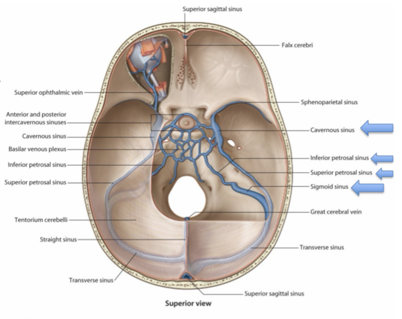
cavernous sinus
venous sinus draining deep cerebral structures + select structures of face via ophthalmic veins
rare → internal carotid artery passing through venous structure
structures passing through/contained within:
internal carotid arteries
pituitary gland
CN III, IV, V1/V2, VI
CN II superior

face danger areas
overlap b/w cerebrum venous drainage + superficial facial venous drainage = potential spread of infection
blood drained by facial vein → into internal jugular vein
also has connections to cavernous sinus via ophthalmic veins
retrograde infection possible
from paranasal sinus, nose, upper teeth → cavernous sinus
affects cerebral structures
eg. meningitis

intercranial bleeds
bleeding b/w spaces in brain
hemorrhage: uncontrolled bleeding - can lead to:
hematoma: collection of blood outside blood vessels, ex. bruise
can cause midline shift + increase in intracranial pressure
4 types:
epidural hemorrhage
subdural hemorrhage
subarachnoid hemorrhage
intracerebral hemorrhage
middle meningeal artery
blood supply to dura mater
branch of maxillary artery enters skull via foramen spinous
maxillary artery = branch of external carotid artery
runs b/w skull + dura mater
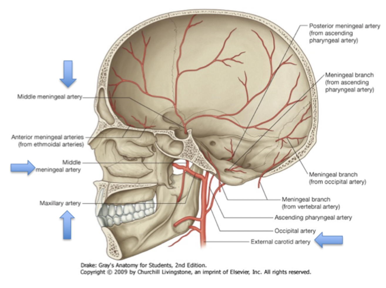
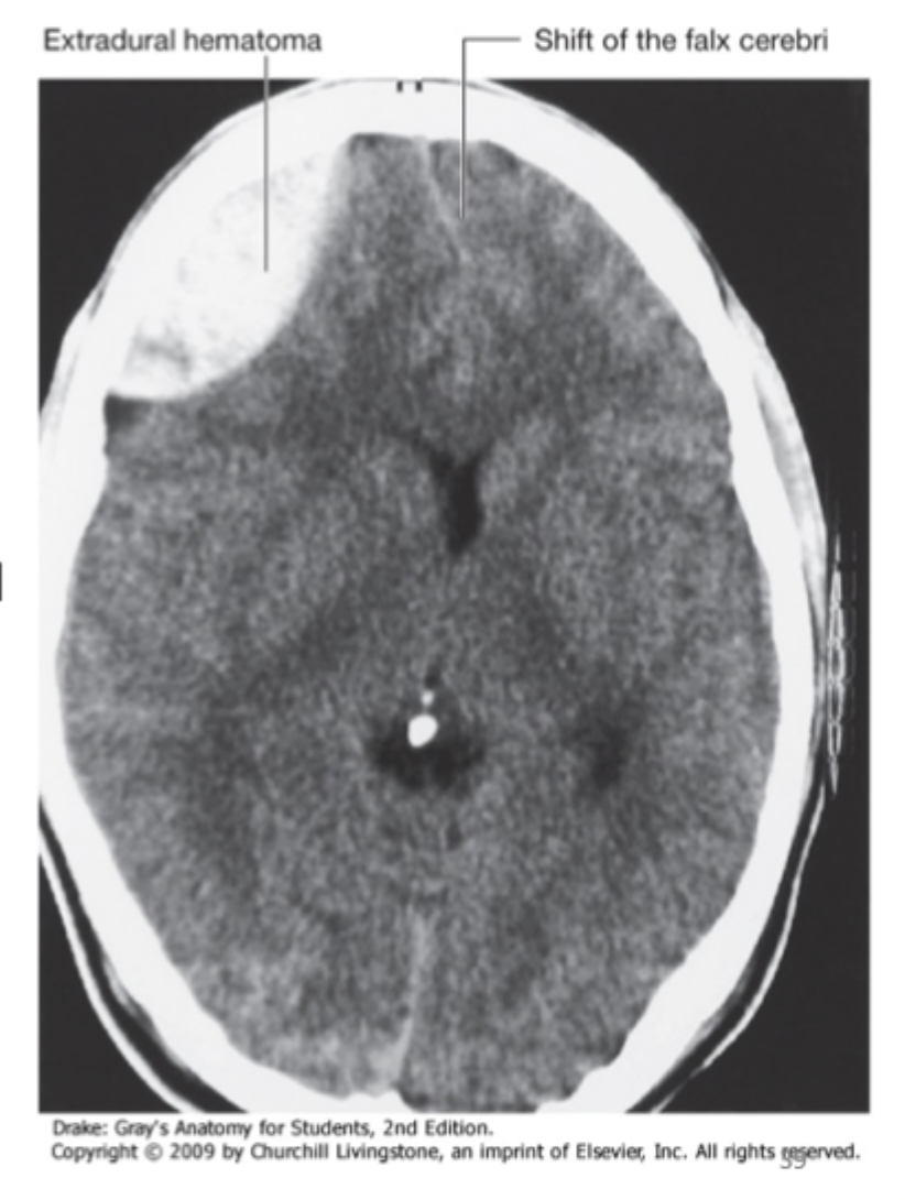
epidural hemorrhage (extradural hemorrhage)
NOTE: epidural space = potential space → dura mater fused to skull
middle meningeal artery branches run within epidural space
vessels tear → blood accumulates b/w dura mater + skulls
resembles convex lens shape
potential epidural space → becomes real space
subdural hemorrhage
NOTE: subdural space = potential space
brain trauma → tear cerebral veins connected w/ superior sagittal sinus
blood accumulates b/w dura mater + arachnoid mater
resembles crescent moon shape
potential subdural space → becomes real space

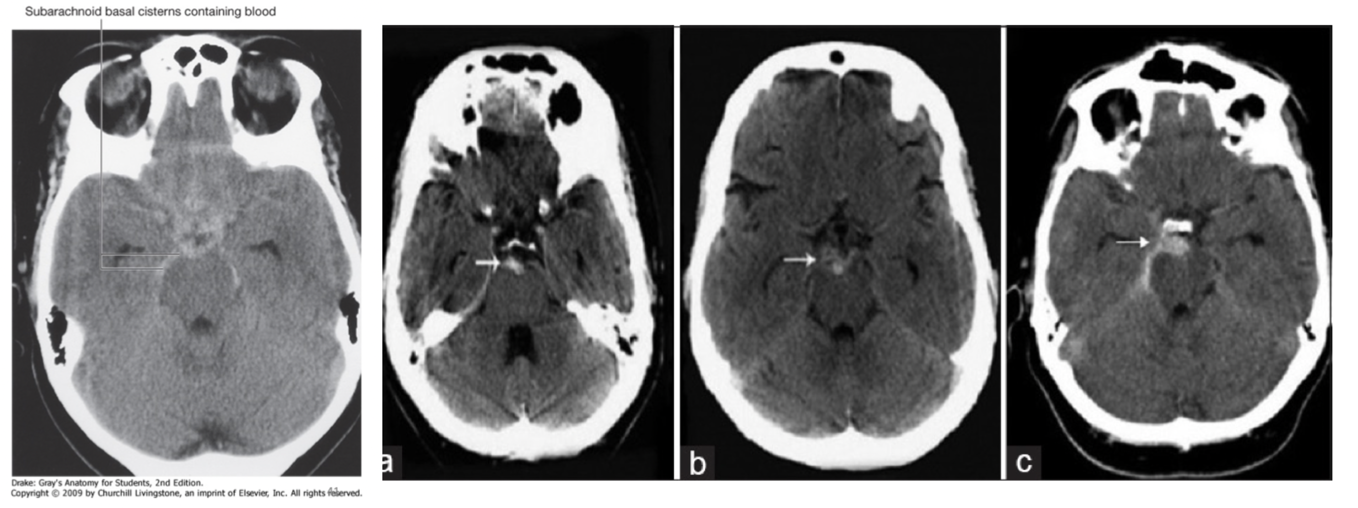
subarachnoid hemorrhage
NOTE: subarachnoid space = real space
cerebral arteries supplying brain run within subarachnoid space
cerebral arteries compromised in brain or cerebrovascular event, ex. ruptured aneurysm
subarachnoid space + cisterns rapidly fill w/ blood
blood CSF from lumbar puncture
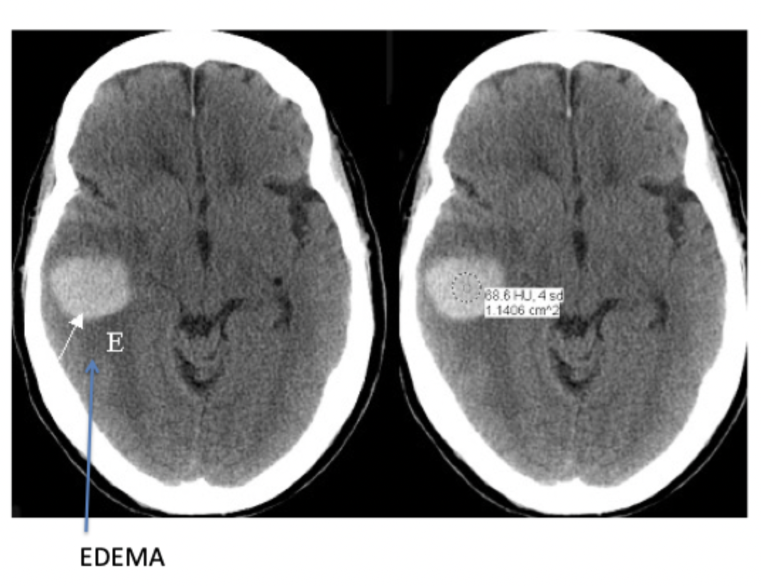
intracerebral hemorrhage (parenchymal hemorrhage)
hemorrhaging vessel in brain from trauma → localized bleeding + surrounding oedema
pressure forces brain downward via foramen magnum
free edge of tentorium cerebelli = sharp → can cut midbrain