Pulmonary Embolism (PE)
 Call Kai
 Learn
 Practice Test
 Spaced Repetition
 Match
 Flashcards
 Knowt Play
1/19

There's no tags or description
Looks like no tags are added yet.
20 Terms
Acute Pulmonary Embolism (PE)
PP: blocks center of vascular lumen→vessel distention
or
occludes a vessel→vessel distention
RF: 2nd m/c sudden death
3rd m/c hospitalized pts death
1/1000 people
DVT (60-80%)
E: virchow’s triad
venous stasis→accumulation of platelets+thrombin in veins
endothelial/venous wall injury
-catheter
-PICC lines
-HTN/smoking
-atherosclerosis
hypercoagulability
-factor v leiden mutation
-antithrombin 3 deficiency
-m/c→antiphospholipid sx
-malignancy
-hormone replacement
-OCPs
CM: asx (50+%)
or
abrupt onset
pleuritic chest pain
dyspnea
unexplained breathlessness
tachycardia
tachypnea
hypoxemia
cough
hemoptysis
PE: jugular venous distention (JVD)
deep vein thrombosis (DVT)
hypotension
fever
crackles
rare:
wheezing
s3
s4
cyanosis
DX: clinical+PE findings+vitals
CTA→strong suspicion
SpO2→normal
CBC→leukocytosis
CMP
BMP
PT
PTT
troponin→elevated
BNP→elevated
ABG
d-dimer
EKG
CXR
well’s score
PERC rule
venous doppler
CTA
pulmonary angiography
v/q scan
echo
P: poor cardiac reserve/large vasoconstriction (massive emboli)→pulmonary HTN+RHF
small embolus→dissolved by fibrinolytic system→lung fxn returns to normal
Chronic Pulmonary Embolism (PE)
PP: blocks artery→reduces artery diameter over 50+%→recanalization in thrombus+arterial web
RF: 2nd m/c sudden death
3rd m/c hospitalized pts death
1/1000 change
DVT (60-80%)
E: virchow’s triad
venous stasis→accumulation of platelets+thrombin in veins
endothelial/venous wall injury
-catheter
-PICC lines
-HTN/smoking
-atherosclerosis
hypercoagulability
-factor v leiden mutation
-antithrombin 3 deficiency
-m/c→antiphospholipid sx
-malignancy
-hormone replacement
-OCPs
CM: asx (50+%)
or
abrupt onset
pleuritic chest pain
dyspnea
unexplained breathlessness
tachycardia
tachypnea
hypoxemia
cough
hemoptysis
PE: jugular venous distention (JVD)
deep vein thrombosis (DVT)
hypotension
fever
crackles
rare:
wheezing
s3
s4
cyanosis
DX: clinical+PE findings+vitals
CTA→strong suspicion
SpO2→normal
CBC→leukocytosis
CMP
BMP
PT
PTT
troponin→elevated
BNP→elevated
ABG
d-dimer
EKG
CXR
well’s score
PERC rule
venous doppler
CTA
pulmonary angiography
v/q scan
echo
P: poor cardiac reserve/large vasoconstriction (massive emboli)→pulmonary HTN+RHF
small embolus→dissolved by fibrinolytic system→lung fxn returns to normal
Pulmonary Embolism PP (General)
emboli→obstruction of pulmonary blood flow→increased physiologic dead space→impaired gas exchange→increased pulmonary resistance+loss of alveolar surfactant→atelectasis (collapsed lung)+shrinking of lung tissue+scarring of lung tissue
Embolus Causes
m/c→thrombus
air
IV infusion
central venous catheters
diving
amniotic fluid entering maternal circulation+pulmonary arteries post-delivery
fat moves to BM post-fracture
foreign bodies
parasites
infxn
tumor cells
DVT Embolism
thrombus that forms in femoral vein
below calf/UE→rare
50-60%→develop PE
-50%+embolic events→asx
-50%+DVT→asx
Provoked vs Unprovoked PE
provoked
RF/E that directly caused PE
-virchow’s triad
unprovoked
PE occurred idiopathically (no RF/E)
-need hypercoagulable dx
-ask FHx+ROS/PE→r/o malignancy
-need cancer screening
increased blood clot RF
PE EKG Findings
70% pts→abnormal
m/c dx pattern→S1Q3T3 pattern
other abnormalities:
sinus tachycardia+non-specific ST+T wave changes (40%)
right ventricular hypertrophy (RVH)
right axis deviation
right bundle branch block (RBBB)
PE CXR Findings
m/c→normal
other findings→engorged pulmonary vasculature:
westermark’s sign
blood vessel collapses→hyperlucency
palla’s sign
enlarged right descending pulmonary artery
hampton’s hump
small PE→wedge-shaped opacity
-edge of the lung
-untxd→pleuritic cor pulmonale
E: hemorrhage
pulmonary infarct
Wells Score
estimates probability of PE
guide for further dx screenings

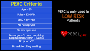
PERC Rule
low probability of PE (wells score u2)
determines if d-dimer is needed→avoids unnecessary testing
all 8 filled→no test
all 8 not filled→d-dimer
D-Dimer
I: low-moderate pretest probability of PE
negative+LOW probability→r/o PE
negative+HIGH probability→no r/o PE
high probability pts→low negative predictive value
high sensitivity+LOW SPECIFICITY (can’t confirm dx)
CT Angiography (CTA)
gold standard+1st line dx→in high-suspicion pts
IV contrast→nephrotoxic
CI: anaphylactic contrast dye allergy
hemodynamic instability
AMI
decompensated CHF
renal impairment
V/Q Scan
CTA not useable/CI to CTA
results equivocal+non-diagnostic
Venous Doppler (Ultrasound)
I: detecting proximal DVT
equivocal for PE V/Q scan→perform
distal/recurrent thrombi+asx pts→less accurate
results:
positive→PE dx
negative→repeat dx x days 1-3+days 7-10→negative→low chance of VTE
Pulmonary Angiography
most accurate
invasive
embolectomy can be done with dx
I: uncertain PE dx
catheter-directed thrombolysis
planned embolectomy
positive→PE dx
negative→r/o PE
Echocardiogram
m/c→normal
I: RV dysfxn
RV strain
pulmonary artery pressure
right chamber emboli
mcconnel’s sign
middle section of RV free wall doesn’t move→right side of heart strain
PE Thrombolytic vs Anticoagulant Mx Tx
thrombolytic
I: hypotension+no high bleeding RF
massive PE+high hypotension RF
high hypotension/hypoxemia death RF
MX: altepase
streptokinase
urokinase
CI: active internal bleeding+stroke x past 2 months
uncontrolled HTN+surgery x past 6 weeks
C: increased major hemorrhagic complications RFs
no difference x 1 week+1 month post-dx compared to heparin/warfarin
P: 24 hrs→resolution of emboli
anticoagulant
I: not tx→secondary prophx
MX:
DOACs
rivaroxaban
apixaban
dabigatran
warfarin “bridge”
heparin→PO warfarin x 6 months
takes ~5-7 days for tx effect
C: doesn’t break down clots→body does that over time
hemorrhage
heparin induced thrombocytopenia (HIT)
PE Duration of Tx
optimal duration→pt specific
considerations
age
RFs
-inherited
-reversibleconsequences of hemorrhage
pt functional status
provoked RFs
trauma/surgery→tx x 3-6 months
proximal leg embolism→tx x 6 months
unprovoked PE episode→tx x extended/indefinite
PE NP Tx
embolectomy
I: massive PE
thombolytic CI
unstable post-thrombolytic tx
bad prognosis
C: high mortality rate→last resort tx
IVC filter
I: anticoagulation CI
recurrent VTE despite OAC
massive PE+high recurrent+fatal embolism RF
ROA: reduce short-term PE incidence
C: 2x increase recurrent DVT RF x first 2 years of insertions
P: clinically safe→add OAC
once stable→remove
PE Prognosis
untxd→30% mortality rate
RV dysfxn→10% mortality rate
recurrent VTE→3% mortality rate (rare)
mortality m/c→progressive RHF
1%→chronic thromboembolic pulmonary HTN develops
dx+tx→favorable
perfusion defects→resolve in most