SS 102 - NERVOUS SYSTEM 1
 Call Kai
 Learn
 Practice Test
 Spaced Repetition
 Match
 Flashcards
 Knowt Play
1/46

There's no tags or description
Looks like no tags are added yet.
Name | Mastery | Learn | Test | Matching | Spaced | Call with Kai | Chat |
|---|
No analytics yet
Send a link to your students to track their progress
47 Terms
2 Main Divisions of the Nervous system
Central nervous system
Peripheral nervous system
Central nervous system
consists of the brain & spinal cord
acts as the boy’s main command center
Peripheral nervous system
comprises the ganglia and nerve
everything except for the spinal cord
serves as the communicstion network connecting the CNS to the rest of the body
Functional divisions of the NS
Sensation
Integration
Response
Sensation (functional division)
external environment
activation of stimulus
change in homeostasis
Integration (functional division)
receives, processes, interprets incoming sensory input
how we give meaning to the stimulus
assosciation
memories, learning, emotions
Response (functional division)
actual reaction
motor
2 divisions: voluntary & involuntary
examples of physical stimuli
hearing - vibrations
touch
taste
Classification of the NS according to type of responses
somatic
autonomic
Somatic response
voluntary movement
Autonomic response
involuntary movement
involves the CNS
external & internal stimulus
example: reflexes
Enteric response
Additional type of NS response
gastro system
controls smooth and glandular tissue
part of the PNS but not dependent on the CNS
2 types of Autonomic reponse
parasympathetic - rest & digest
sympathetic - fight/flight
Synaptic valve
its not a physcial structure but a concept to describe how a synaose functions as a one-way valve
release of neurotransmitters
Basic cells & tissues of the NS
neurons - communication; glue; electrically active
glial cells - supporting role to nervous tissue; cannot funtion w/o neurons
soma
known as the cell body
bulbous part of a neuron that contains the nucleus
Processes/Extensions of a neuron
Axon - fiber connecting neurons to target (exit)
Dendrite - receiver of input to soma
Regions of the CNS
White matter - white due to lipid rich myelin/axons
Grey matter - many cell bodies and dendrites
Respective CNS & PNS Cell body & Axon
CNS cell body - nucleus
CNS axon - tract
PNS cell body - ganglion
PNS- nerve
Polarity
the electrical difference across its cell membrane when it is at rest
Unidirectional flow
dendrites → soma → axon

Parts of a neuron
soma/cell body - contains the nucleus
processes: axon & dendrites
initial segment/axon hillock - tapering of the cell body towards the axon fiber; cytoplasm→ axoplasm
myelin sheath
node of Ranvier - gaps in the myelin covering
axon segment - length of axon between each gap that is covered in myelin
axon terminal - end of the axon; has several branches extending toward the target cell
synaptic end bulb - enlarged ends of terminal branches which make the connection with the target cell at synapse
Types of neurons
unipolar (in vertebraes)/psuedo-unipolar (in humans)
bipolar
multipolar
Unipolar/Psuedo-unipolar Neuron
1 process
exclusively sensory (touch)
cell bodies always found in ganglia
dendrites receive info soemtimes directly from the stimulus
near the spinal cord
Bipolar Neuron
2 processes (1 from each side)
1 is an exon, 1 is a dendrite
olfactory + retina/vision
Multipolar Neuron
2 and more process
everything else that is not unipolar/bipolar
Types of Glial Cells (CNS)Micro
Astrocyte
multiple processes that interact w/ neurons/blood vessels
maintains concentrations of chemicals in the extracellular space, removes excess signaling molecules
contributes to blood-brain barrier
Oligodendrocyte
cell of a few branches
surrounds an exon to insulate it in myelin
Microglia - CNS resident michrophages and can ingest them
Ependymal cells
produce CSF; have cilia at the surface
filters blood in the choroid plexus
ine ventricles in the brsin
look like epithilial cells
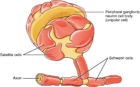
Types of Glial Cells (PNS)
Satellite - found in sensory and autonomic ganglia; performs functions like the astrocytes in the CNS, but do not contribute to the BBB
Schwann - insulates axons with myelin; supplies myelin to one axon segment only
Myelin Sheath in Glial Cells
1-2mm thick;wrapped around multiple times
provided insulation for axons by the glial cells
Lipid-rich
what are the cells that construct and wrap that insulation around the nerves
oligodendrocytes (CNS) and Schwann cells (PNS)
What is the function of the nervous tissue
sensory/sensation input
integration
response/motor input
Enumerate the Sensation/Sensory Input (e.g. via thermoreceptor)
A physical stimulus triggers a sensory cell, changing its electrical voltage. the strength of the voltage change matches the intensity of the stimulus
if the stimulus is strong enough, the voltage change hits a critical point called the threshold. Crossing this threshold generates a non-stop electrical pulse called an action potential
the action potential travels down the length of the nerve fiber (axon)
when the electrical signal reaches the very end of the nerve (synaptic end bulbs), it triggers the release of chemical messengers called neurotransmitters.
this chemical binding creates a new graded potential in the 2nd neuron. if this voltage change is strong enough at the axon hillock, it triggers a fresh action potential to keep the message moving.
the signal travels up to the brain to the thalamus, which is the central post office, sorting sensory data and directing it to the right destination
the thalamus delivers the msg to the cerebral cortex (outer layer). this is the exact moment you register the feeling.
Enumerate the integration stage (nervous tissue function)
Integration is how your brain converts raw info into a meaningful decision
Once sensory signals arrive at the cerebral cortex, neurons analyze what the input actually means
the brain merges this fresh data with past experiences
using this context, the cerebral cortex creates a strategy on how to react
the cortex fires an outgoing motor signal down through the nervous system to your muscles, triggering the physical movement
Enumerate the motor/response output stage (nervous tissue function)
the signal begins in the precentral gyrus (a strip in the frontal cortex). nerve cells called Upper motor neurons, extend axons all the way from the brain into the spinal cord
inside the spinal cord, the upper motor neuron hands off the message to a Lower Motor Neuron. this creates an electrical charge (graded potential) that fires a new signal out of the spinal cord and directly toward the target muscle
the lower motor neuron meets the muscle at a gap called the neuromuscular junction. When the electrical signal reaches this end point, the nerve releases a chemical messenger called acetylcholine.
Acetylcholine attaches to receptors on the muscle, causing a spike in voltage called an end plate potential (a muscle action potential). This electrical signal spreads through the muscle cell, triggering the muscle fibers to contract and generate movement.
what is a graded potential
a temporary, localized change in a neuron's membrane potential (voltage) that varies in size
Associated with dendrites of a neuron
Can either be depolarizing or hyperpolarizing (changes above or beyond -70 mV)
How Do graded potentials Respond to Stimuli?
A small stimulus causes a small depolarization, whereas a larger stimulus causes a larger depolarization
A longer-lasting stimulus causes a longer-lasting depolarization, but does not increase the overall strength of depolarization
A large stimulus that depolarizes the membrane above threshold (-55 mV) may trigger an action potential in a postsynaptic neuron.
Some stimuli cause hyperpolarization instead of depolarization, depending on which specific ion channels are activated in the membrane.
What Are Generator Potentials and Receptor Potentials?
Generator Potential: A graded potential that develops in the dendrites of unipolar sensory neurons and directly influences the generation of an action potential in the axon.
Receptor Potential: A graded potential in specialized sensory cells (such as taste cells or retinal photoreceptors) that results in the release of neurotransmitters at synapses with sensory neurons.
What Are Postsynaptic Potentials (EPSPs and IPSPs)?
Postsynaptic Potential (PSP): A graded potential occurring in the dendrites of a neuron receiving synaptic signals from other cells.
Excitatory Postsynaptic Potential (EPSP): A depolarizing PSP that moves the membrane potential closer to threshold.
Inhibitory Postsynaptic Potential (IPSP): A hyperpolarizing PSP that moves the membrane potential further away from threshold.
What Is Summation and How Does It Function at the Axon Hillock?
Summation is the process of adding multiple graded potentials together to reach the threshold required to fire an action potential
summation takes place at the axon hillock (initial segment of the neuron)
2 types
Spatial Summation: The association and combined effect of signals arriving simultaneously from multiple different input neurons/synapses.
Temporal Summation: The combined effect of multiple successive action potentials firing rapidly from a single input cell over time.

Explain the picture
Net Voltage Impact: Multiple EPSPs add up to produce a large depolarization (Point A), while a combination of EPSPs and IPSPs balances out to determine the final membrane potential (Point B).
How Do Chemical Synapses and Electrical Synapses Differ?
Chemical Synapse: A junction where a chemical signal (neurotransmitter) is released from one cell and diffuses across a gap to affect another cell.
Electrical Synapse: A junction with a direct physical connection between two cells allowing ions to pass directly, so depolarization of one cell immediately depolarizes the joined cell.
What Are the Key Structural Components of a Chemical Synapse?
Presynaptic Element: The motor neuron's axon terminals.
Neurotransmitter: Acetylcholine (ACh) stored inside presynaptic vesicles.
Synaptic Cleft: The microscopic fluid-filled space between cells through which neurotransmitters diffuse.
Receptor Proteins: Nicotinic acetylcholine receptors located on the postsynaptic side.
Postsynaptic Element: The sarcolemma (cell membrane) of the target muscle cell at a neuromuscular junction.
Neurotransmitter Elimination: Acetylcholinesterase enzyme breaks down acetylcholine to terminate the signal.
What Are the Step-by-Step Mechanisms of Neurotransmitter Release and Clearance?
An action potential reaching the synaptic end bulb opens voltage-gated Ca^2+ channels.
Increased Ca^2+ concentration inside the end bulb causes Ca^2+ ions to associate with proteins on the outer surface of neurotransmitter vesicles.
Exocytosis: Vesicles merge with the presynaptic membrane, releasing neurotransmitters into the synaptic cleft via exocytosis.
Receptor Binding: Neurotransmitters diffuse across the synaptic cleft and bind to specific receptor proteins on the postsynaptic membrane via a lock-and-key mechanism.
What Are the Four Major Neurotransmitter Systems
cholinergic System
amino acid System
Biogenic Amine System
Neuropeptide System
explain the Cholinergic System (Neurotransmitters, receptors, elimination)
Neurotransmitter: Acetylcholine (ACh).
Receptors: Nicotinic receptors (bind nicotine; ionotropic cation channels causing depolarization) and Muscarinic receptors (bind muscarine; metabotropic receptors causing either depolarization or hyperpolarization depending on subtype).
Elimination: Enzymatic degradation by acetylcholinesterase.
explain the amino acid System (Neurotransmitters, receptors, elimination, postsynaptic effects)
Neurotransmitters: Glutamate (Glu), Glycine (Gly), and GABA (γ-aminobutyric acid).
Receptors: Glutamate receptors, Glycine receptors, and GABA receptors.
Elimination: Reuptake by neurons or glial cells for repackaging.
Postsynaptic Effects: Glutamate is excitatory (causes depolarization); Glycine and GABA are inhibitory (cause hyperpolarization).
Explain the Biogenic Amine System (Neurotransmitters, receptors, elimination, postsynaptic effects)
Neurotransmitters: Amino acids without carboxyl groups, including Serotonin (5-HT), Dopamine, Norepinephrine (NE), and Epinephrine.
Receptors: 5-HT receptors, D1 and D2 receptors, alpha-adrenergic receptors, and beta-adrenergic receptors.
Elimination: Reuptake by neurons.
Postsynaptic Effects: Mixed effects that can influence gene transcription or other neuronal processes; for example, Dopamine D1 receptors cause depolarization (excitatory) while D2 receptors cause hyperpolarization (inhibitory).
Explain the Neuropeptide System (Neurotransmitters, characteristics, receptors, elimination, postsynaptic effects)
Neurotransmitters: Chains of amino acids such as Met-enkephalin, beta-endorphin, Vasoactive Intestinal Peptide (VIP), and Substance P.
Characteristics: Frequently released alongside another transmitter and often act as hormones.
Receptors: Specific peptide receptors (too numerous to list).
Elimination: Enzymatic degradation by peptidases.
Postsynaptic Effects: Causes depolarization or hyperpolarization depending on the specific receptor.