Protein Techniques Oral
 Call Kai
 Learn
 Practice Test
 Spaced Repetition
 Match
 Flashcards
 Knowt Play
1/54

There's no tags or description
Looks like no tags are added yet.
55 Terms

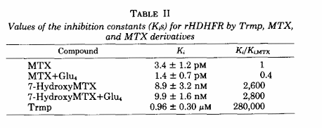
1. The abstract states: "7-HydroxyMTX, another major metabolite of MTX, is a weak inhibitor of rHDHFR (Ki = 8.9 nM), and a polyglutamate form of this metabolite is an equally weak inhibitor (Ki = 9.9 nM), so that the addition of glutamate residues to MTX or 7-hydroxyMTX has little effect on their binding."
Which of these is a weaker inhibitor, how do you know?
7-HydroxyMTX polyglutamate form is a much weaker inhibitors than MTX. We can determine this by comparing their Ki values. A higher Ki means weaker inhibition because more inhibitor is required to inhibit the enzyme.

1. The abstract states: "7-HydroxyMTX, another major metabolite of MTX, is a weak inhibitor of rHDHFR (Ki = 8.9 nM), and a polyglutamate form of this metabolite is an equally weak inhibitor (Ki = 9.9 nM), so that the addition of glutamate residues to MTX or 7-hydroxyMTX has little effect on their binding."
B. How do these glutamate metabolites compare to MTX without the changes?
MTX and MTX+Glu4 are both strong inhibitors of human DHFR. The polyglutamate form has a slightly lower Ki than MTX, approximately 1.4 µM compared with 3.4 µM, suggesting a modest increase in inhibitory strength.
However, 7-hydroxyMTX is much weaker than MTX, and adding glutamate residues does not significantly improve its inhibitory activity. This demonstrates that polyglutamation does not automatically make every MTX metabolite a stronger DHFR inhibitor.
1. The abstract states: "7-HydroxyMTX, another major metabolite of MTX, is a weak inhibitor of rHDHFR (Ki = 8.9 nM), and a polyglutamate form of this metabolite is an equally weak inhibitor (Ki = 9.9 nM), so that the addition of glutamate residues to MTX or 7-hydroxyMTX has little effect on their binding." (Below would be three different possible questions.)
Why do the scientists care about testing these metabolites?
The scientists wanted to understand how MTX metabolism affects inhibition of human DHFR. MTX is converted into metabolites, including 7-hydroxyMTX, and these metabolites may accumulate in the body.
They wanted to determine whether the metabolites could also inhibit DHFR and whether adding glutamate residues changed their inhibitory strength. This helps explain how the original drug and its metabolites contribute to the overall biological and therapeutic effects of MTX.

2. Looking at the data presented in table II, particularly the last column. What is the purpose of presenting the data in this way (dividing the Ki's by MTX's Ki). What does this last column tell you?
The purpose of the last column is to compare the inhibitory strength of each compound relative to MTX, which is used as the reference inhibitor.
The researchers divide each compound's Ki by the Ki of MTX. This makes it easier to see how much stronger or weaker each inhibitor is without having to compare the raw Ki values individually.
A ratio of 1 means the compound has the same Ki as MTX. A ratio below 1 means it has a lower Ki and is a stronger inhibitor. A ratio above 1 means it has a higher Ki and is a weaker inhibitor.
For example, MTX+Glu4 has a relative ratio of approximately 0.4, meaning its Ki is lower than MTX's. In contrast, 7-hydroxyMTX has a ratio of approximately 2,600, meaning its Ki is approximately 2,600 times higher than MTX's. Therefore, it is a much weaker inhibitor.

2. Looking at the data presented in table II, particularly the last column. What is the purpose of presenting the data in this way (dividing the Ki's by MTX's Ki).
The purpose of the last column is to compare the inhibitory strength of each compound relative to MTX, which is used as the reference inhibitor.
The researchers divide each compound's Ki by the Ki of MTX. This makes it easier to see how much stronger or weaker each inhibitor is without having to compare the raw Ki values individually.
2. Looking at the data presented in table II, particularly the last column. What does this last column tell you?
A ratio of 1 means the compound has the same Ki as MTX. A ratio below 1 means it has a lower Ki and is a stronger inhibitor. A ratio above 1 means it has a higher Ki and is a weaker inhibitor.
For example, MTX+Glu4 has a relative ratio of approximately 0.4, meaning its Ki is lower than MTX's. In contrast, 7-hydroxyMTX has a ratio of approximately 2,600, meaning its Ki is approximately 2,600 times higher than MTX's. Therefore, it is a much weaker inhibitor.
3. Figure 2: Describe the different experimental set-up in panels A and B and how this may have lead to the different results seen in the graphs shown.
The main differences are the enzyme concentration, MTX concentration range, and order of addition.
Panel A uses a higher enzyme concentration, so more MTX can become bound to the enzyme. Since MTX is a tight-binding inhibitor, the free inhibitor concentration may be significantly lower than the total inhibitor concentration.
Panel B uses less enzyme and preincubates the enzyme with MTX before adding DHF. This allows MTX to bind and begin forming the tighter enzyme-inhibitor complex before the substrate is introduced.
These differences can affect the observed inhibition curves. However, the researchers obtained similar Ki values in the two panels, approximately 3.4 µM for Panel A and 3.6 µM for Panel B. This suggests their results were reasonably consistent despite the different experimental conditions.

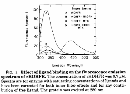
Q1. What is the purpose of Figure 1?
The purpose is to demonstrate how binding of different ligands changes the fluorescence of DHFR. The researchers used these fluorescence changes to study interactions between the enzyme, NADPH, MTX, and trimethoprim.

Q2. What happens to the fluorescence when NADPH binds DHFR?
The intrinsic protein fluorescence decreases, and a second peak appears around 435 nm due to energy transfer from the protein to NADPH.

Q4. Why did the researchers excite the protein at 280 nm?
They used 280 nm excitation to measure intrinsic protein fluorescence, which allowed them to monitor changes associated with ligand binding.

Q1. What is the overall purpose of Figure 2?
The purpose is to determine the inhibition constant, Ki, for MTX binding to recombinant human DHFR under different experimental conditions.

Q2. Describe the experimental setup in Panel A.
The enzyme was preincubated with NADPH. Then DHF and MTX were added together. The researchers used a relatively high enzyme concentration and varied MTX from 0 to 15 µM.

Q3. Describe the experimental setup in Panel B.
The enzyme was preincubated with both NADPH and MTX. DHF was added afterward. This allowed MTX to bind to the enzyme before the substrate was introduced.

What is the purpose of Panel C?
Panel C examines how changing DHF concentration affects enzyme activity in the presence of a fixed concentration of MTX. It demonstrates how substrate concentration and competitive inhibition influence the reaction rate.

Q1. What is the purpose of Figure 3?
The purpose is to examine how inhibition develops over time and identify the different kinetic steps involved in MTX binding to human DHFR

What happens during Phase 3? fig 3
The initially formed enzyme-NADPH-MTX complex undergoes isomerization. This creates a more stable complex in which MTX binds more tightly, reducing the amount of enzyme available for catalysis.

Q6. What causes Phase 4? fig 3
Phase 4 is a slower phase associated primarily with product inhibition by NADP+ and depletion of DHF. It was not depicted in Figure 3.

Which phase is most directly associated with the conformational change? figure 3
Phase 3, because it represents isomerization of the initial enzyme-inhibitor complex.

Q15. What is the overall conclusion from Figure 3?
MTX inhibition is not a single-step process. It involves initial binding followed by isomerization, which significantly strengthens inhibitor binding.

What is the purpose of Table I?
It summarizes fluorescence changes associated with ligand binding and provides dissociation constants for selected enzyme-ligand complexes.

Q3. Which inhibitor binds more tightly to human DHFR: MTX or trimethoprim? table 1
MTX binds more tightly. Its Kd was too small to accurately measure using the fluorescence method, while trimethoprim had a Kd of approximately 480 nM.

Q5. Why did the researchers measure fluorescence at 330 nm and 435 nm? Table 1
The 330 nm signal represents intrinsic protein fluorescence, while the 435 nm signal reflects energy transfer to NADPH.

Q10. Why is the NADPH-bound enzyme important in this table? table 1
The researchers wanted to understand inhibitor binding to the enzyme-cofactor complex, which is relevant to the enzyme's catalytic cycle.

Q1. Which compound is the strongest inhibitor in Table II?
MTX+Glu4 has the lowest Ki in the table, approximately 1.4 µM, so it is the strongest by the reported Ki values. MTX is also a strong inhibitor, with a Ki of 3.4 µM.

Q2. Which compound is the weakest inhibitor? table ii
Trimethoprim is the weakest inhibitor of human DHFR in this table because it has the highest Ki and a relative ratio of approximately 280,000.

Q6. What does a ratio of 2,600 mean? Table ii
It means the inhibitor has a Ki approximately 2,600 times higher than MTX. It is therefore much weaker.

Q8. Why did the researchers test 7-hydroxyMTX? Table ii
7-hydroxyMTX is a major metabolite of MTX that can accumulate in patients receiving high-dose MTX. The researchers wanted to determine whether this metabolite could also inhibit human DHFR.

Q9. Why did they test a polyglutamate form of 7-hydroxyMTX? table 2
Previous studies suggested that adding glutamate residues might make 7-hydroxyMTX a stronger inhibitor. The researchers tested this possibility and found that the polyglutamate form remained weak.

Q15. What is the main conclusion of Table II?
MTX and its polyglutamate form are strong inhibitors, while 7-hydroxyMTX and trimethoprim are much weaker inhibitors of human DHFR. Polyglutamation has only a modest effect on MTX inhibition and little effect on 7-hydroxyMTX inhibition.
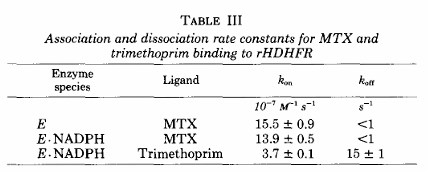
Q1. What is the purpose of Table III?
It compares the association and dissociation rates of MTX and trimethoprim binding to recombinant human DHFR.

Q4. Which inhibitor has the faster association rate? table iii
MTX has a faster association rate than trimethoprim, although the difference is relatively small.

Q5. Which inhibitor dissociates more rapidly? table iii
Trimethoprim dissociates much more rapidly, with a koff of approximately 15 s⁻¹.

Q11. What would happen if an inhibitor had a high kon but also a high koff? table iii
It would bind quickly but also dissociate quickly. It may not remain bound long enough to produce strong sustained inhibition.

Q15. What is the major conclusion from Table III?
MTX and trimethoprim have relatively similar association rates compared with their much larger difference in dissociation behavior. The slow release of MTX contributes to its stronger binding.

Q1. What is the purpose of Table IV?
It describes the kinetic steps involved in MTX and MTX+Glu4 binding to human DHFR, including initial association, dissociation, and the subsequent isomerization step.

Q5. Which inhibitor undergoes faster isomerization? table iV
MTX has a slightly higher kiso of approximately 0.4 s⁻¹ compared with 0.3 s⁻¹ for MTX+Glu4.

Q7. Why is the isomerization step important? Table Iv
It significantly increases the stability of the inhibitor-enzyme complex. This means MTX becomes a much stronger inhibitor after the conformational change.

Q9. How does MTX compare with MTX+Glu4 in Table IV?
Both undergo similar binding and isomerization processes. MTX+Glu4 has a somewhat faster initial association rate, but both show substantial improvements in binding after isomerization.

Q10. Why does Table IV provide more information than Table III?
Table III focuses on association and dissociation. Table IV provides additional information about the conformational change that occurs after initial binding.

Q14. What does Table IV tell us about the effect of glutamate residues?
The polyglutamate form has somewhat different kinetic constants, but it undergoes the same general two-step binding mechanism. The additional glutamate residues do not fundamentally change the process.

Q17. What is the most important conclusion from Table IV?
MTX binding involves an initial association followed by isomerization. This second step strengthens binding approximately 60-fold and contributes significantly to overall inhibition.

Q1. What is the purpose of Table V?
It compares observed and calculated rates of inhibitor release from recombinant human DHFR complexes.

Q3. Why did the researchers use DAMP? table v
DAMP is a competing ligand that can displace MTX or MTX+Glu4 from the enzyme complex. The researchers used it to study inhibitor release.

Q11. Why is Table V important when considered alongside Table IV?
Table IV proposes the kinetic steps involved in binding and isomerization. Table V provides an independent way to examine release and test whether the proposed model is consistent with the experimental data.

Q12. What is the main conclusion from Table V?
The agreement between observed and calculated release rates supports a model in which MTX and MTX+Glu4 undergo one isomerization after binding, while trimethoprim does not undergo the same isomerization.

Q1. What is the purpose of Table VI?
The purpose is to compare the kinetic properties and inhibitor sensitivity of bacterial DHFR with recombinant human DHFR.

Q3. Which enzyme is more sensitive to trimethoprim? table vi
E. coli DHFR is much more sensitive to trimethoprim because its Ki is substantially lower than that of human DHFR.

Q5. What does a 12,000-fold difference in Ki mean? table vi
It means the Ki for bacterial DHFR is approximately 12,000 times lower than for human DHFR, indicating much stronger inhibition of the bacterial enzyme.

Q14. What is the main conclusion from Table VI?
Human and bacterial DHFR have similar overall functions but important differences in inhibitor binding and kinetics. These differences are especially important for trimethoprim and support the need to study human DHFR directly.

Q9. What does Table VI tell us about trimethoprim and antibiotic selectivity?
Trimethoprim preferentially inhibits bacterial DHFR over human DHFR. This allows it to interfere with bacterial folate metabolism while having substantially weaker inhibition of the human enzyme.

Q6. Does MTX inhibit bacterial and human DHFR equally?
No. The table shows differences in their Ki values and kinetic behavior. MTX has an overall Ki of approximately 1.0 µM for E. coli DHFR and 3.4 µM for human DHFR.
Q5. Compare Figure 2 and Figure 3.
Figure 2 investigates how MTX concentration, enzyme concentration, substrate concentration, and order of addition affect inhibition. Figure 3 examines the different kinetic phases through which inhibition develops over time, including the isomerization step.
Why use MTX as the reference?
It is the original drug being compared with its metabolites and derivatives.
Q7. Compare Table IV and Table V.
Table IV describes the kinetic model for inhibitor binding and isomerization. Table V tests the model by comparing predicted and experimentally observed inhibitor release rates.
Q6. Compare Table III and Table IV.
Table III focuses on association and dissociation rates. Table IV adds the isomerization step and shows how it contributes to the final binding strength of MTX.