Unit 8: Advanced Imaging
 Call Kai
 Learn
 Practice Test
 Spaced Repetition
 Match
 Flashcards
 Knowt Play
1/17

Earn XP
Description and Tags
Flashcards covering key advanced ultrasound imaging techniques, processing stages, advantages, and operational principles.
18 Terms

B-Color
post-processing, that takes information from a grayscale image and assigns color shades using different color maps to assist the human eye in discerning subtle details
improves contrast resolution (known as Tissue Color on GE systems and Chroma Map on Philips systems).
Human Gray Scale Perception Limit
Threshold wherein human eyes can perceive fewer than less than 64 shades of gray at any given point in time.
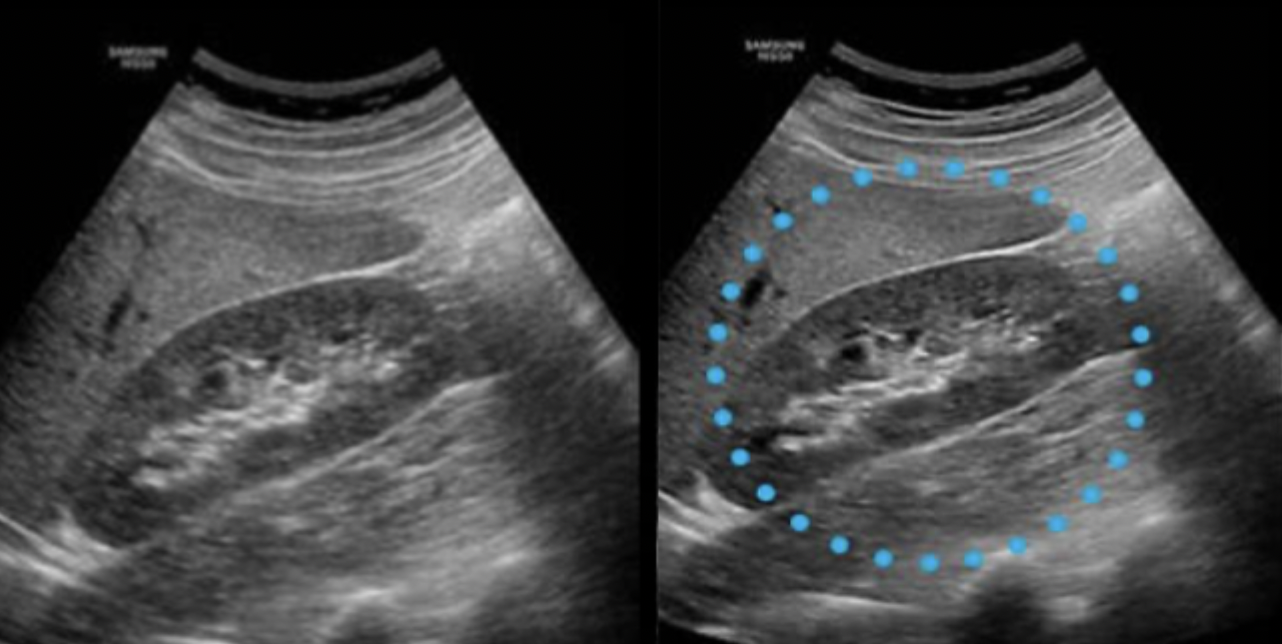
Edge Enhancement
pre-processing feature that sharpens boundary interfaces in order to improve contrast resolution
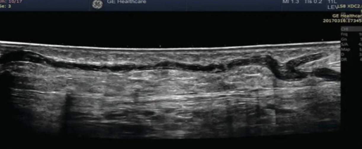
Panoramic Imaging
pre-processing method, operated by sliding the transducer across a scan plane, adding new data continuously as the transducer moves to create an expanded or wider field of view (FOV).
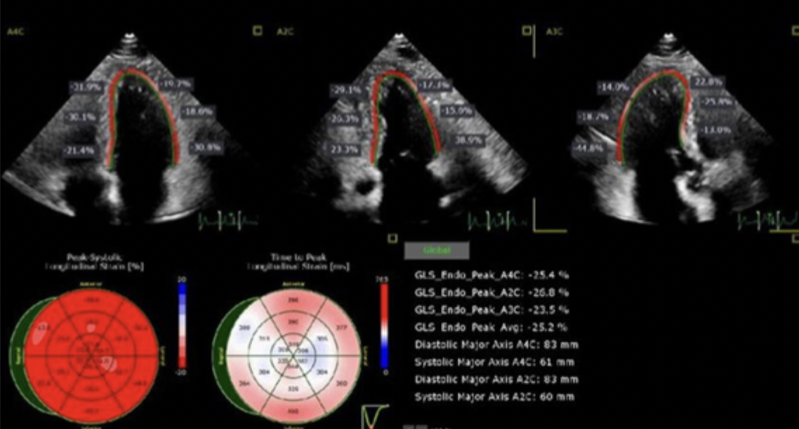
Cardiac Strain Imaging
An advanced cardiac imaging method that measures changes in tissue length (strain) and strain rate to assess the effectiveness of myocardial contraction, facilitating the early detection of heart failure.
Speckle Tracking
A component of cardiac strain imaging that tracks acoustic speckle tissue motion over time to document the physical movement of specific regions of the myocardium.
Elastography
An ultrasound imaging modality that evaluates and assesses the relative stiffness or elasticity of tissues.
Static (Strain) Elastography
A form of elastography that assesses tissue stiffness through manual transducer compression or palpation.
Shear Wave Elastography
A form of elastography that evaluates tissue stiffness utilizing mechanical vibration or acoustic radiation force impulse (ARFI).

Spatial Compounding
A pre-processing technique requiring a phased array transducer that steers and averages multiple scan frames from different angles to improve spatial resolution and reduce artifacts, while degrading temporal resolution.
Temporal Compounding
pre-processing technique, also called persistence, temporal averaging, or color sensitivity, acquires frames at different times and layers them over one another; it increases SNR and contrast resolution but degrades temporal resolution.
Frequency Compounding
pre-processing technique (also called frequency fusion) that averages images created from different frequencies within each pulse to improve spatial and contrast resolution and reduce speckle artifact without degrading temporal resolution.
Dynamic Frequency Tuning
A technique (also known as frequency tuning or sliding receive filters) requiring a wide bandwidth during transmit that adjusts receive filters to layer frequencies across image depth: high frequencies for the near field, medium for the mid field, and low for the far field.

Coded Excitation
A beamformer feature that transmits complex, elongated voltage pulses repeating a specific pattern to impart a unique signature on the sound beam; it improves axial resolution, spatial resolution, contrast resolution, penetration, and SNR, while reducing acoustic speckle artifact.
2D Array Transducer
A transducer comprising a checkerboard configuration of approximately 2,000 to 9,000 elements used to collect ultrasound data across three dimensions simultaneously.
Voxel
A volume element representing a value on a regular grid in three-dimensional space, gathered across three planes in 3D ultrasound imaging to measure volume.
4D Imaging
Three-dimensional (3D) ultrasound imaging acquired and presented continuously during real-time scanning.
3D Rendering
A computer-generated post-processing technique (also called 3D reconstruction or 3D reformation) that reconstructs thin image slices into realistic surface images with tissue overlays to optimize anatomical and pathological visualization.