PNB UNIT 4 (AXIAL SKELETON + APPENDICULAR MUSCULOSKELETON) STUDY SET
 Call Kai
 Learn
Learn Practice Test
 Spaced Repetition
 Match
Match Flashcards
 Knowt Play
1/51

There's no tags or description
Looks like no tags are added yet.
Name | Mastery | Learn | Test | Matching | Spaced | Call with Kai | Chat |
|---|
No analytics yet
Send a link to your students to track their progress
52 Terms
The axial skeleton contains:
The skull, thoracic, and vertebral columns
TMJ stands for? What bones does it involve?
Temporomandibular joint, the temporal bone (mandibular fossa) and mandible
Where is the temporomandibular joint located?
Between the temporal bone and ear canal.

What mechanism / joint movement is the TMJ?
Combined hinge and gliding.
What are the muscles that move the TMJ? What do they do and where are they located?
1) Digastricus (opens the jaw), in either side of the neck
2) Temporalis and masseters (closes jaw), sides of head and cheeks
3) Medial and lateral pterygoids
Describe the spinal vertebrae:
26 bones total: 7 cervical, 12 thoracic, 5 lumbar, sacrum (5 fused), coccyx (4 fused)

Function of the vertebral column:
Support the weight of trunk and head, protect the spinal cord
What are the movements of the vertebral column?
1) Flexions and Extension
2) Lateral Flexion
3) Circumduction
4) Rotation
Between vertebrae, there are slightly moveable symphysis joints. How would you functionally and structurally classify these?
Cartilaginous, amphiarthrosis
What connects vertebral bodies?
Intervertebral discs
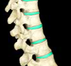
Intervertebral discs are made of 2 parts...
1) Outer annulus fibrosus
2) Inner nucleus pulposus: Soft, elastic gel which allows disc to absorb shock.

What are articular facets?
Slightly moveable gliding joints that allow adjacent vertebrae to articulate with each other
Draw and label a vertebra:
--->

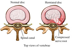
Name and describe a disc complication:
Herniated "slipped" disc: A tear in the annulus fibrosus that allows the protrusion of the nucleus pulposus --> presses on the spinal nerve causing pain. It can go away with time or with surgical intervention
Name and describe our atypical vertebrae:
C1 (atlas): Supports our skull and has no body
C2 (axis): Body has tooth-like projections "dens", contains a pivot joint to turn our head in a "no" motion
Where / what is the atlanto-axial joint?
Articulation between C1 and C2
Where / what is the atlanto-occipital joint?
Articulation: The superior facets of the atlas connect to the occipital condyles (x2) on the underside of the skull. Known as a double condyloid joint. Allows for extended ROM of flexion/extension of the head and neck.
Name the important muscles (and their location + function) of the axial skeleton (head, neck, thorax):
1) Sternocleidomastoid: (From the lower back of the skull to the clavicle) tilts the skull
2) Rectus abdominus: (6 pack) flexes the vertebral column
3) Erector spinae: (Behind the spine) extends the vertebral column
What is the thorax? What is its main function?
Contains the sternum, ribs, and T vertebrae. Protects the heart, lungs, and allows for breath and ventilation.
What is the Costovertebral joint? Where is it located and what kind of joint is it?
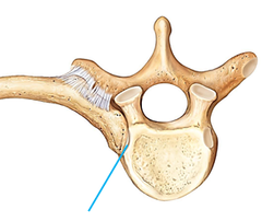
Where the head of the rib (inner end) connects to the vertebral body, and the tubercule of the rib connects to the transverse process of the vertebrae. These are gliding joints (synovial)
What are the two types of postural curves, and which sections of the vertebrae are in which?
Primary curves: Curves that protrude backwards (thoracic + sacral vert)
Secondary curves: Curves that push the body forward (cervical + lumbar vert)
Which kind of postural curves are newborns born with?
Primary, meaning our T + S curves are present at birth. C + L occur when babies learn to hold their head and sit on their own.
Name 3 irregular postures:
Kyphosis, lordosis, scoliosis
Review: What is Kyphosis?
Exaggerated thoracic curvature. Common in older women, or those with osteoporosis.

Lordosis:
Exaggerated lumbar curve. Common in pregnant people

Scoliosis:
A lateral curvature of the spine, usually in a "c" or "s" shape

What are the 3 muscle roles?
Agonist: Directly performs desired movement
Antagonist: Opposes agonist movement, performs opposite action
Synergist: "Helper" recruited for extra strength
What are the 4 sections of the appendicular musculoskeleton?
Pectoral girdle, elbow / wrist, pelvic girdle, the knee
What is the main function of the pectoral girdle, and what are the three bones that it is comprised of?
Connects the arms to the axial skeleton, made up of the clavicle, scapula, and upper sternum (manubrium)
(Pectoral girdle) Sternoclavicular joint:
Connects the Manubrium to the proximal end of the clavicle. (gliding joint)

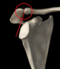
(Pectoral girdle) Acromioclavicular joint:
Connects the distal end of the clavicle to the acromion process of the scapula. (gliding joint)
(Pectoral girdle) Glenohumeral joint:
Connects the head of the humerus to the glenoid cavity on the scapula. The most freely moveable joint (ball-and-socket)

Name all of the important pectoral girdle muscles, where they are located, and what actions they perform. (hint: there are 7, and these muscles mainly move the shoulder)
1) trapezius: upper back muscle (upper region ELEVATES, lower region DEPRESSES, middle region RETRACTS)
2) deltoid: back of the shoulders (ABDUCTS, EXTENDS)
3) latissimus dorsi: lower back muscle (ADDUCT, EXTENDS)
4. supraspinatus: above the scapular spine (ABDUCTS)
5. infraspinatus: below the scapular spine (EXTERNAL ROTATION)
6. teres minor: below the infraspinatus (EXTERNAL ROTATION)
7. triceps brachii: back of the upper arm (EXTENDS)
What 4 important bones are the elbow and wrist comprised of?
1. Humerus
2. Radius
3. Ulna
4. Carpals
What three bones form the elbow joint? What is the elbow joint called and how is it classified / what type is it?
The radius, ulna, and humerus. Known the Humero-ulnar joint. It is a hinge joint (uniaxial) that can flex and extend.

(Wrist) Radiocarpal joint:
Connects the carpals to the radius. Condyloid joint (biaxial, synovial)

Name all of the important elbow + wrist muscles, where they are located, and what actions they perform. (hint: there are 8)
1.) Brachialis: deep inside, slightly to the right (when in anatomical pos) just above the elbow (FLEXES)
2. Brachioradialis: thumb side of the forearm (FLEXES)
3. Biceps brachii: front of the upper arm (FLEXES & SUPINATES)
4. Triceps brachii: back of the upper arm (EXTENDS)
5. Flexor carpi ulnaris: on the pinky side of the forearm (FLEXES)
6. Flexor carpi radialis: in the middle/thumb side of the forearm (FLEXES)
7. Extensor carpi ulnaris: back of the forearm, pinky side (EXTENDS)
8. Extensor carpi radialis: back of the forearm, thumb side (EXTENDS)
Ulnar Flexor + Extensor =
Adduction
Radial Flexor + Extensor =
Abduction
Combine the ulnar flexor, extensor, radial flexor, and extensor. What movement/action do you get?
Circumduction
What is the main function of the pelvic girdle, and what are the three bones that it is comprised of?
Supports the weight of the upper body. Made up of the ilium, ischium, and pubis. The three together are known as the "Os Coxae"
(Pubic girdle) Sacroiliac joint:
Connects the ilium's and the sacrum (gliding joint)

(Pubic girdle) Pubic symphysis:
Divot between both pubi and ischium's (symphysis joint)

(Pubic girdle) Lumbarsacral joint:
Connects L5 and the sacrum (gliding and symphysis joint)

Name all of the important pubic muscles, where they are located, and what actions they perform. (hint: there are 3)
1.) Rectus abdominus: 6 pack (FLEXION)
2.) Erector spinae: behind the spine (EXTENSION)
3.) Quadratus lumborum: sides of the back (LATERAL TILTING)
Where does the femoral head articulate? What type/classification of joint does it create?
The acetabulum, ball-and-socket

Name all of the important hip muscles, where they are located, and what actions they perform. (hint: there are 9)
1.) Psoas major: deep inside, on the sides of the lumbar vertebrae (FLEXION)
2.) Gluteus medius: side glutes / hip area (ABDUCT & INTERNAL ROTATION)
3.) Gluteus maximus: buttcheek (lol) (EXTENSION & EXTERNAL ROTATION)
4.) Adductors: inner thighs (ADDUCTION)
5.) Rectus femorus: middle front of thigh (FLEXION)
6.) Semitendinosus: middle back of thigh (EXTENSION)
7.) Semimembranosus: inner back of thigh (EXTENSION)
8.) Bicep femorus: outer back of thigh (EXTENSION)
9.) Sartorius: inner front of thigh (FLEXION, ABDUCTION, EXTERNAL ROTATION)
What are the three bones that make up the knee?
Femur, tibia, patella (in the photo: B, A, and C)

What kind of special joint is the knee? Explain why using articulations:
The knee is a double condyloid joint, NOT a hinge. This is because the medial condyles of the femur connect to the tibia, AND the lateral condyles of the femur connect to the tibia.

The ACL and PCL are called _______ ligaments.
Cruciate
What is the function of the patella?
Protection, mechanical advantage of knee extension
What is an ACL tear? What is it caused by and how is it treated?
A tear or rip in the Anterior Cruciate Ligament. It can be caused by a direct blow to the knee, sudden stopping or jumping, sudden changes in direction when running, etc. It can be treated with surgical ligament reconstruction.
