UNT Anatomy & Physiology 1 Lab Exam 1
 Call Kai
 Learn
 Practice Test
 Spaced Repetition
 Match
 Flashcards
 Knowt Play
1/170

There's no tags or description
Looks like no tags are added yet.
171 Terms

Regional Terminology
Anterior abdominal

Regional Terminology
Anterior Acromial

Regional Terminology
Anterior Antebrachial

Regional Terminology
Anterior Antecubital

Regional Terminology
Anterior Axillary
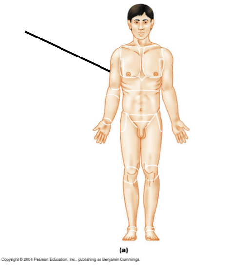
Regional Terminology
Anterior Brachial

Regional Terminology
Anterior Buccal
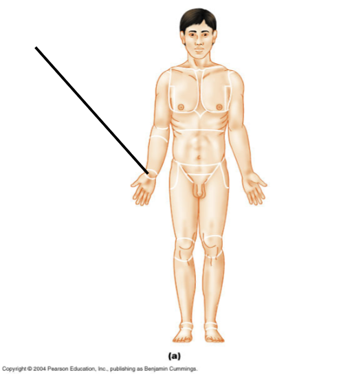
Regional Terminology
Anterior Carpal
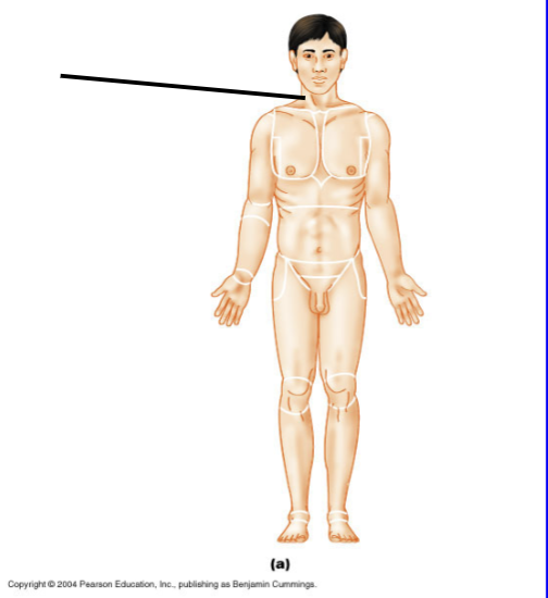
Regional Terminology
Anterior Cervical

Regional Terminology
Anterior Coxal

Regional Terminology
Anterior Crural

Regional Terminology
Anterior Digital

Regional Terminology
Anterior Femoral

Regional Terminology
Anterior Frontal

Regional Terminology
Anterior Hallux
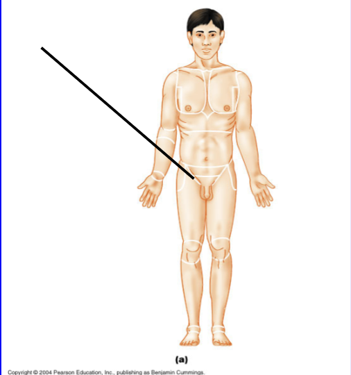
Regional Terminology
Anterior Inguinal

Regional Terminology
Anterior Mammary
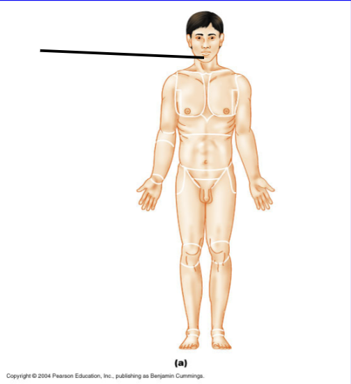
Regional Terminology
Anterior Mental

Regional Terminology
Anterior Nasal

Regional Terminology
Anterior Oral

Regional Terminology
Anterior Orbital

Regional Terminology
Anterior Palmar
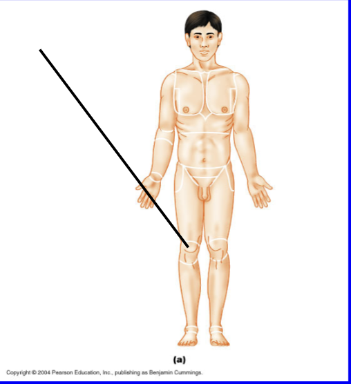
Regional Terminology
Anterior Patellar
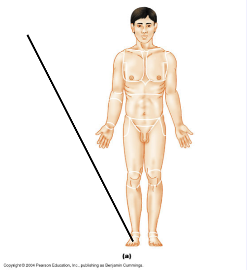
Regional Terminology
Anterior Pedal

Regional Terminology
Anterior Pelvic
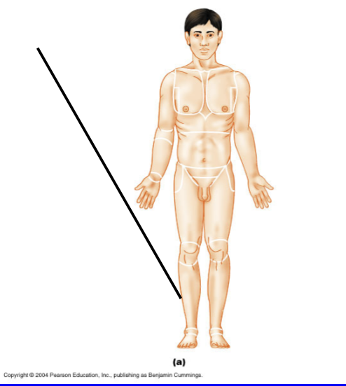
Regional Terminology
Anterior Fibular (peroneal).

Regional Terminology
Anterior Pollex

Regional Terminology
Anterior Pubic

Regional Terminology
Anterior Sternal

Regional Terminology
Anterior Tarsal

Regional Terminology
Anterior Thoracic

Regional Terminology
Anterior Umbilical

Regional Terminology
Posterior Acromial

Regional Terminology
Posterior Brachial
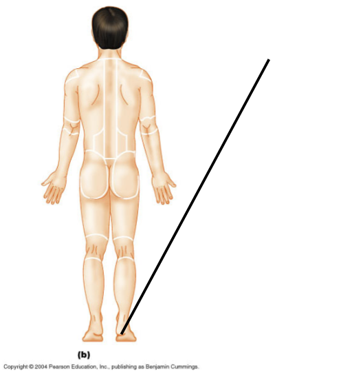
Regional Terminology
Posterior Calcaneal

Regional Terminology
Posterior Cephalic
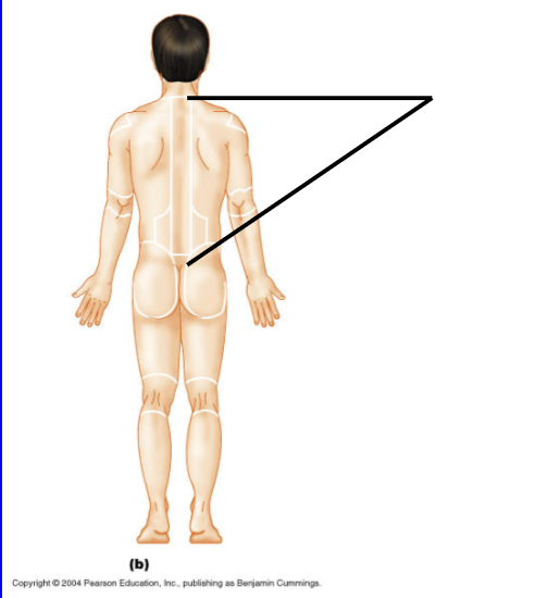
Regional Terminology
Posterior Dorsum

Regional Terminology
Posterior Femoral

Regional Terminology
Posterior Gluteal

Regional Terminology
Posterior Lumbar

Regional Terminology
Posterior Manus

Regional Terminology
Posterior Occipital

Regional Terminology
Posterior Olecranal
Regional Terminology
Posterior Otic

Regional Terminology
Posterior Perineal

Regional Terminology
Posterior Plantar

Regional Terminology
Posterior Popliteal
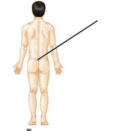
Regional Terminology
Posterior Sacral

Regional Terminology
Posterior Scapular

Regional Terminology
Posterior Sural

Regional Terminology
Posterior Vertebral

Planes of Section
Longitudinal frontal; divides body into anterior and posterior sections.
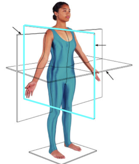
Planes of Section
Longitudinal sagittal; divides body into right and left portions.
Parasagittal Section
Longitudinal; divides body into unequal left and right portions.
Midsagittal Section
Longitudinal; divides body into equal left and right portions.

Planes of Section
Cross section or transverse section; lie perpendicular to the long axis of the body.

Planes of Section
Oblique section; lies at an angle to the long axis of the body.

Body Cavities
Cranial and Spinal

Body Cavities
Ventral cavity; has multiple sub-cavities.

Body Cavities
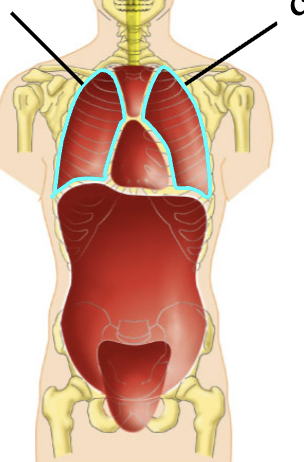
Thoracic cavity; includes two pleural cavities and the mediastinum
Body Cavities
Pleural cavities; enclose the lungs, bounded by serous membranes called the pleura.

Body Cavities
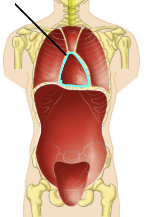
Mediastinum; includes both superior mediastinum and pericardium.
Body Cavities
Pericardial cavity; encloses the heart and is bounded by a serous membrane called the pericardium.
Super mediastinum
Encloses the esophagus and trachea.

Body Cavities
Abdominopelvic cavity; includes abdominal cavity and pelvic cavity, bounded above by the diaphragm.

Body Cavities
Abdominal cavity; encloses most of the digestive organs, bounded by a serous membrane called the peritoneum.
Body Cavities
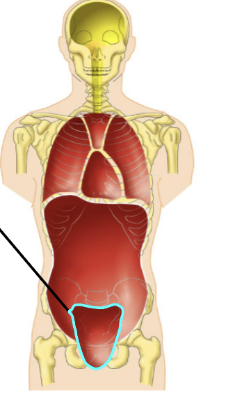
Pelvic cavity; encloses the reproductive organs, bounded by the bony pelvis.
Serous Membranes
Continuous membranes that fold to produce two portions, the parietal serosa and visceral serosa.
Parietal serosa
line the walls of a cavity.
Visceral serosa
line the organs within a cavity.
Types of Serosa
Pleura, pericardium, and peritoneum.
Primary Tissue Types
Epithelial, connective, muscle, and nervous.
Eptihelial Tissues
Line body surfaces; primary function in protection, absorption, filtration, excretion, secretion, and sensory reception. They are avascular and regenerative.
Simple Epithelia
Composed of single layer of cells.
Stratified Epithelia
Composed of multiple layers of cells, from two layers to many layers.
Squamous Cells
Flat; individual cells are wider than they are tall.
Cuboidal Cells
Cube shaped. Individual cells are same width as their height.
Columnar Cells
Column shaped. Individual cells are taller than they are wide.

Histology
Simple Squamous Epithelium
Simple Squamous Epithelium
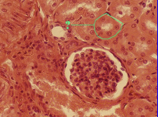
single layer of flattened cells; necessary on surfaces where diffusion is important. Found in kidney glomeruli, alveoli of the lungs, endothelial lining of blood vessels and heart, and serosae.
Histology
Simple Cuboidal Epithelium
Simple Cuboidal Epithelium
Single layer of cuboidal cells; necessary in absorption and secretion. Found in kidney tubules, ducts and secretory portions of small glands, ovary surface.

Histology
Simple Columnar Epithelium
Simple Columnar Epithelium
Single layer of tall cells, may be ciliated and may contain goblet cells. Necessary for absorption and extensive secretion. Lines digestive tract, gall bladder, ducts of some glands, small bronchi, uterine tubes.
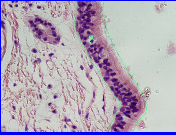
Histology
Pseudostratified Columnar Epithelium
Pseudostratified Columnar Epithelium
Single layer of tall cells that have differing heights. Appears stratified but with indistinct layers. May bear cilia and goblet cells. Found in trachea, male sperm carrying ducts, and ducts of large glands.

Histology
Stratified Squamous Epithelium
Stratified Squamous Epithelium
Multiple layers of flat cells, may be keratinized or non-keratinized. Functions in protection from abrasion. Found in epidermis of skin, mouth, esophagus, and vagina.

Histology
Stratified Cuboidal Epithelium
Stratified Cuboidal Epithelium
Usually two layers of cuboidal cells; functions in protection. Found in the largest ducts of sweat glands, mammary glands, and salivary glands.

Histology
Stratified Columnar Epithelium
Stratified Columnar Epithelium
Several layers of tall cells; function in protection and secretion. Rare, found in male urethra and in large ducts of some glands.

Histology
Transitional Epithelium
Transitional Epithelium
Changes from stratified cuboidal to stratified squamous, depending on the straight of stretch of the organ. Functions to allow the organ to stretch. Found in ureters, urinary bladder, part of the urethra.
Connective Tissues
Composed of one or more specific cell types embedded within a non-living (acellular) matrix.
Ground Substance
composed of interstitial fluid and globular proteins.
Collagen
Most abundant connective tissue fiber; subunits form long, unbranched strands. Very strong, does not stretch or break easily, bends readily when deformed perpendicular to the long axis. Used to provide strength when arranged in parallel bundles.
Elastin
Found in tissue that must deform then resume resting shape; subunits form moderately branched fibers, not as strong as collagen but extremely stable.
Reticulin
Found in tissues where a network of overlapping fibers is necessary; subunits form fine, heavily-branched strands. Functions to create a sieve-like net structure to trap debris or support other tissues where strength is unnecessary.
Loose Connective Tissues
Proper connective tissue; areolar, adipose, and reticular.