Diagnostic Imaging 2- Exam 3- Tumor 1 Malignant Notes- part 3
 Call Kai
 Learn
Learn Practice Test
 Spaced Repetition
 Match
Match Flashcards
 Knowt Play
1/90

There's no tags or description
Looks like no tags are added yet.
Name | Mastery | Learn | Test | Matching | Spaced | Call with Kai | Chat |
|---|
No analytics yet
Send a link to your students to track their progress
91 Terms
Localized form of plasma cell proliferation
Solitary plasmacytoma
Favorite spots for solitary plasmacytoma listed in most to least common
Vertebra
Pelvis
Skull
Sternum
Ribs
Typical presentation of solitary plasmacytoma
Soap bubbly, highly Expansile lesion
Solitary plasmacytoma tends to do what with the vertebra
Spare the posterior elements
Most solitary plasmacytoma`s develop into
Multiple myeloma
Solitary plasmacytoma example in the ilium

Another example of a solitary plasmacytoma example in the ilium

Multiple myeloma looks like what
Circumscribed osteolyic defects (punched out)
-they're lesions are often of similar size
If you do a bone scan of multiple myeloma it looks
Typically normal
MRI examination for multiple myeloma is sensitive for
Marrow changes
What are the 3 different radiologic features that multiple myeloma can have
Osteoporosis/osteopenia
Osteolyic defects "rain drop skull"
Osteoblastic lesions
What is the most common radiologic presentation of multiple myeloma
Osteoporosis/osteopenia
What is it called when multiple myeloma is in the skull
Rain drop skull
Multiple lytic defects results in a punched out appearance in the skull and this is called_____ during what disease
Rain drop skull during multiple myeloma
Rain drop skull

Another example of rain drop skull

Pathological fracture are most likely from what 3 things
Osteoporosis
Lytic Mets
Multiple myeloma
If you get a pathological fracture and then X ray of the skull and it has a rain drop appearance than what most likely caused the pathological fracture
Multiple myeloma

Examples of holes in the skull and what they are from slide

Myeloma is hard to differentiate form what?
Lytic metastasis
Myeloma has what on T1 and T2 MRI
Diffuse low T1
ISO (no change) to increased T2
Myeloma has what occur in the spine
Multiple pathological fractures/multiple wedge compression fractures
Myeloma has what else occur along side bone changes
Little soft tissue mass
What does myeloma look like in the spine on a T1 MRI
Bunch of little dark spots

MRI T1 rule of thumb? So what does this mean for pathological fractures in the spine
White is right on T1
-old fractures look bright
-new fractures look dark
What is the most likely cause of a pathological fracture in a child
Eosinophilic granuloma (EG)
A primary malignant tumor of undifferentiated connective tissue which forms neoplastic osteoid (bone)
Osteosarcoma
What is the 2nd most common primary malignant bone tumor overall
Osteosarcoma
What it's eh most common primary malignant bone tumor in children and young adults
Osteosarcoma
Age for osteosarcoma
Mainly 10-25 years
Over 60
Patients tend to be what with osteosarcoma
Tend to be taller than their peers
Clinical presentation of osteosarcoma
Painful swelling
-antecedent history of trauma (traumatic determination)
What does it mean for osteosarcoma to commonly have antecedent history of trauma (traumatic determination)
Patient usually comes in for trauma then its found or a trauma "sets off" the growth of the tumor
What type of pain does a patient feel with osteosarcoma
Initially insidious and transient becoming severe and persistent
Osteosarcoma can cause what in the joint
Minor resection of ROM
Examples of osteosarcoma just from observation

Osteosarcoma comes in what forms
Central
Parosteal (outside bone)
Secondary (from other pathology)
Multicentric (multiple locations)
Extraosseous
What is the most common form of osteosarcoma
Central
Osteosarcoma most commonly occur where
Around knee and proximal humerus
Most osteosarcomas occur where in the bone
Metaphysics next to the growth plate
-areas of growing bone
Osteosarcoma occurs in what type of bones before the age of 30
Cylindrical bones
Osteosarcoma occurs in what type of bones after the age of 50
Flat bones
Metastasis is common in osteosarcoma to which location
Lungs (On chest films or CT)
When you find osteosarcoma spread to the lungs, where do you typically see it as well
In the knee
Most osteosarcomas are what type
Sclerotic
Osteosarcoma typically occur where in bones
Metaphyseal region
-knee and proximal humerus
How doe osteosarcomas look
Ill defined dense ivory or sclerotic lesion, moth eaten, Permeative destructor with a long zone of transition
Spiculated/sunburst periosteal reaction/codmans
What occurs along side the bone damage with osteosarcoma
Large soft issue mass
Osteosarcoma

Another example of osteosarcoma

Example of osteosarcoma again, more subtle this time

Osteosarcoma in the proximal humerus

What is it called when there is bone produced in the soft tissue mass along side a tumor
Osteoid matrix
14 year old boy with pain and a mass above the knee
Young, pain, near knee, so osteosarcoma

What is the name for the imaging finding with osteosarcoma in the lungs
"Cannonball" osteosarcoma pulmonary metastasis

What are some things that could fall under secondary osteosarcoma
Paget's disease
Olliers disease
Ionizing radiation
Polyostotic fibrous dysplasia
Hereditary mutiple exostosis
Treatment for osteosarcoma
Multidrug therapy
What do you see on nuclear imaging with osteosarcoma
Increased activity (hypervasculatirty, new bone formation)
What do you see on SPECT with osteosarcoma
Soft tissue extension
What do you see on T1 and T2 MRI images of osteosarcoma
T1 shoes marrow involvement
T2 shows soft issue
Primary malignant tumor of chondrogenic origin
Chondrosarcoma
3rd most common primary malignant bone tumor and 2nd most common in adult
Chondrosarcoma
Chondrosarcoma arises from
Chondroblasts and collagenoblasts
Chondrosarcoma has 2 different forms, primary and secondary. Describe what a secondary Chondrosarcoma is
Complication of preexisting skeletal abnormality such as Osteochondroma or echondroma
Age for Chondrosarcoma
Usually over 50 (40-60)
Chondrosarcoma clinical features
Large soft tissue mass
Late development of pain
Duration of symptoms less than 2 yrs (have symptoms for awhile before discovered)
Cho on location for Chondrosarcoma
Pelvis
Radiographic features of Chondrosarcoma
Round or oval area of demineralization with ill defined margins
Endosteal scalloping
Popcorn matrix calcification in 2/3
Large soft tissue mass
What physical features of a tumor mean that it is cartilage based
Stippled
Floccluent
Ring and arc

Chondrosarcoma in the proximal humerus

Lytic destruction, some calcification, cortical thinning, all point to cartilage based tumor that looks aggressive... so this is what type of tumor most likely
Chondrosarcoma

What are the differential diagnosis for a Chondrosarcoma? What is the thing that would set it apart from the other 2?
Enchondroma or medullary infarct
-If there is pain then most likely a Chondrosarcoma cause the other 2 don't have pain associated with them

People with benign cartilage tumors (________ or __________) rarely have pain
Enchondroma or osteochondroma
People with __________ will have pain and many have swelling
Chondrosarcoma
Chondrosarcoma in the pelvis

Echondorma can turn into a
Chondrosarcoma (secondary Chondrosarcoma)

Primitive primary malignant tumor composed of tumor cells derived form connective tissue framework of bone marrow
Ewings sarcoma
4th most common primary malignant bone tumor and 2nd most common primary malignant bone tumor in children
Ewings sarcoma
Ewings sarcoma looks like and like?
Round cell tumor and like diaphysis
Ewings sarcomas are caused by
Chromosome translation, 11 and 22 swap
Ewings sarcoma age
10-25, peak around 15
Clinical features of Ewings sarcoma
Severe localized pain
The only primary malignant tumor that stimulates an infection
Leukocytes is
What is the only primary malignant tumor that stimulates an infections
Ewings sarcoma
Skeletal location of Ewings sarcoma
Long tubular bones
Diaphyseal is classic location
What is the classic radiographic feature of a Ewings sarcoma
Onion skin periosteal reaction
What is the most common primary malignancy to Mets to bone
Ewings sarcoma
Ewings sarcoma likes to spread to bone and where else
Lungs
Ewings sarcoma

What is another radiographic feature that can occur with Ewings sarcoma
Cortical saucerization
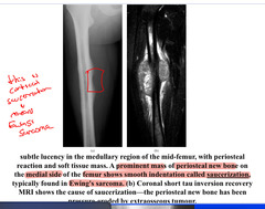
Another Ewings sarcoma

One more Ewings sarcoma
