Jenna: ANAT-225 Lab Block 5
 Call Kai
 Learn
Learn Practice Test
 Spaced Repetition
 Match
Match Flashcards
 Knowt Play
1/215

There's no tags or description
Looks like no tags are added yet.
Name | Mastery | Learn | Test | Matching | Spaced | Call with Kai | Chat |
|---|
No analytics yet
Send a link to your students to track their progress
216 Terms
Oral Cavity
mouth; first part of the gastrointestinal tract
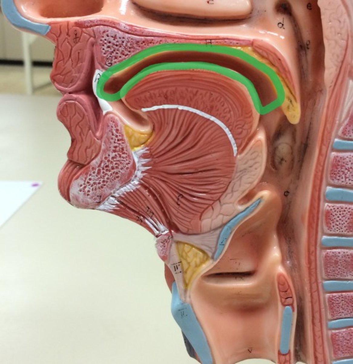
Oropharynx
central portion of the pharynx between the roof of the mouth and the upper edge of the epiglottis
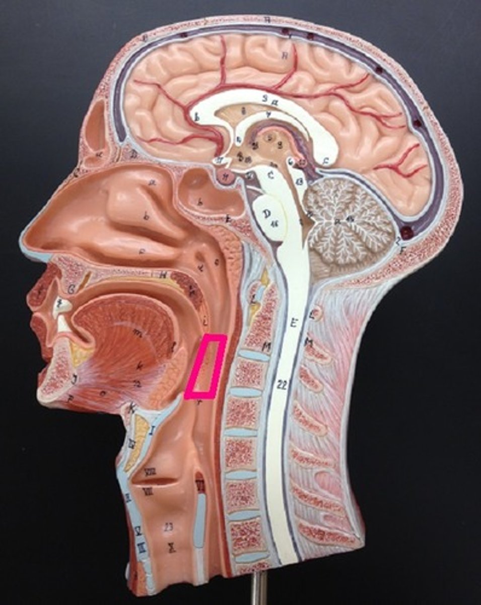
Vestibule
space between teeth and lips or cheek
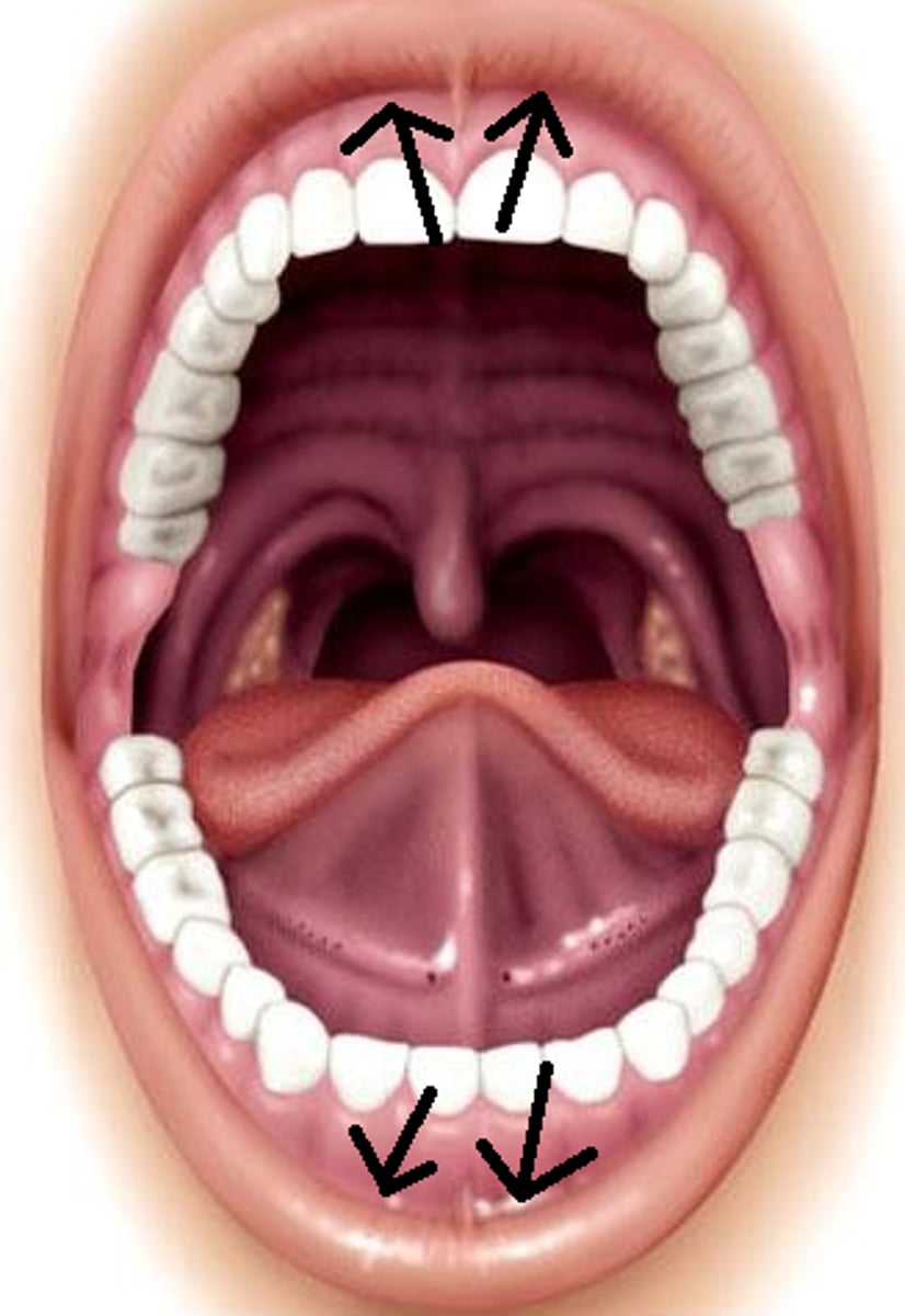
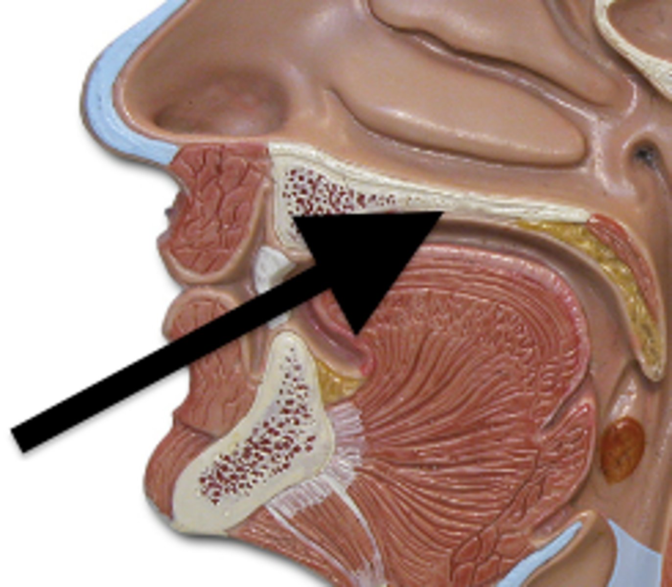
Hard Palate
anterior portion of the roof of the mouth and is formed by the maxillary and palatine bones
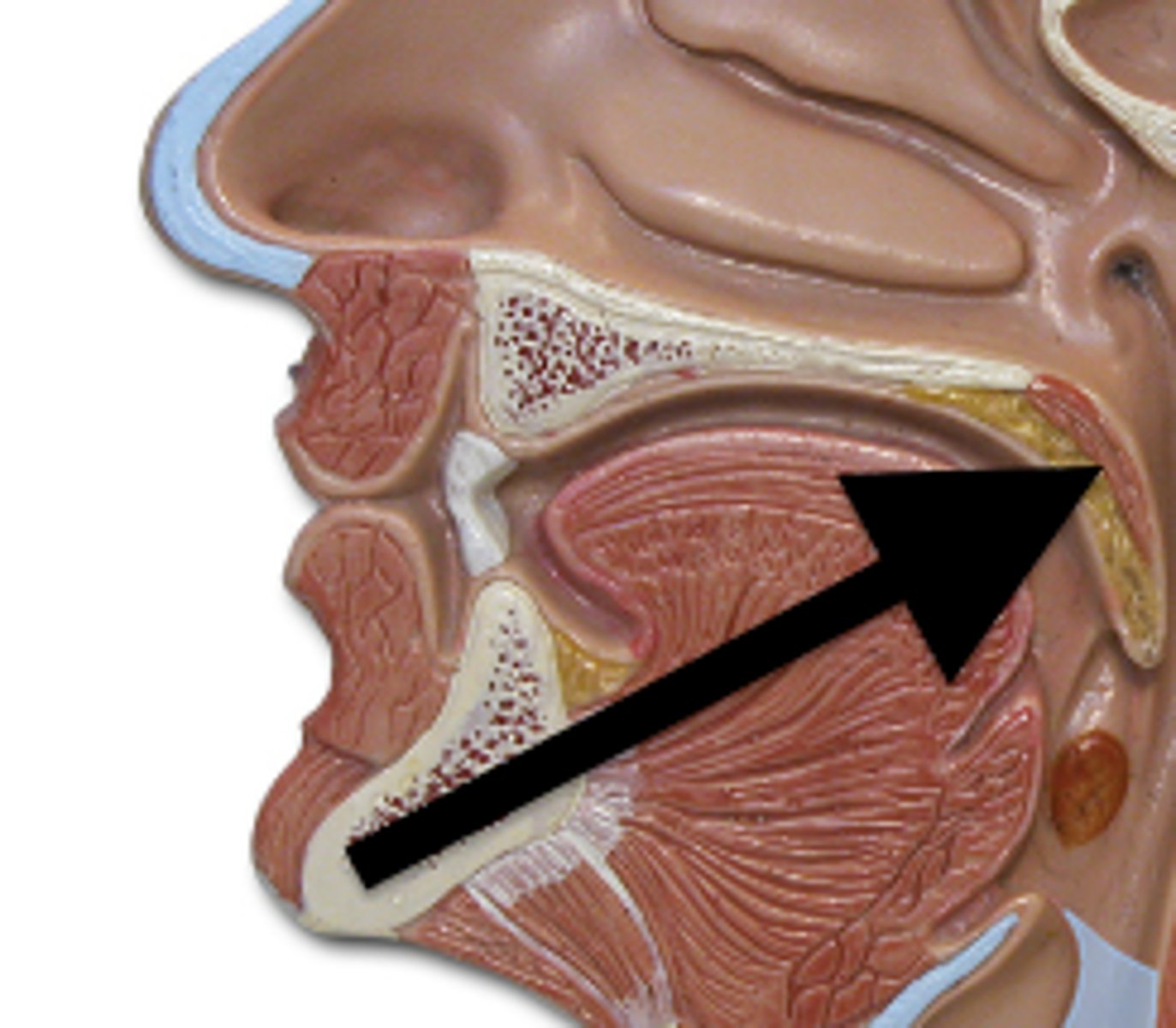
Soft Palate
extends posteriorly from the hard palate and is formed primarily from skeletal muscle. The soft palate separates the oral cavity from the nasopharynx
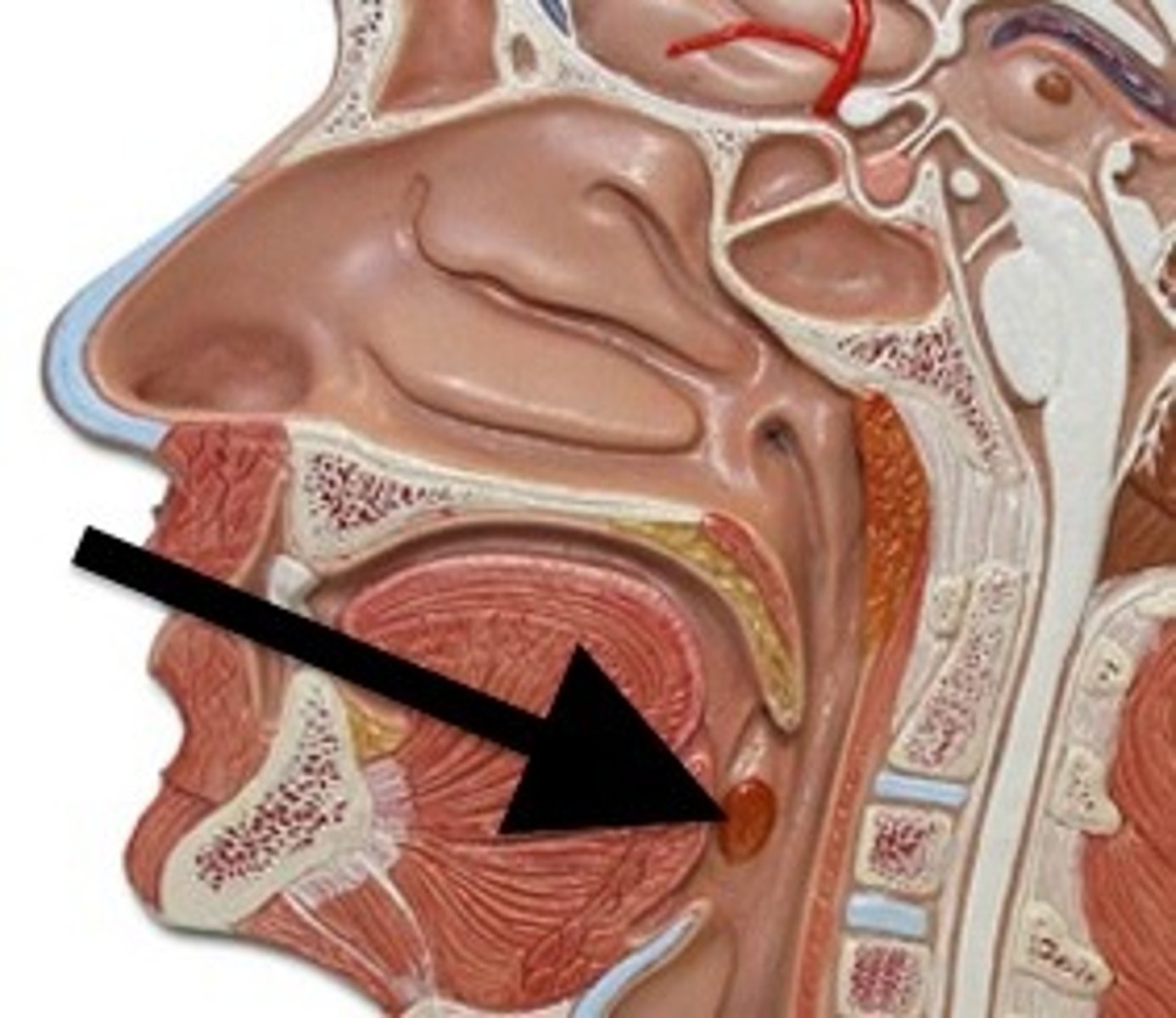
Palatine tonsils
paired lateral masses of lymphatic nodules at the border between the oral cavity and the oropharynx
Tongue
lies on the floor of the mouth; manipulates food for chewing and swallowing; a taste organ
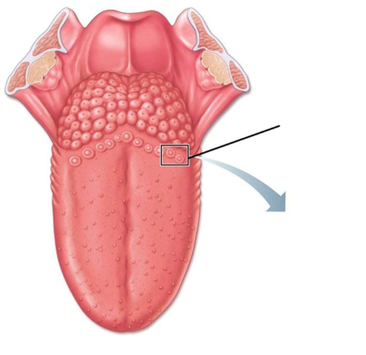
Vallate papillae
largest taste buds with 8-12 forming "V" at back of tongue
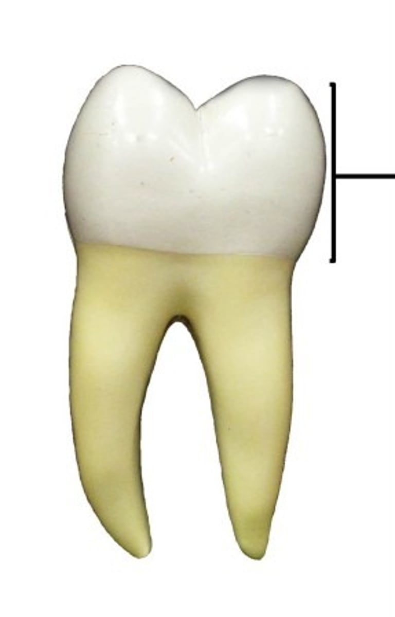
Crown of tooth
extending into the oral cavity; covered by enamel
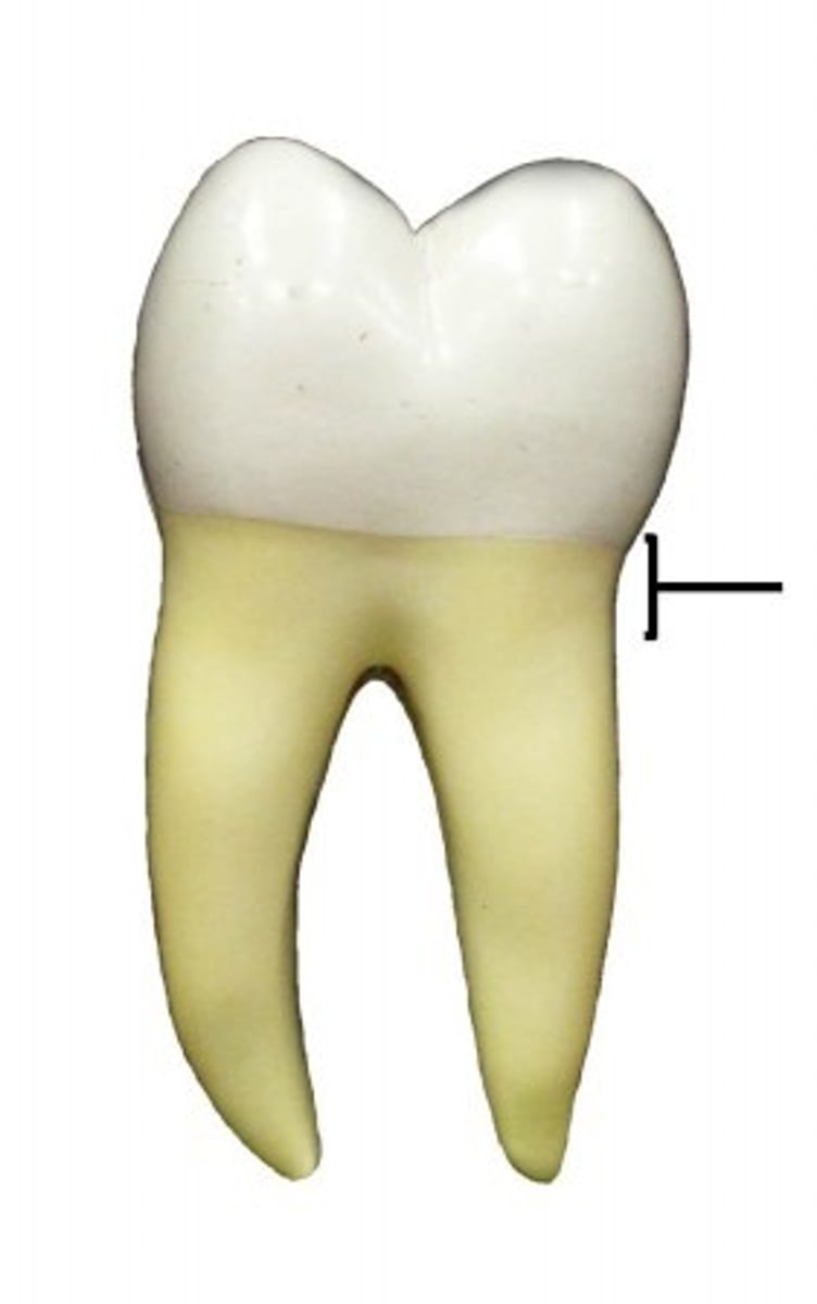
Neck of tooth
narrow, slightly constricted region below the crown, in contact with gums that cover alveolar processes of the mandible and maxillae
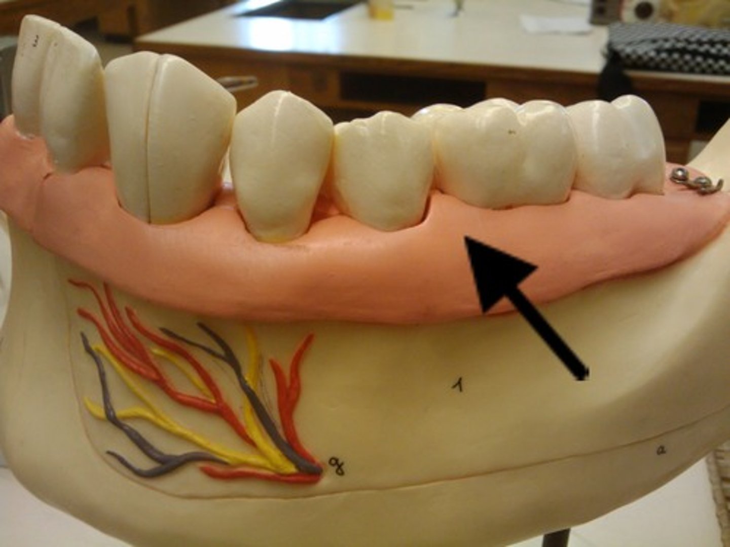
Gums
tissue covering the processes of the jaws
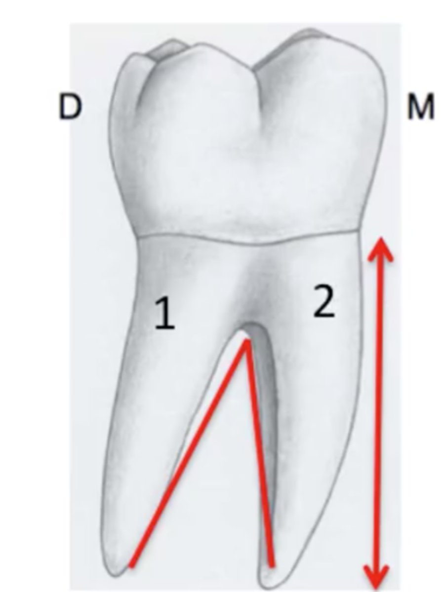
Root of tooth
portion below the gum line; anchor to the tooth into its alveolus
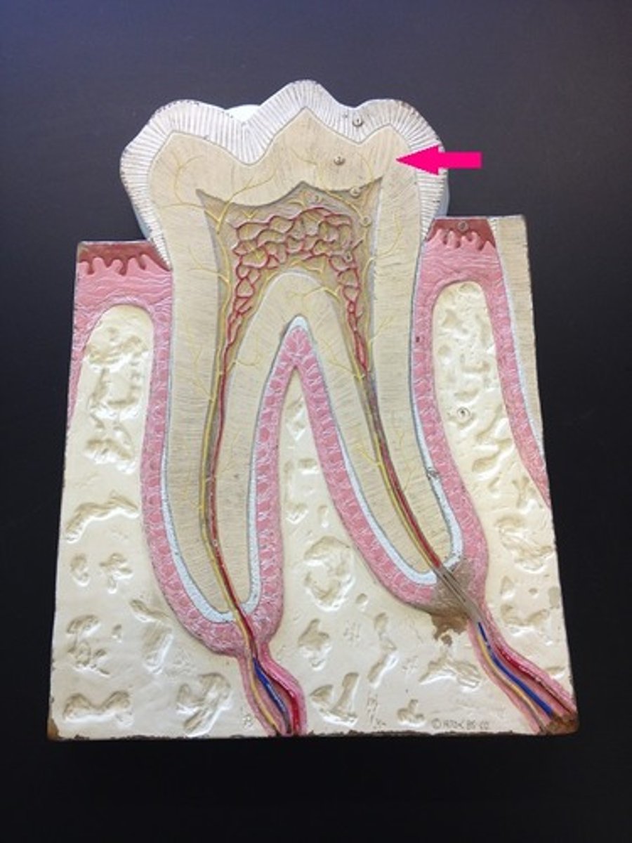
Dentin
Dense tissue forming the bulk of a tooth; hard, yellowish, calcified substance
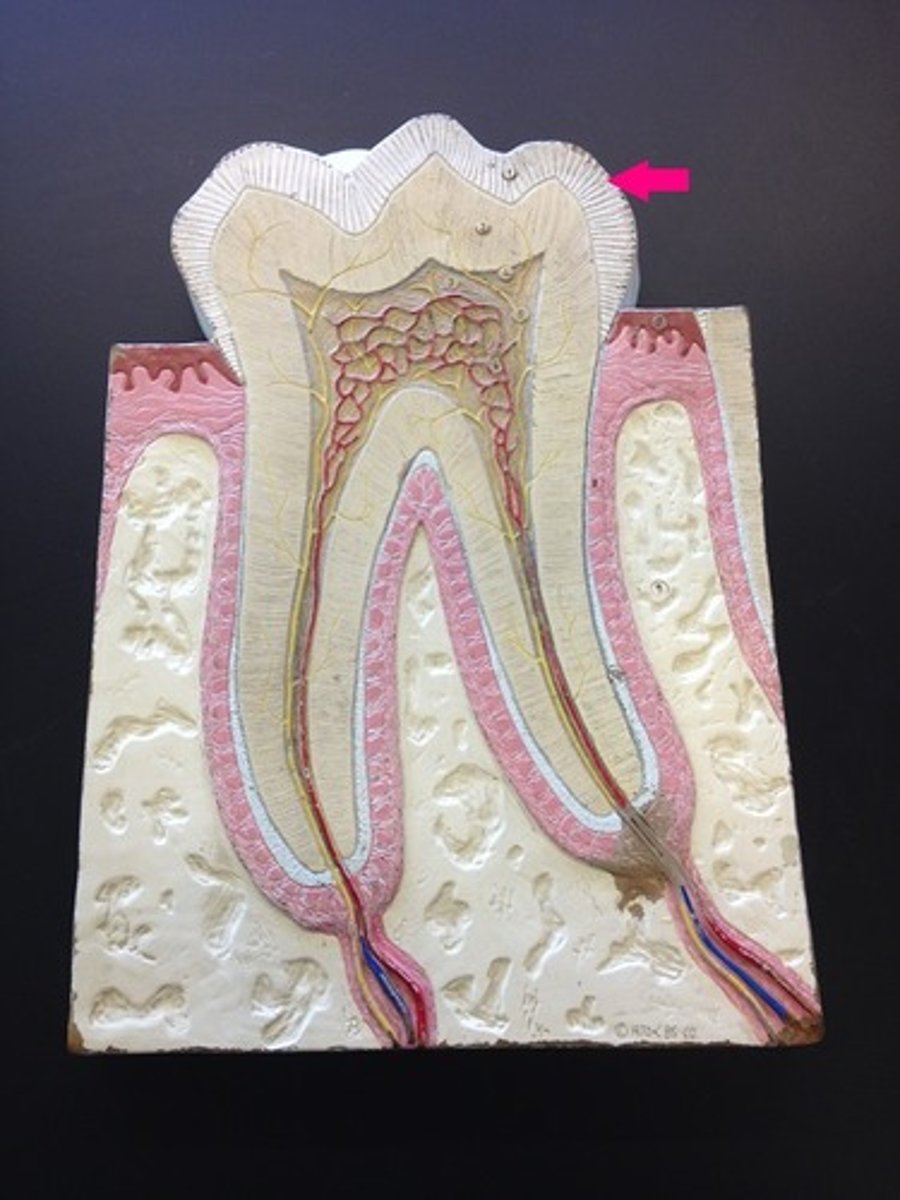
Enamel
the hardest substance in the body and is composed largely of inorganic salts; covers the dentin of the crown
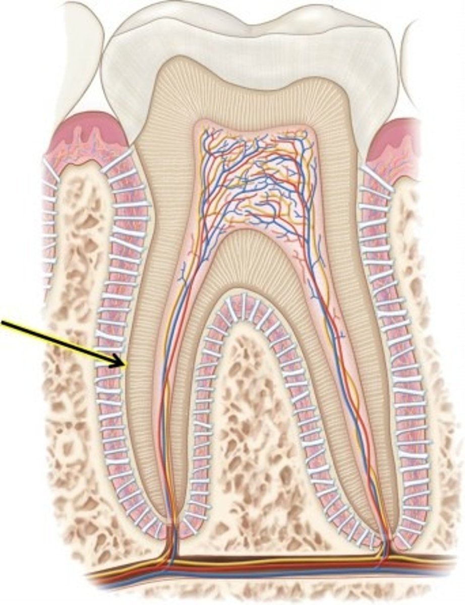
Cementum
bonelike substance which surrounds the dentin of the root and helps anchor the tooth
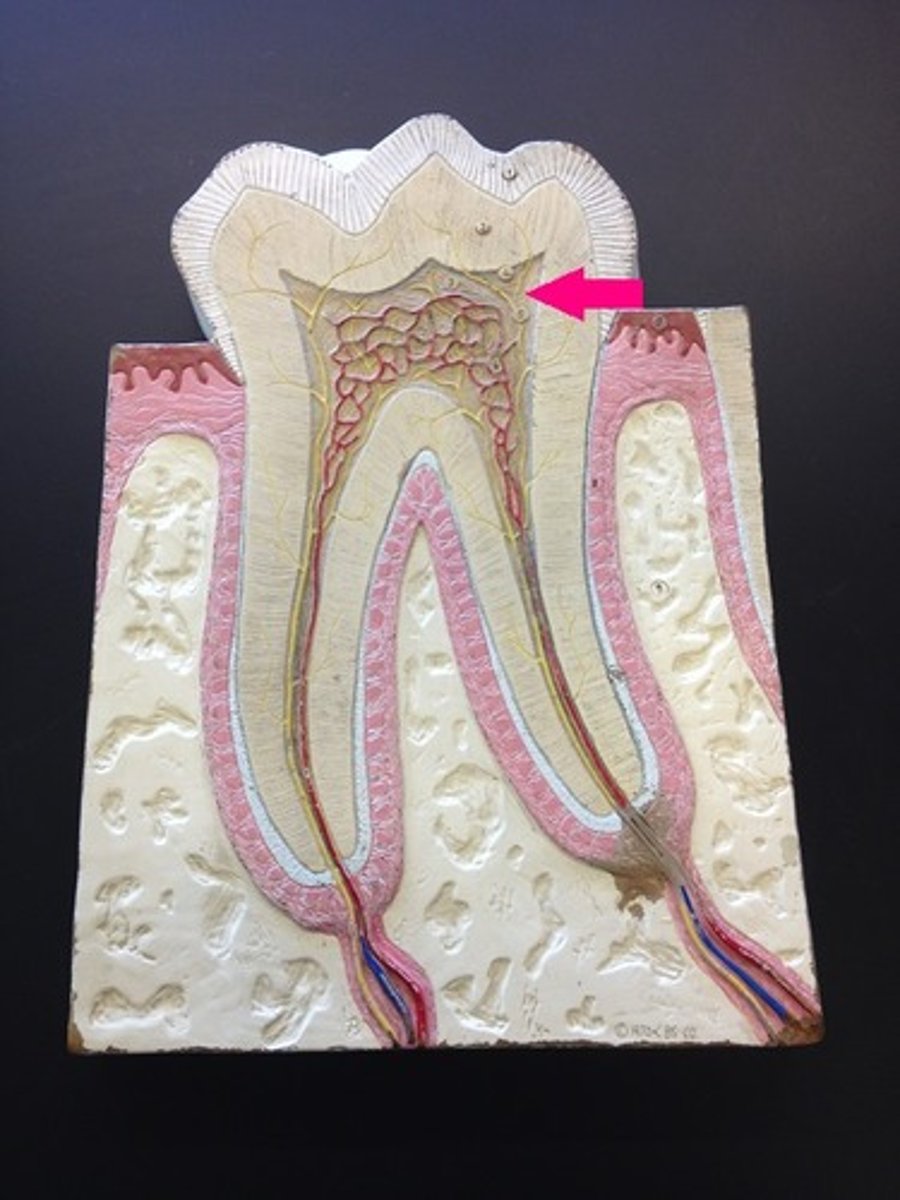
Pulp
soft tissue within a tooth, containing nerves and blood vessels
Pulp Cavity
contains blood vessels and nerves
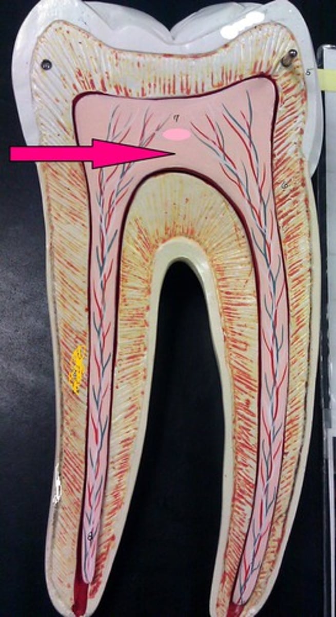
Root Canal
where the pulp cavity extends into the root
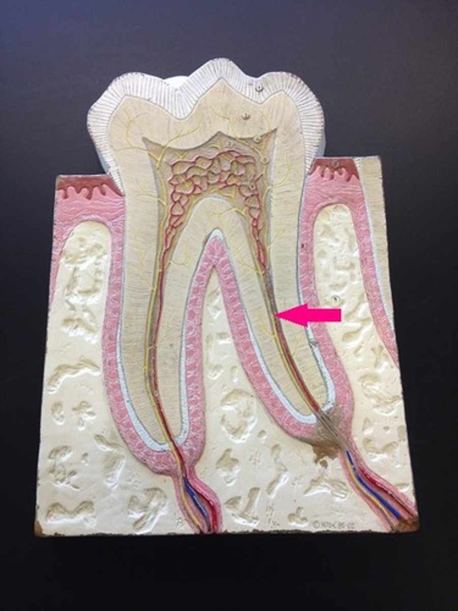
Periodontal ligaments
composed of dense regular connective tissue and run between the bone of the alveolus and the cementum to anchor the tooth
Parotid glands
large glands at the angle of the jaw which produce saliva
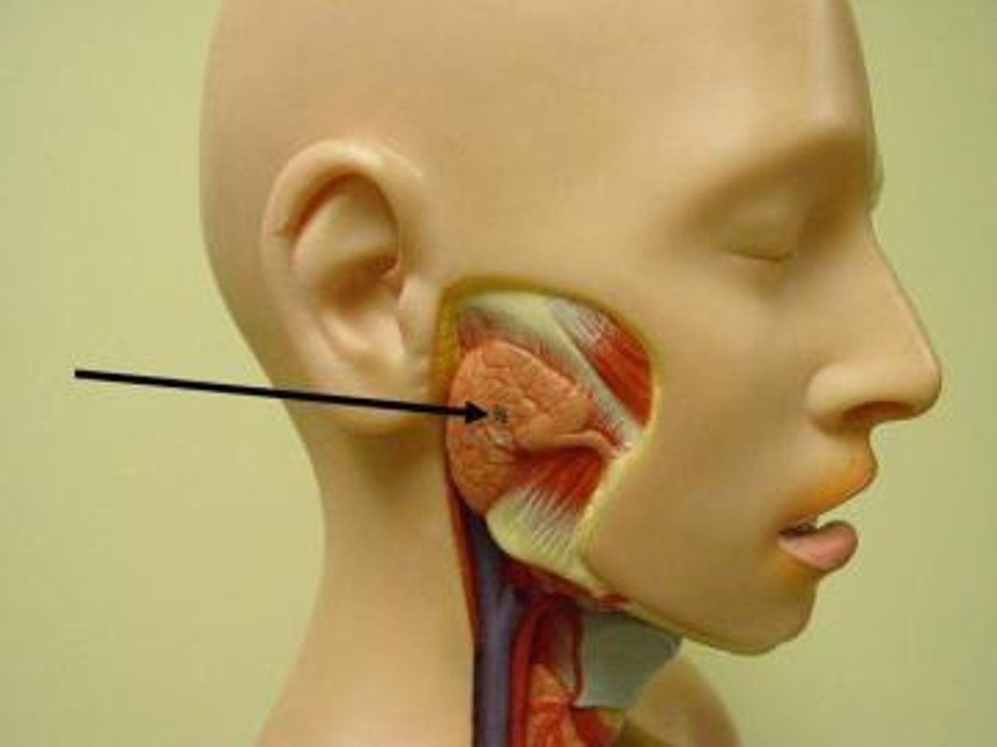
Parotid duct
passes over the masseter muscle, pierces the buccinator muscle, and enters into the vestibule of the oral cavity opposite the second upper molar tooth
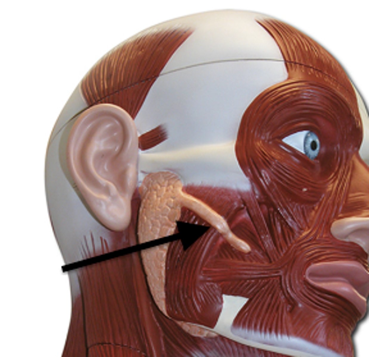
Submandibular glands
located along the medial aspects of the mandible, each with a superficial portion which is continuous posteriorly around the mylohyoid muscle with a larger deep portion. the duct of each is formed in the deep posterior portion and run anteriorly to enter the oral cavity at the base of the tongue
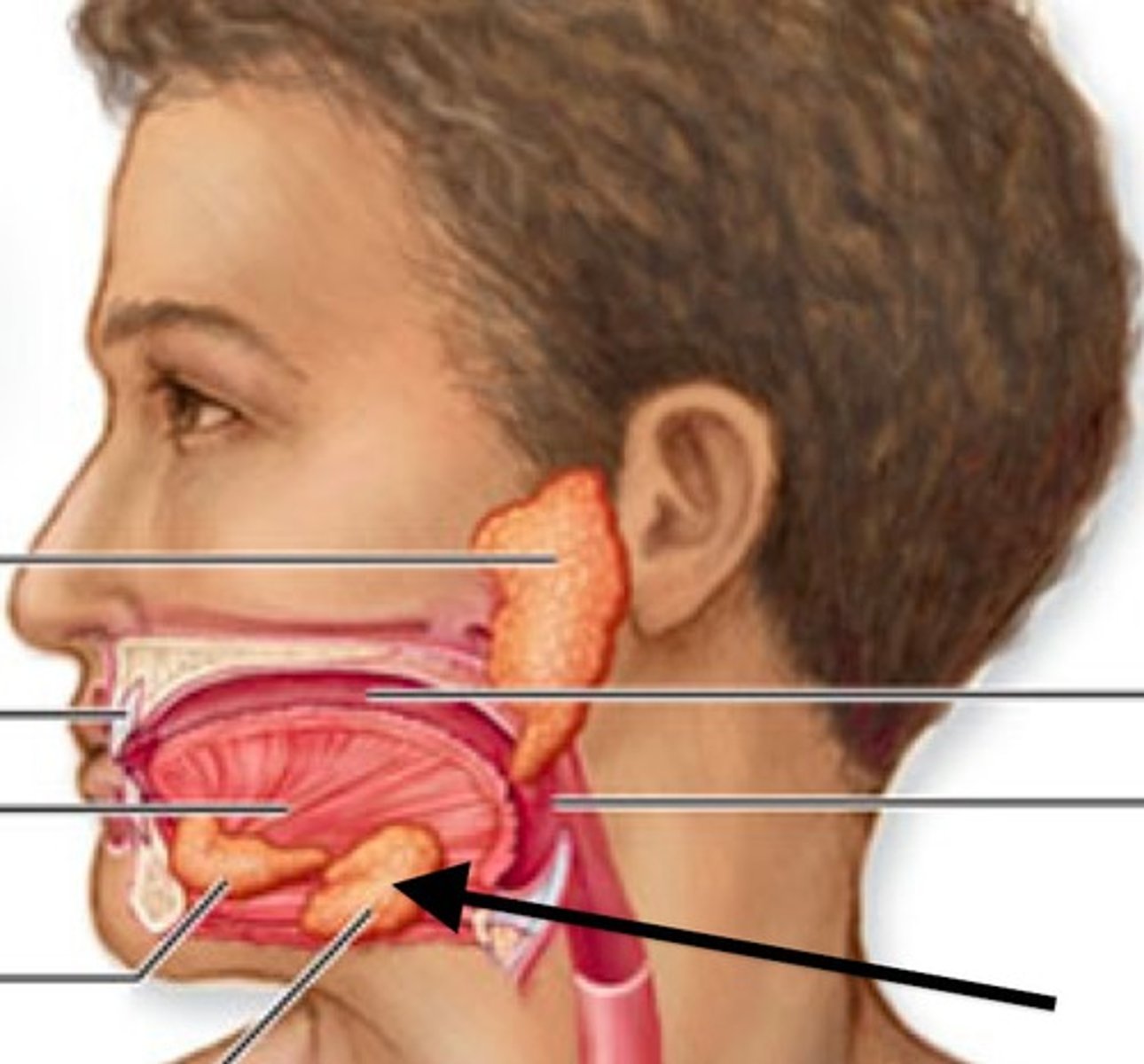
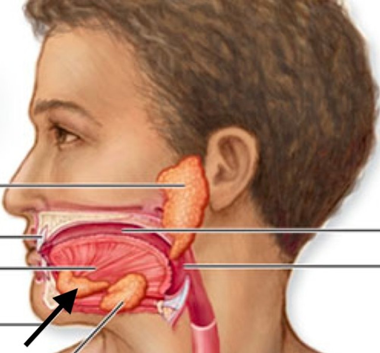
Sublingual glands
located on the anterior floor of the mouth deep to the oral cavity mucosa. They are the smallest of the salivary glands. Each gland has several small ducts which open separately along the floor of the mouth
Esophagus
A muscular tube that connects the mouth to the stomach.
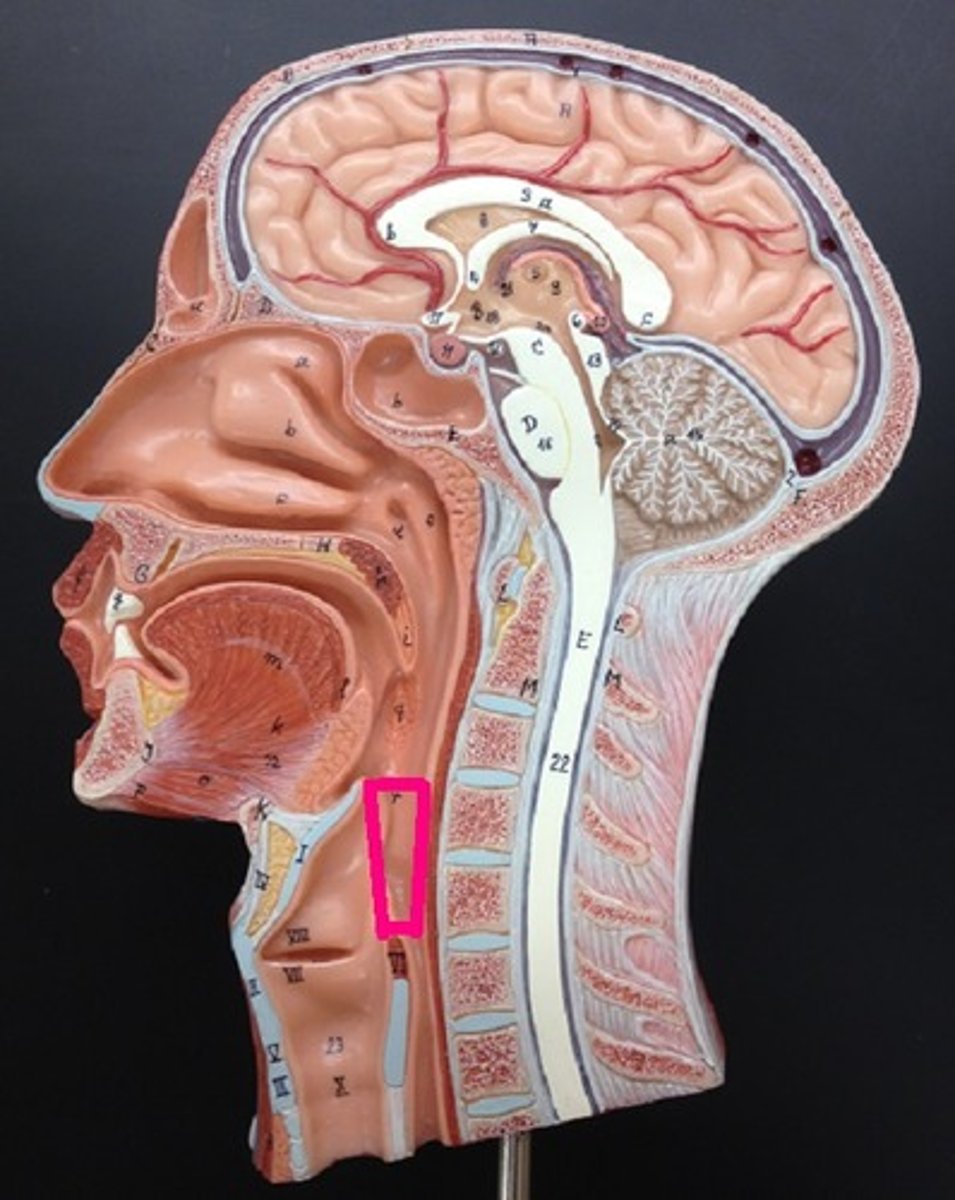
Laryngopharynx
lower part of the pharynx, just below the oropharyngeal opening into the larynx and esophagus
Stomach
large muscular sac that continues the mechanical and chemical digestion of food
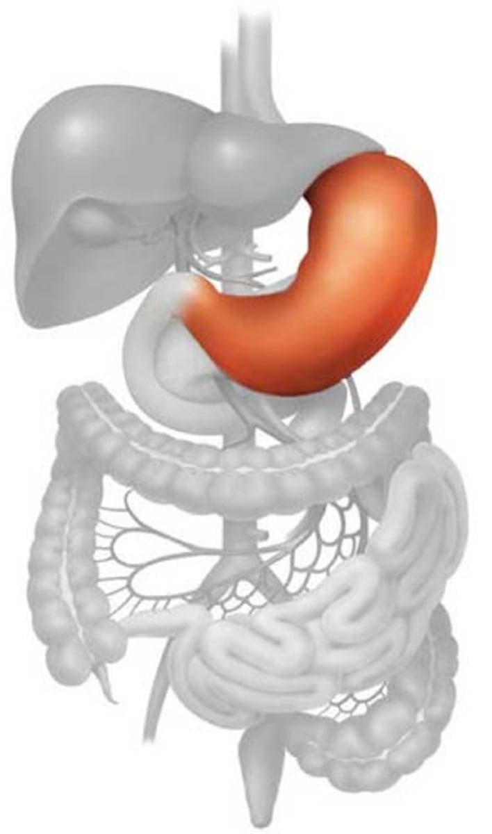
Greater curvature
convex lateral surface of the stomach
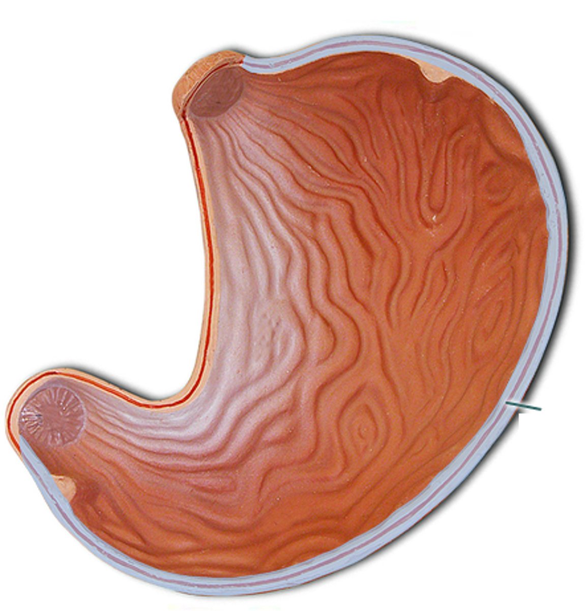
Lesser curvature
concave medial surface of the stomach
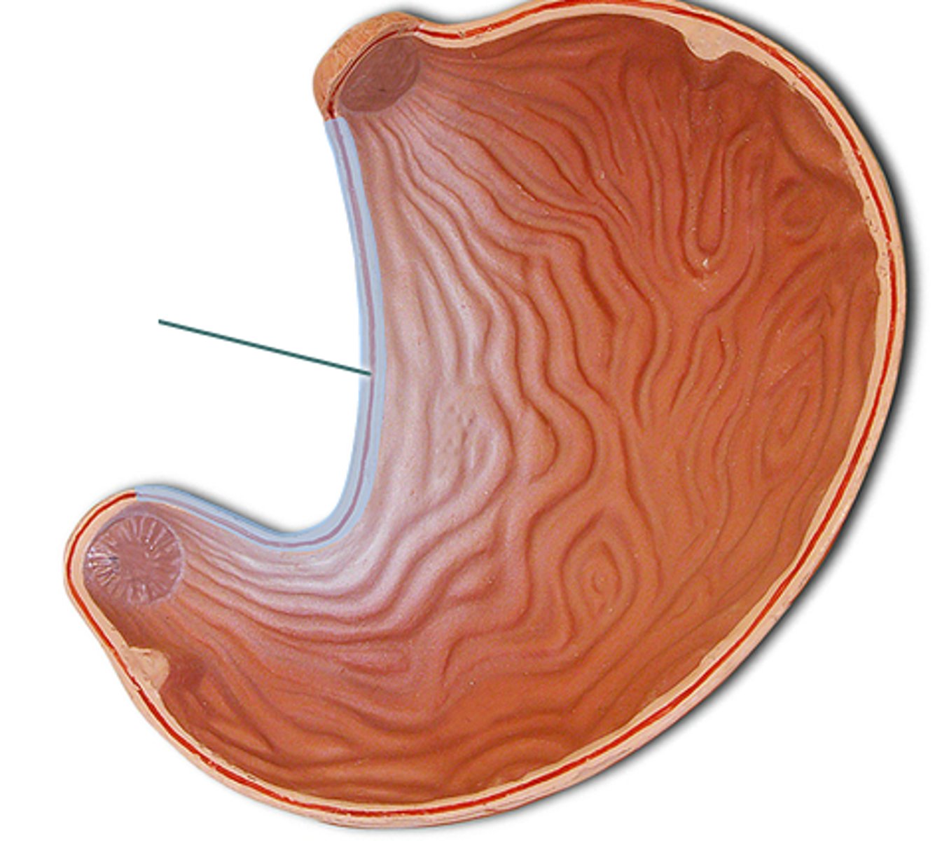
Pyloric sphincter
Controls passage of food from stomach to small intestine
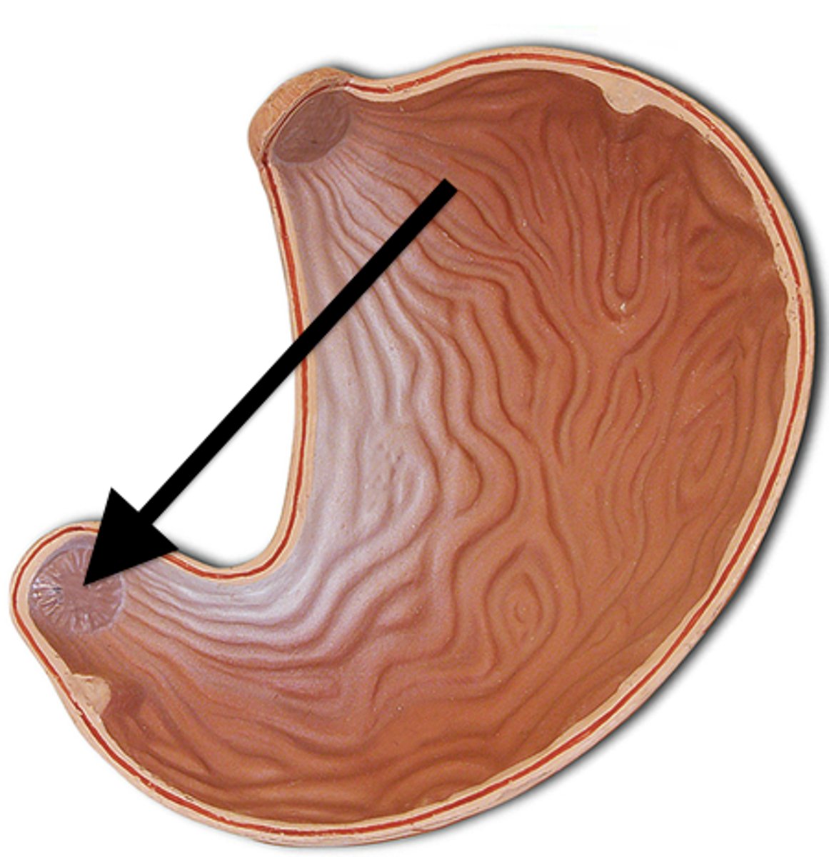
Regions of the stomach
cardia, fundus, body, pylorus
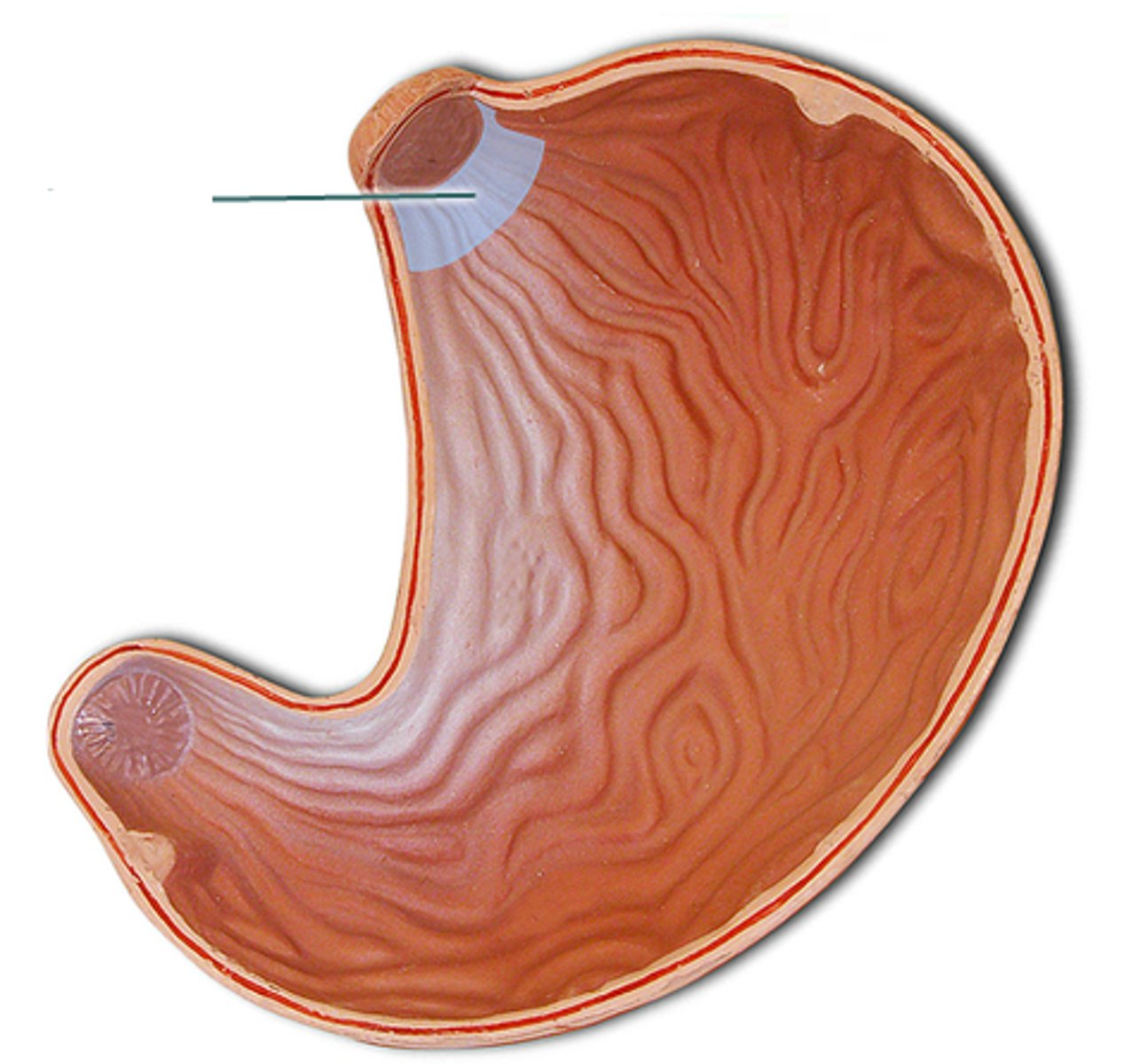
Cardiac region
region of the stomach attached to the esophagus surrounding the cardiac orifice
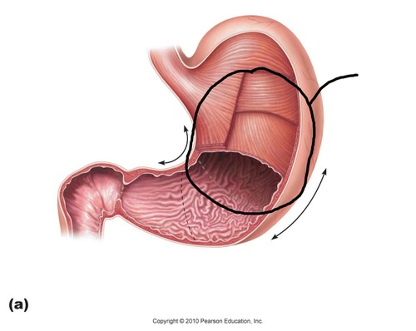
Body region
main part of stomach
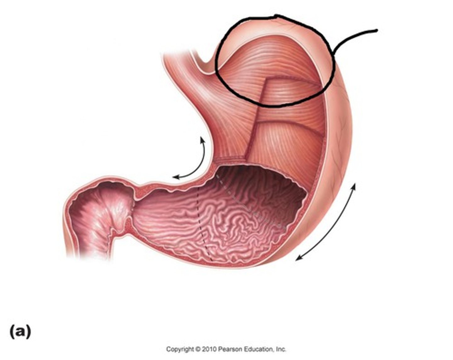
Fundus region
a rounded portion at the top of the stomach
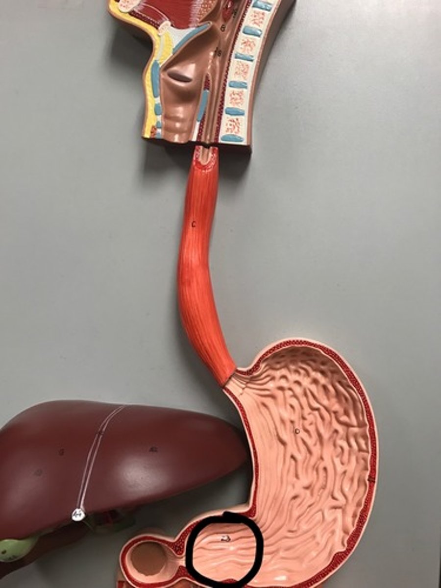
Pylorus region
funnel shaped terminal end of the stomach; tapers toward the pyloric sphincter at the distal end of the stomach
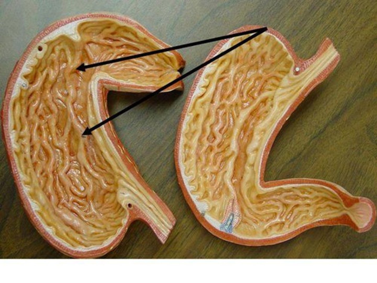
Gastric folds
-rugae, found on internal stomach lining when stomach empty
-allow stomach to expand greatly when fills
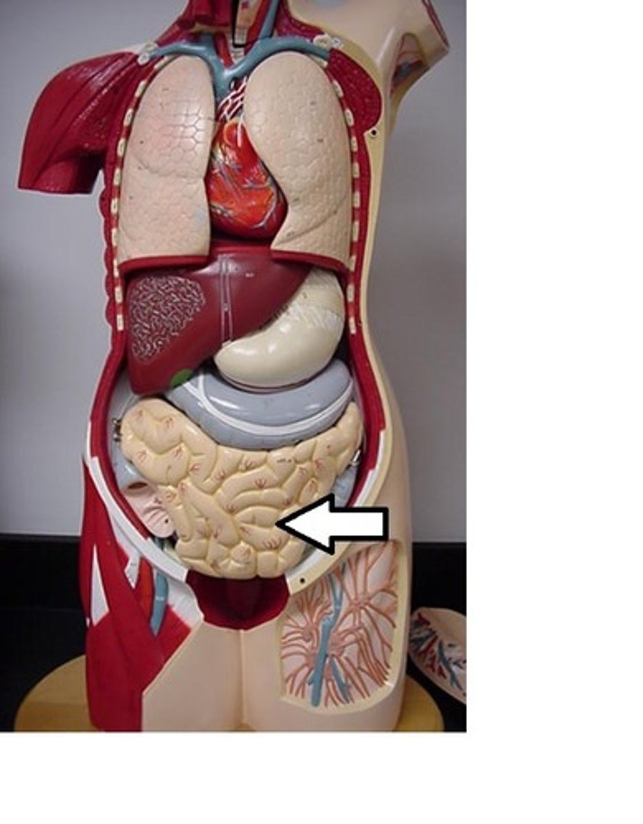
Small Intestine
Digestive organ where most chemical digestion and absorption of food takes place
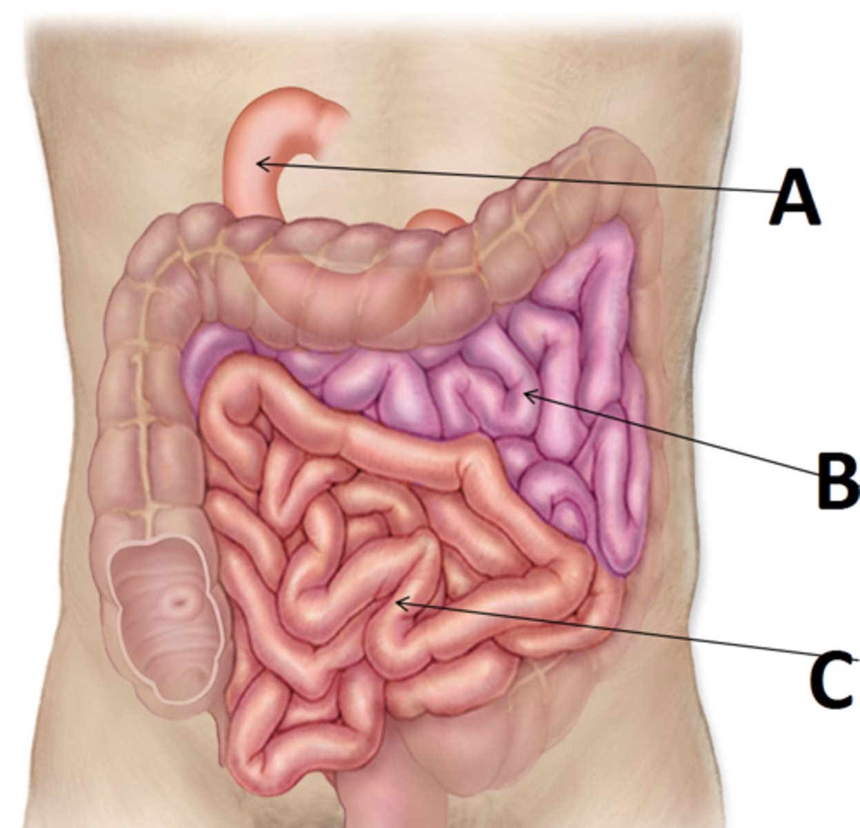
Duodenum
(A) first portion of the small intestine
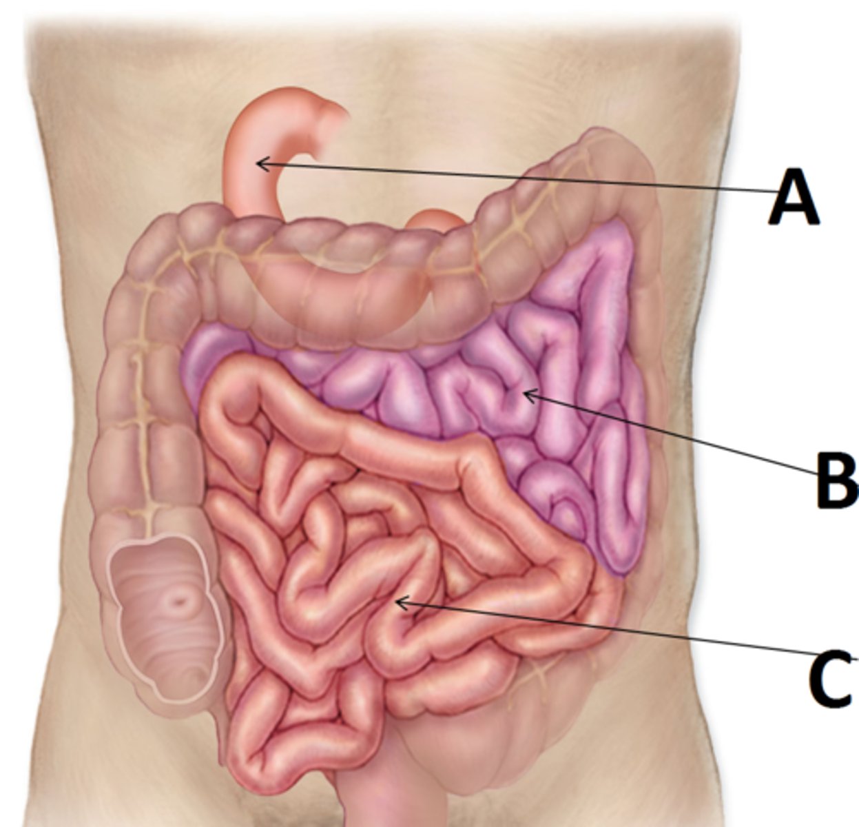
Ileum
(C) the last and longest portion of the small intestine
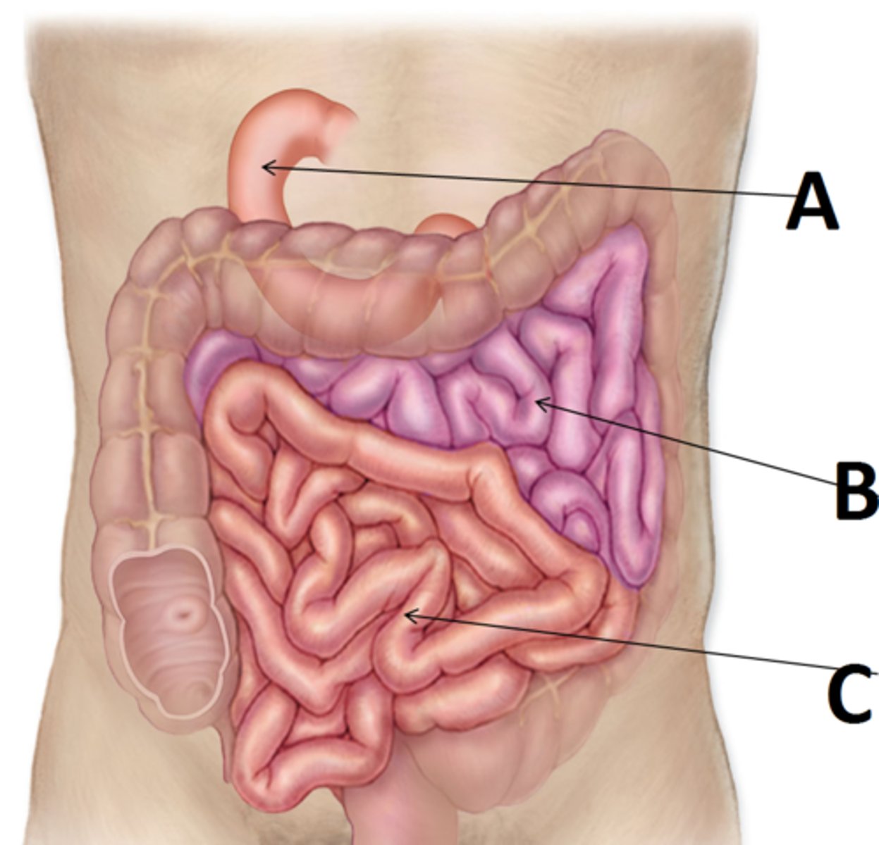
Jejunum
(B) Middle portion of the small intestine
Mesentery
a fused double layer of the parietal peritoneum that attaches parts of the intestine to the interior abdominal wall
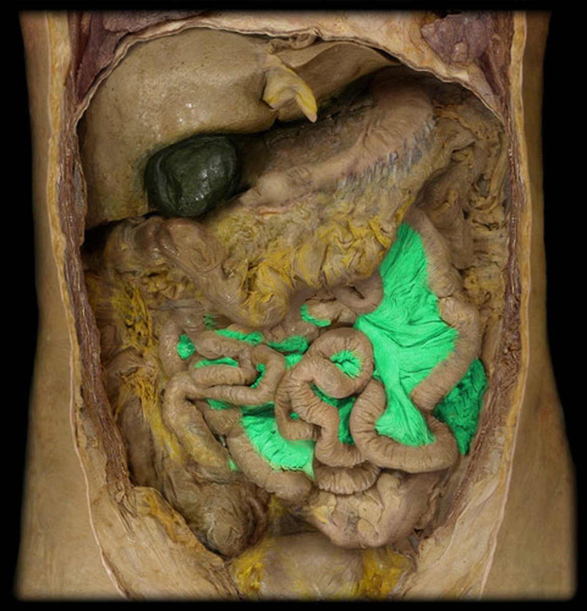
Circular folds
folds in the duodenum that are permanent and are not obliterated when the intestine is full
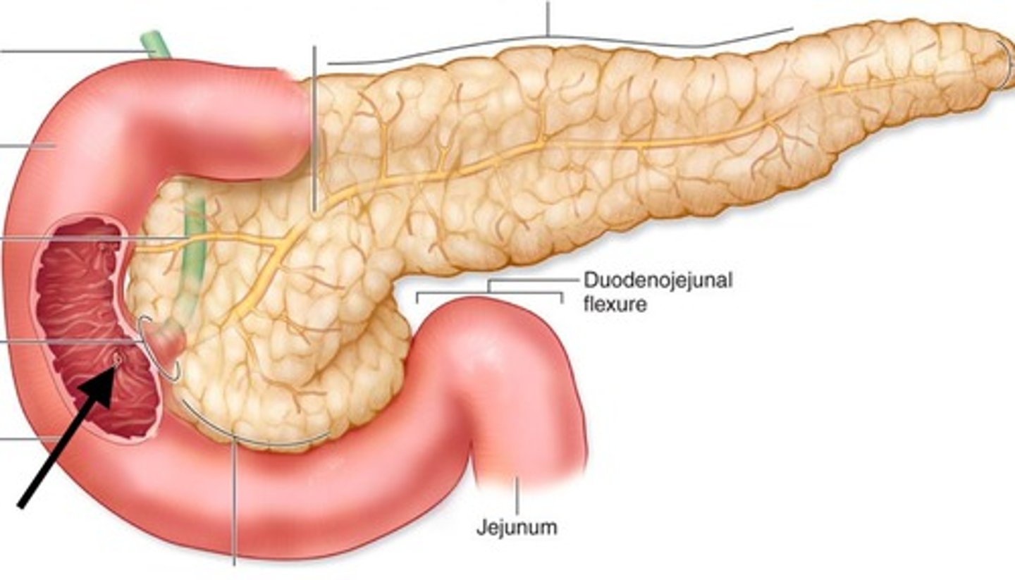
Duodenal papilla
opening of the ampulla into the duodenum
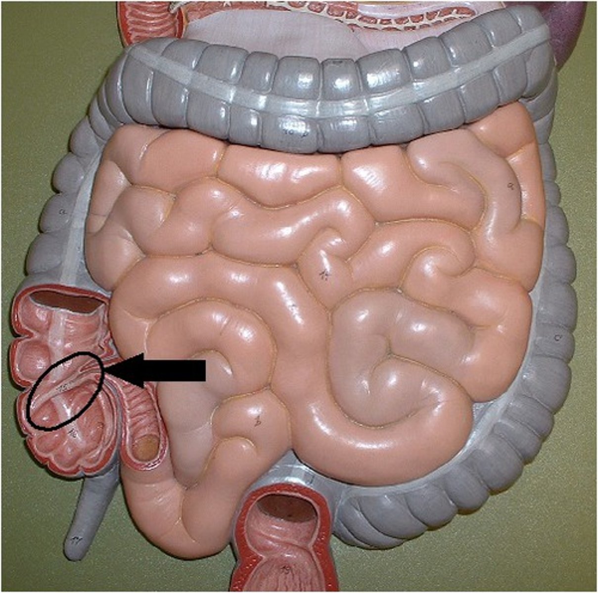
Ileocecal valve
Valve at the junction of the small and large intestines
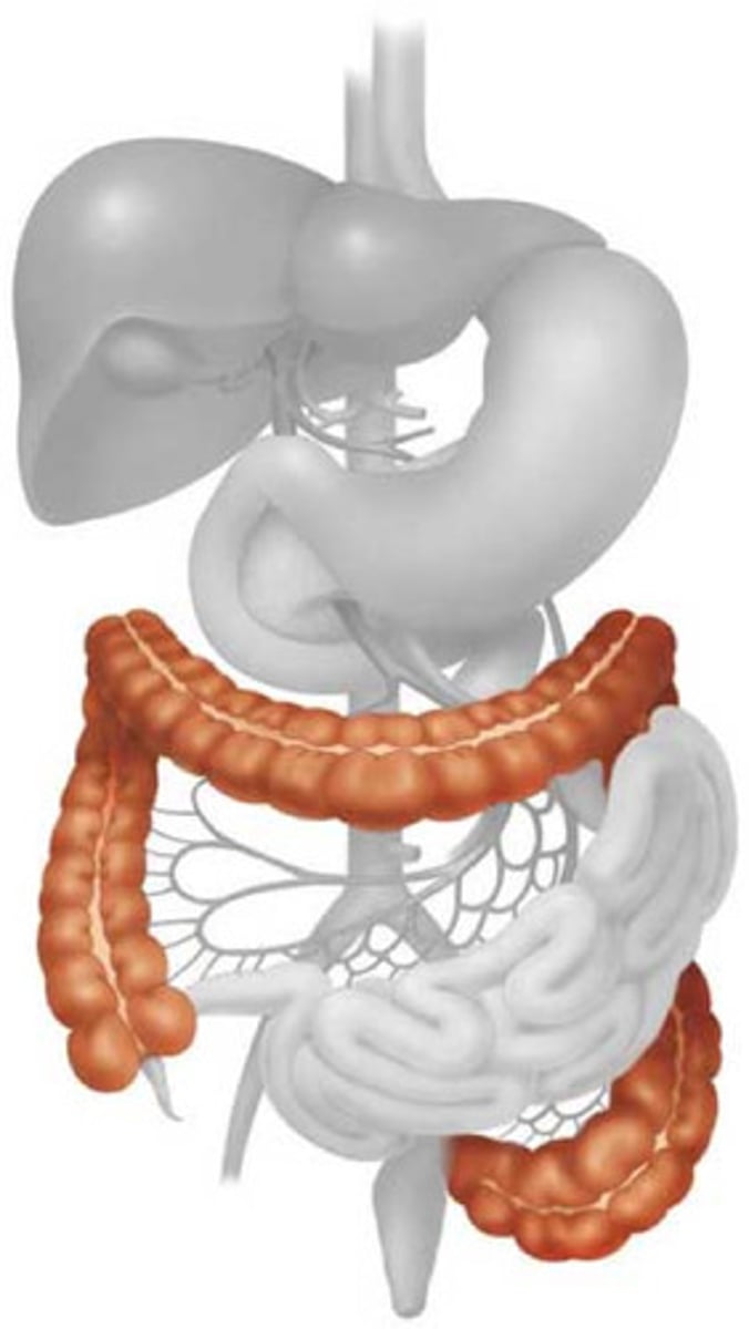
Large Intestine
The last section of the digestive system, where water is absorbed from food and the remaining material is eliminated from the body
Teniae Coli
three bands of longitudinal smooth muscle in muscularis
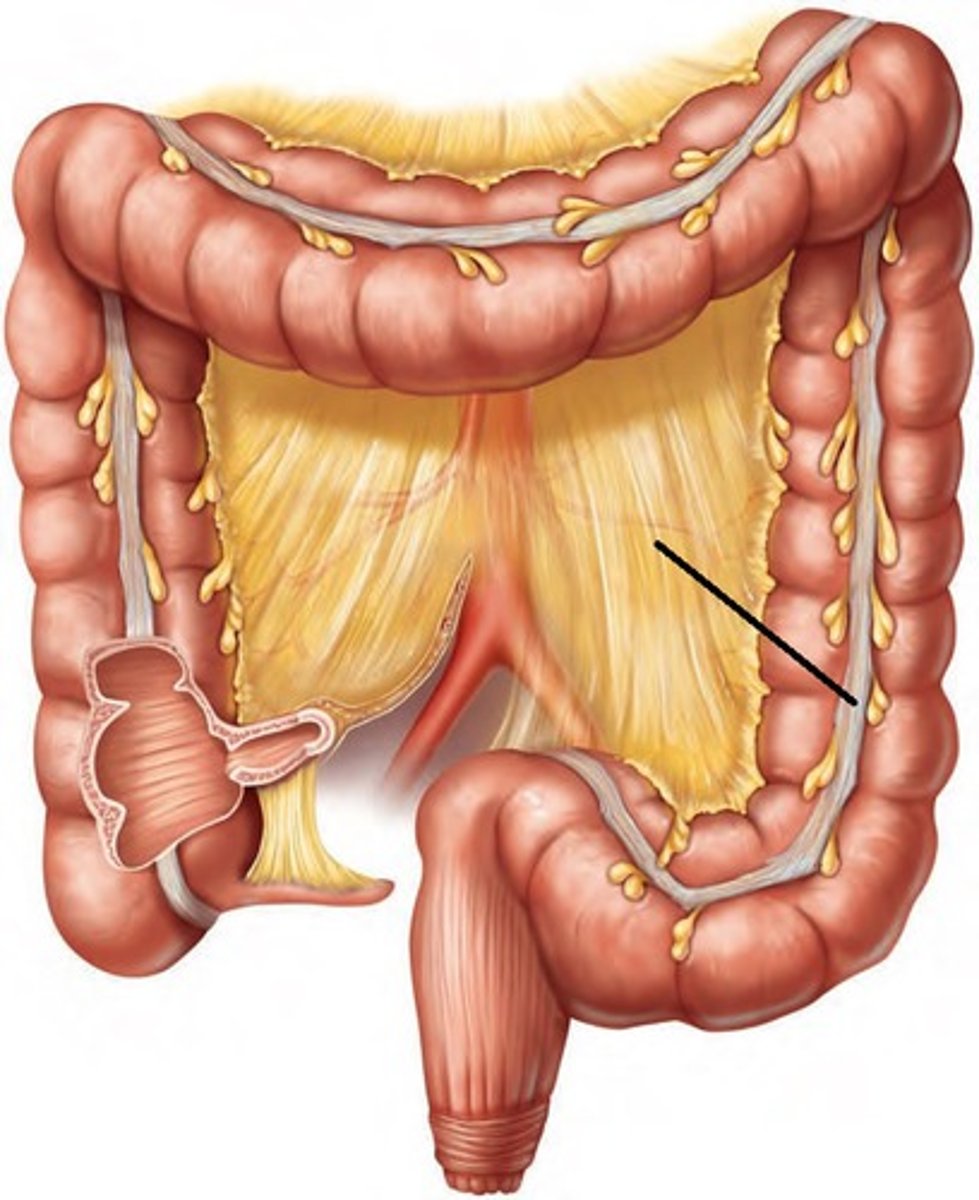
Haustrum
small pouches caused by sacculation, which give the colon its segmented appearance
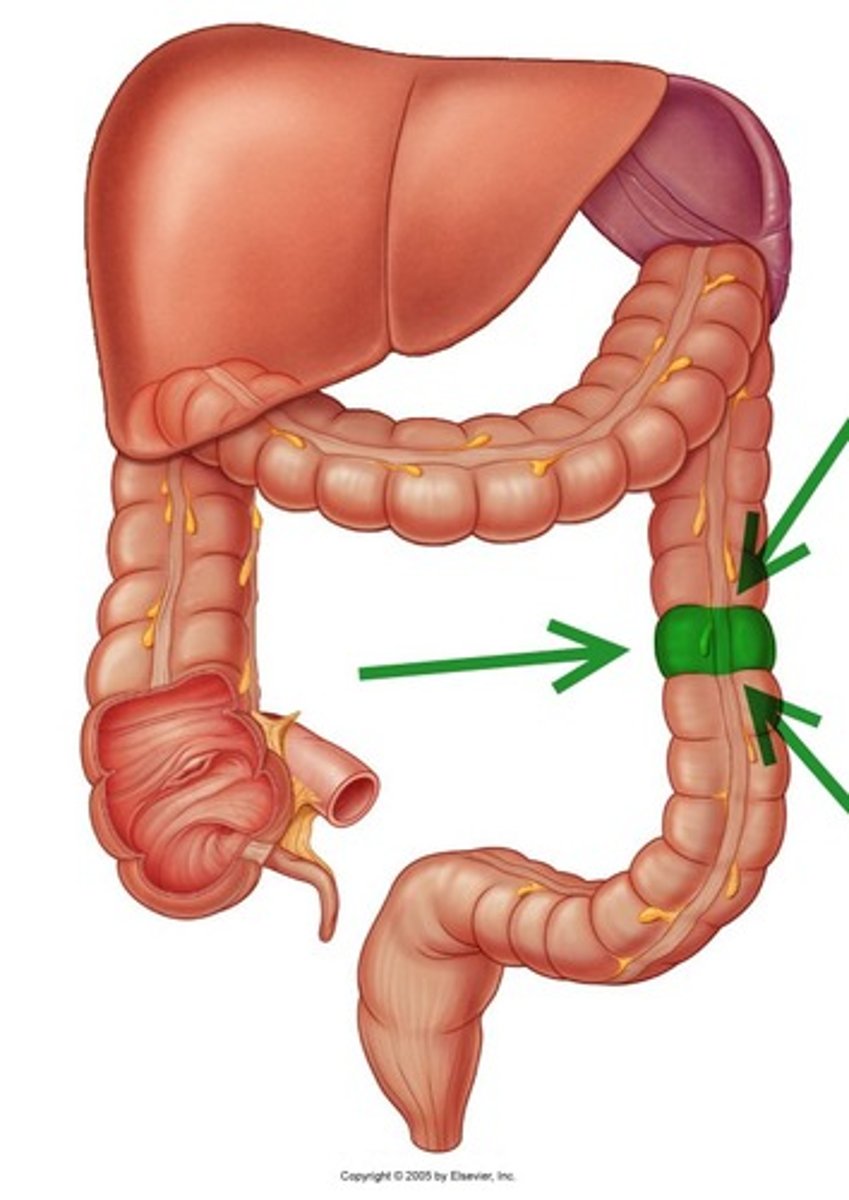
Omental appendices
Lobules of fat hanging off external surface of haustra
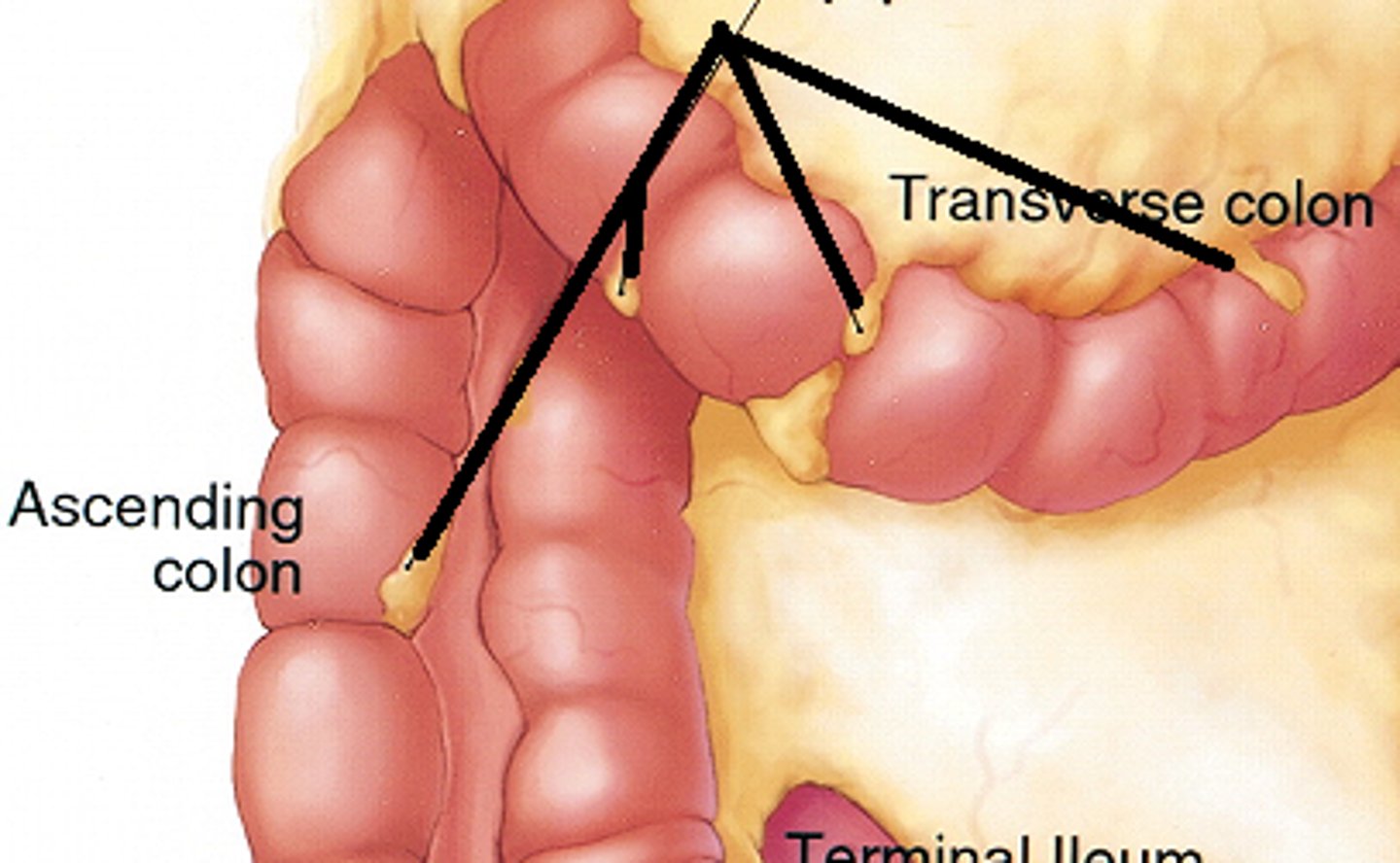
Cecum
a pouch connected to the junction of the small and large intestines.
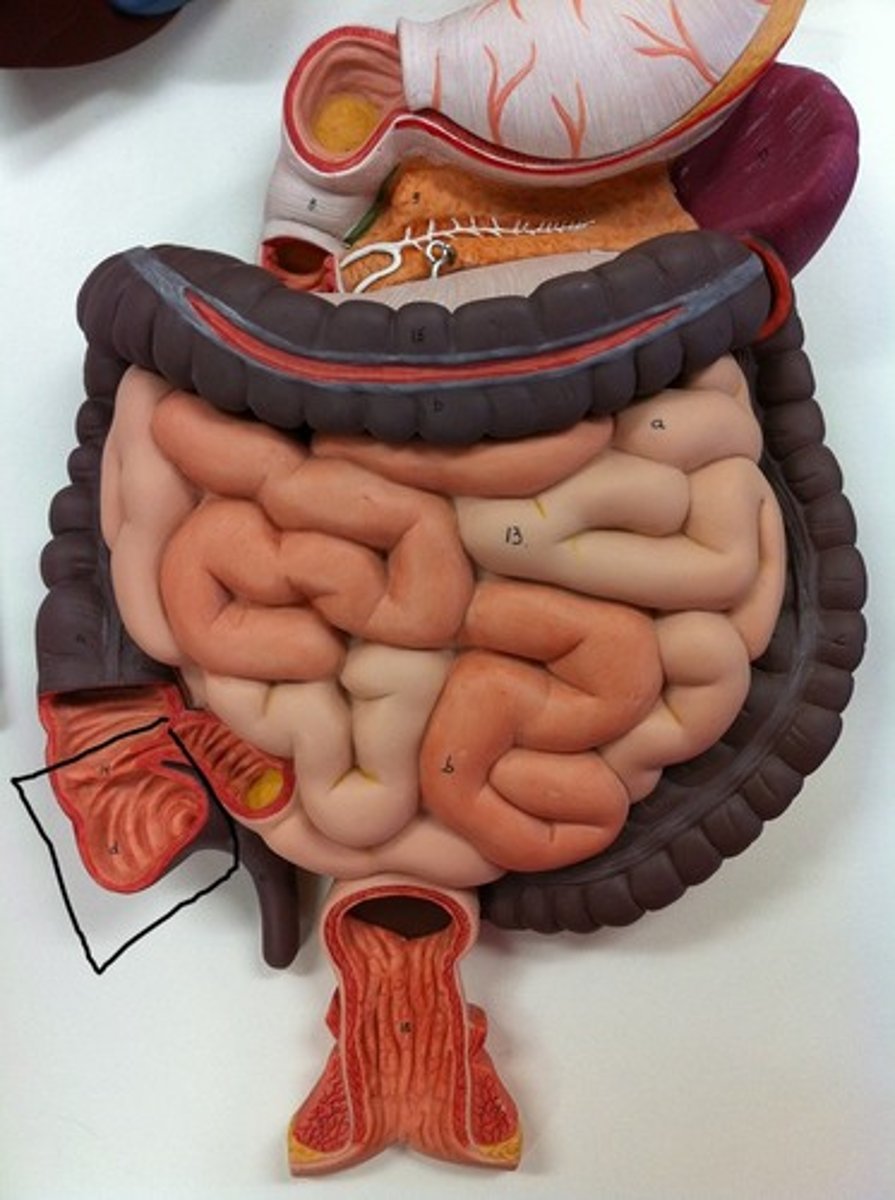
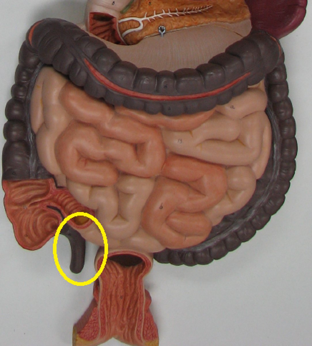
Vermiform appendix
worm-like projection of lymphatic tissue hanging off the cecum with no digestive function; may help to resist infection
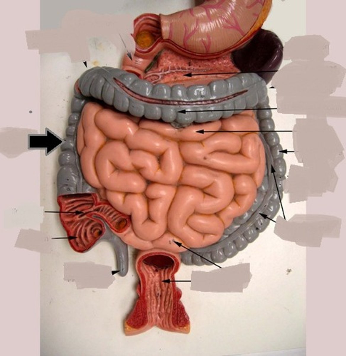
Ascending colon
travels upward from the cecum to the undersurface of the liver
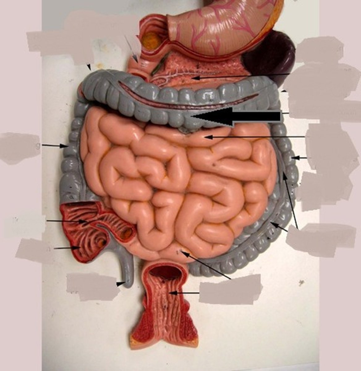
Transverse colon
passes horizontally from right to left toward the spleen
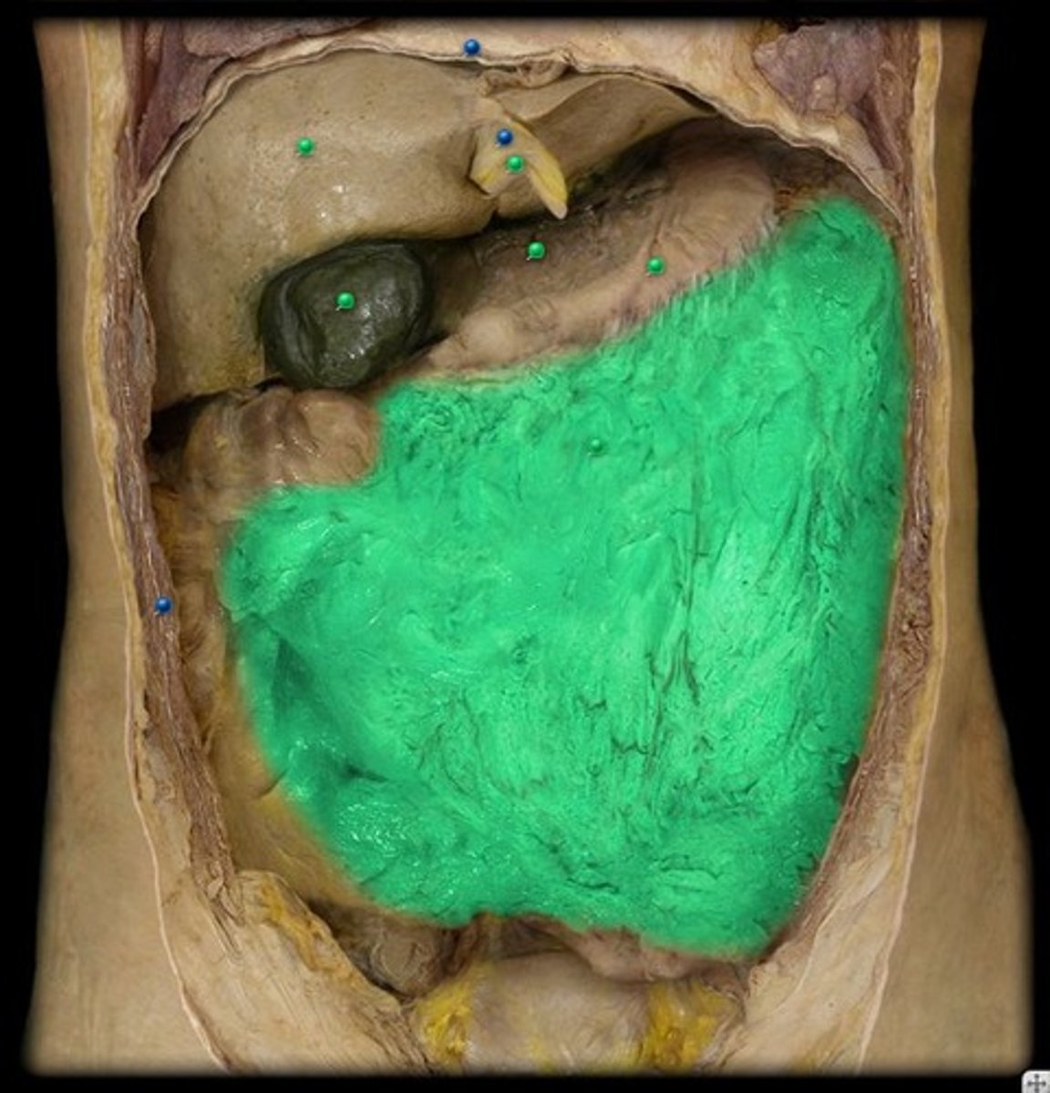
Greater omentum
a large apron-like fold of visceral peritoneum that hangs down from the stomach
Descending colon
portion of the colon that extends downward from the transverse colon
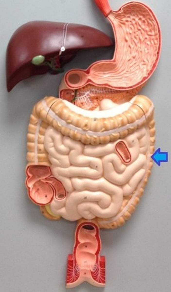
Sigmoid colon
an S-shaped structure that continues from the descending colon above and joins with the rectum below
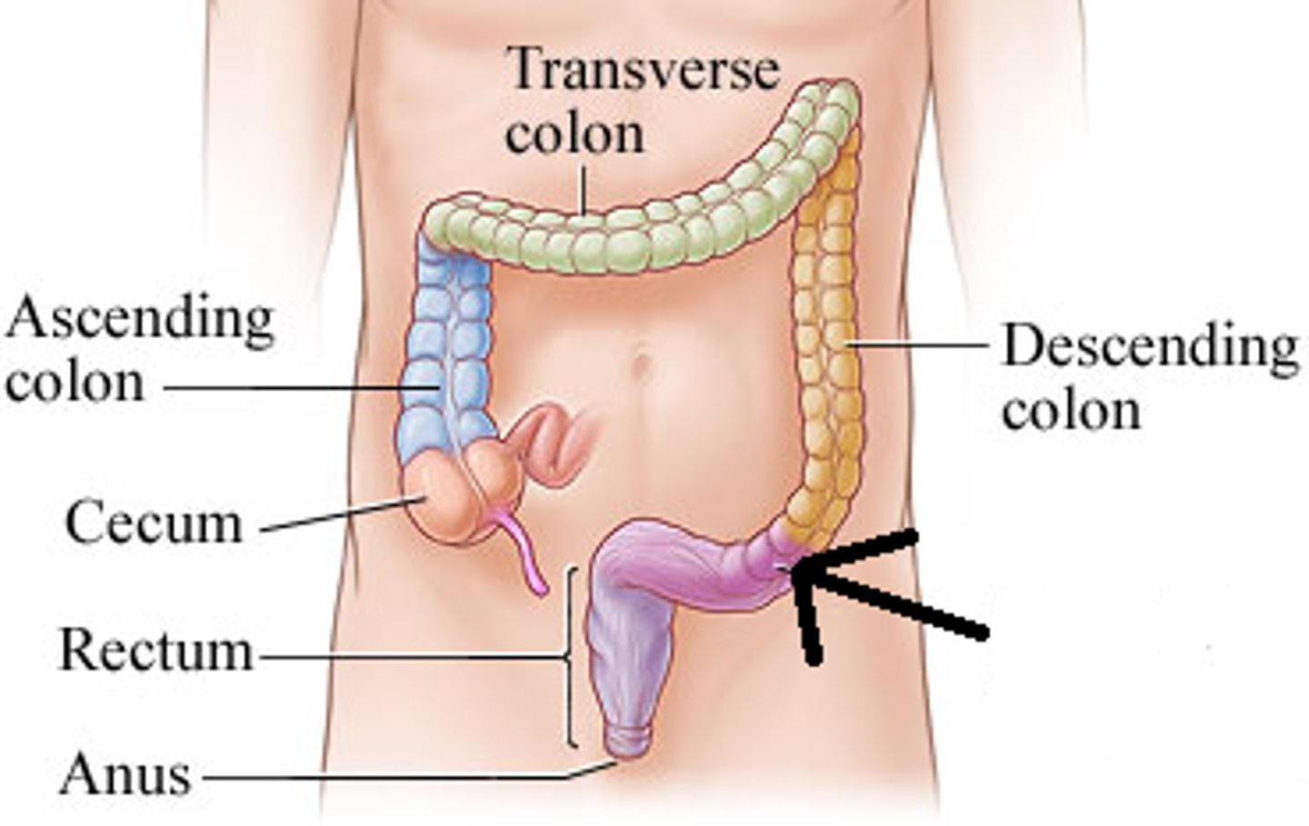
Rectum
A short tube at the end of the large intestine where waste material is compressed into a solid form before being eliminated
Anal canal
region, containing two sphincters, through which feces are expelled from the body
Anal sphincter
The valve that controls the release of feces from the recturm. It has an internal part made of smooth muscle (thus involuntary) and an external part made of skeletal muscle (thus voluntary).
Liver
largest internal gland and is situated under the diaphragm in the upper right part of the abdominal cavity
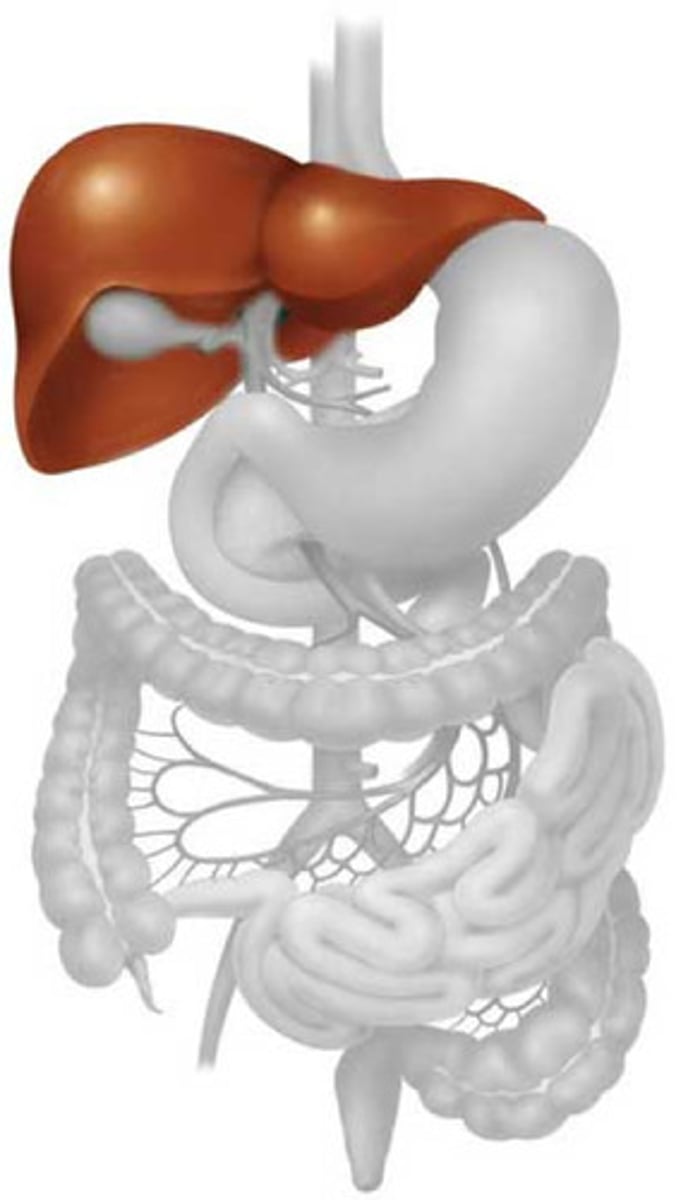
Gallbladder
A muscular sac attached to the liver that secretes bile and stores it until needed for digestion; located on the inferior surface of the liver at the medial border of the right lobe. Attached to liver by CT
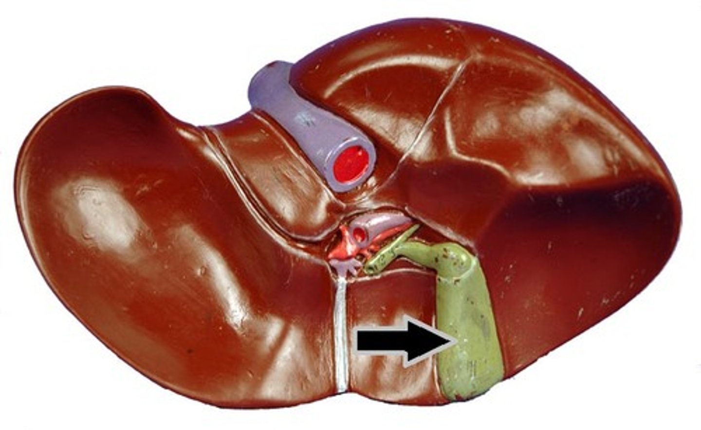
Common Hepatic Duct
large bile duct leading from liver; joins with the cystic duct to form the common bile duct
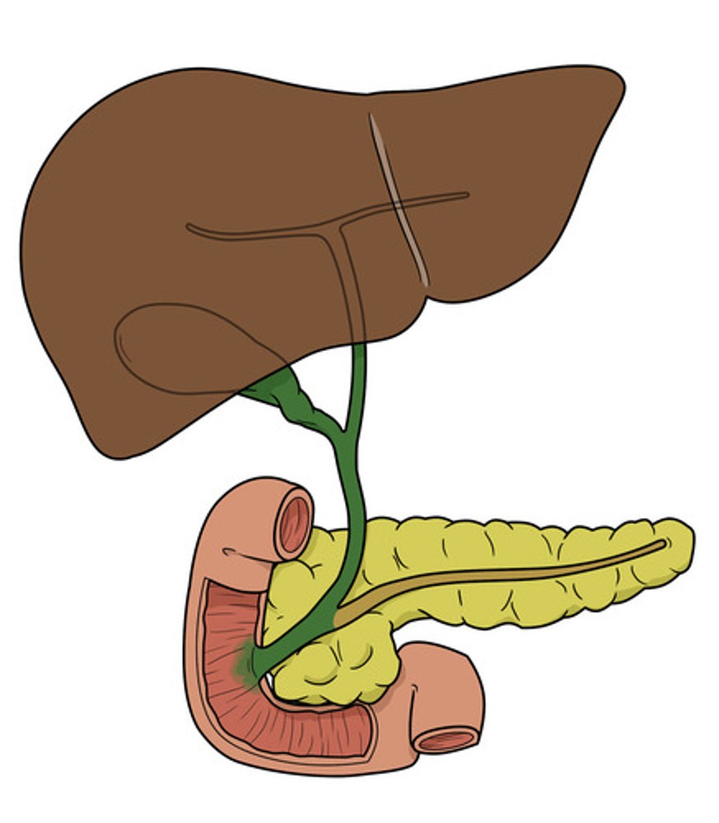
Cystic duct
(D) Duct leading from the gallbladder to the common bile duct; carries bile
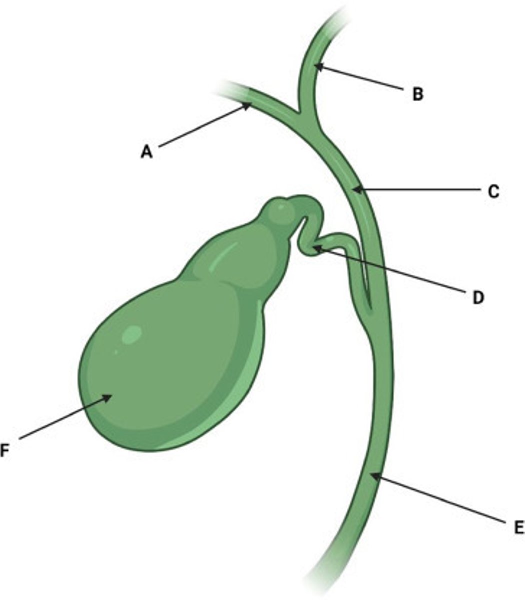
Common bile duct
carries bile from the liver and gallbladder to the duodenum; runs from the junction of the common hepatic duct and cystic duct
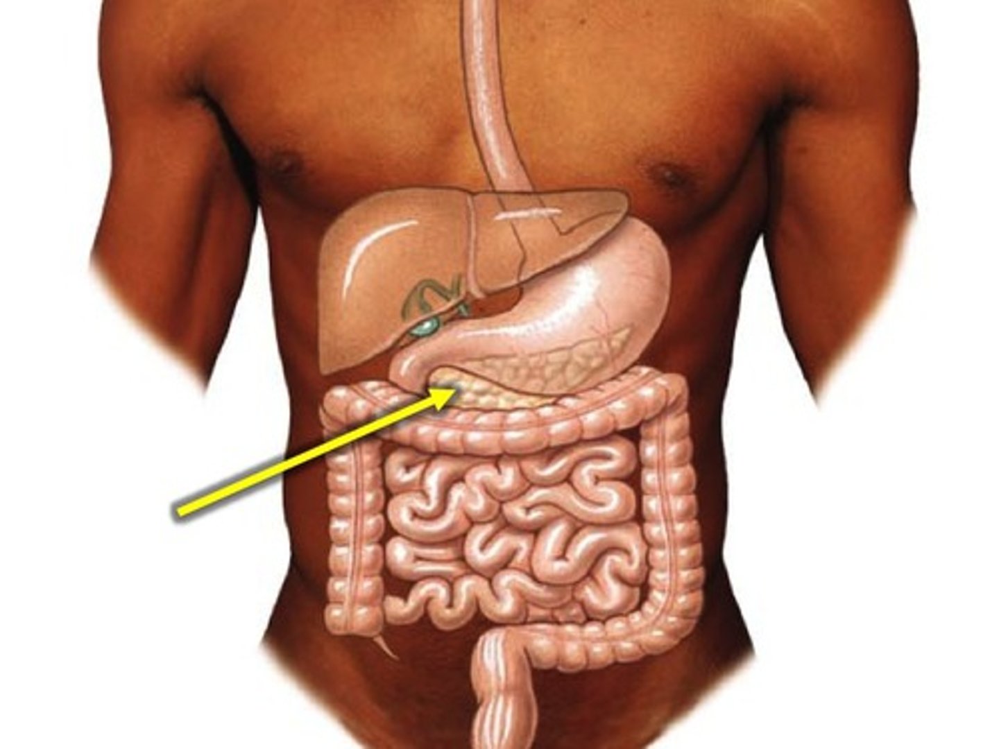
pancreas
located behind the stomach and runs from the inner curve of the duodenum to the vicinity of the spleen
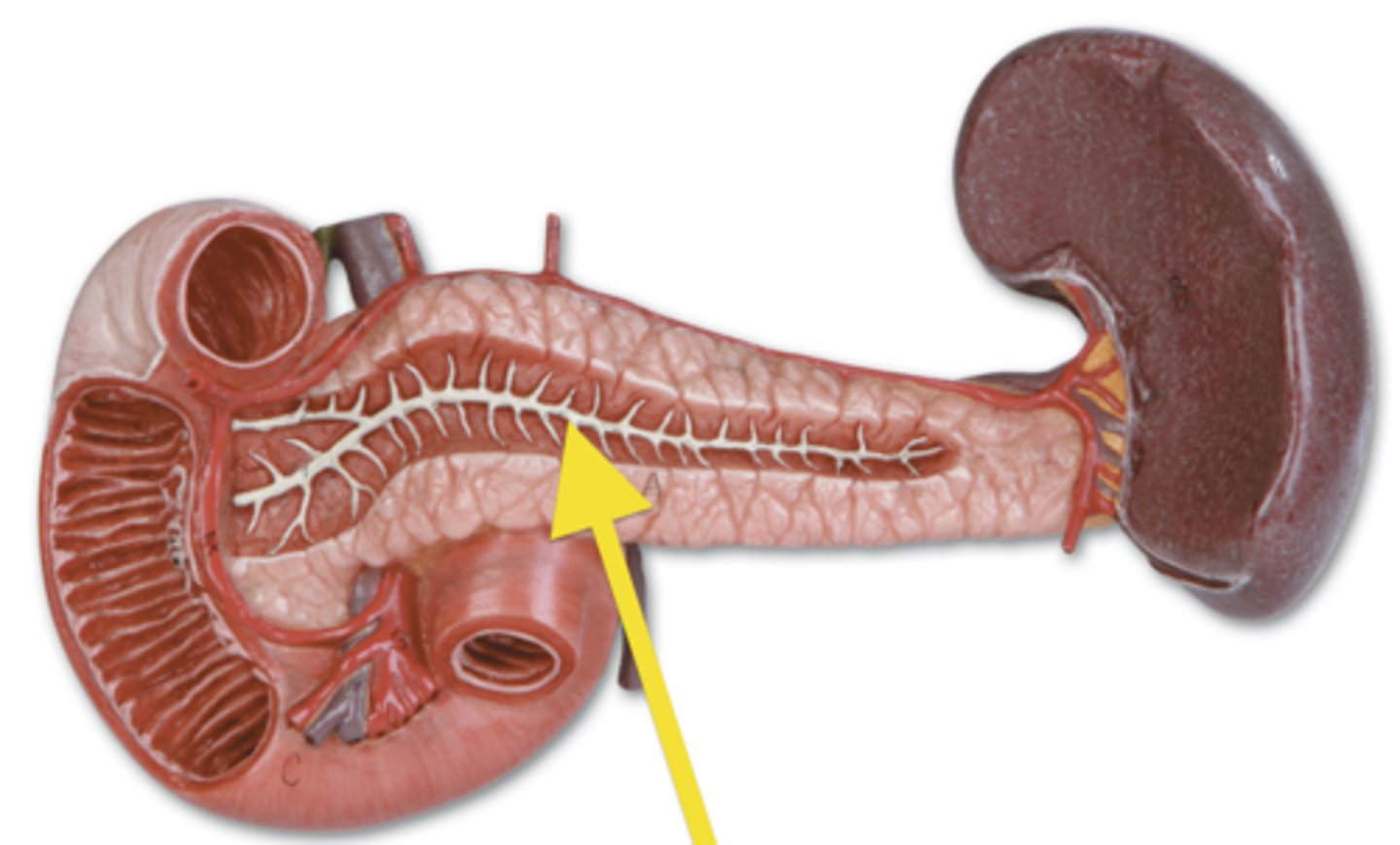
pancreatic duct
running the length of the pancreas to join the common bile duct at the duodenum
Celiac trunk
Large unpaired branch of the abdominal aorta that supplies the liver, stomach, and spleen.
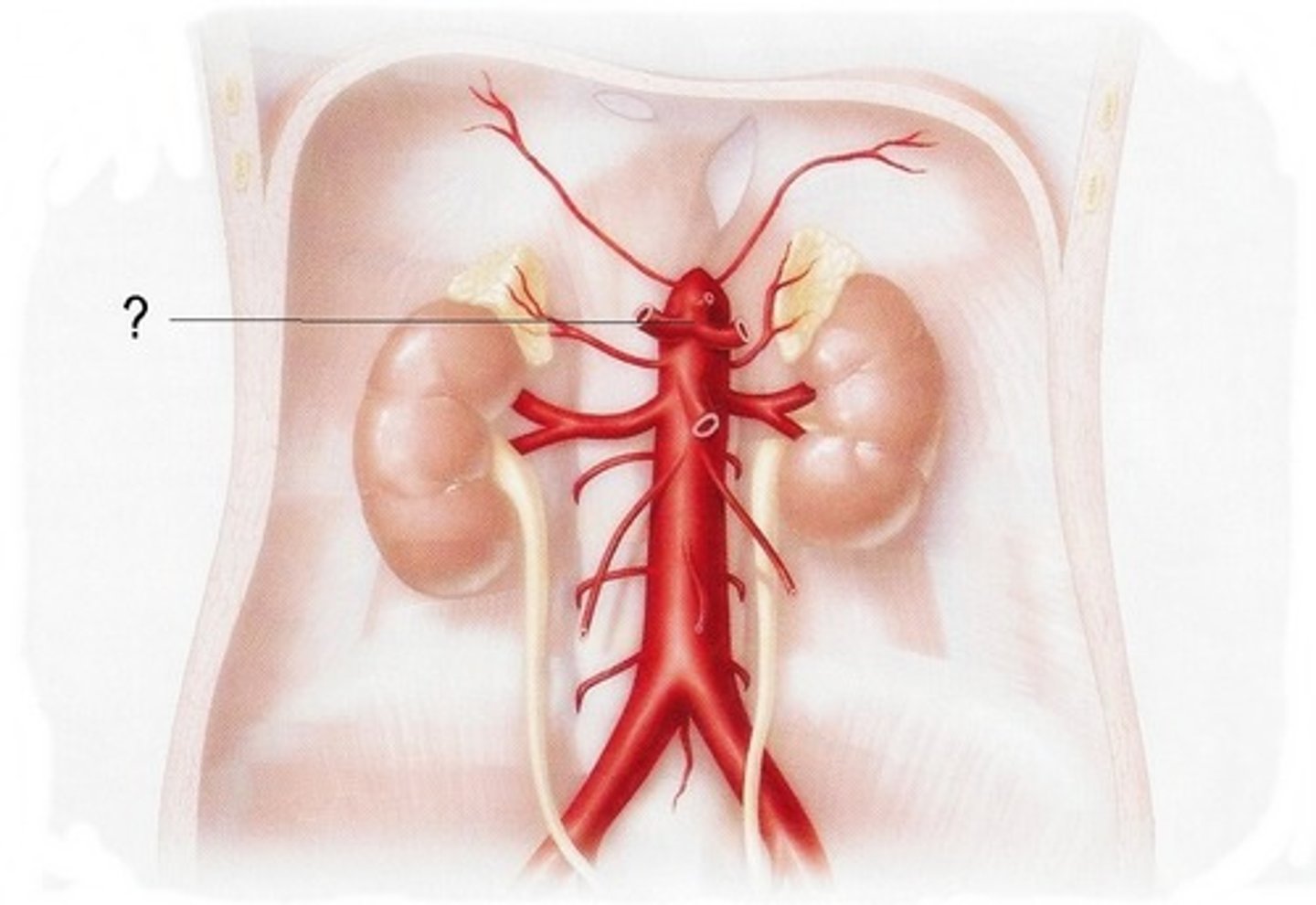
Left Gastric Artery
artery that leads from the celiac trunk to the stomach (on the left)
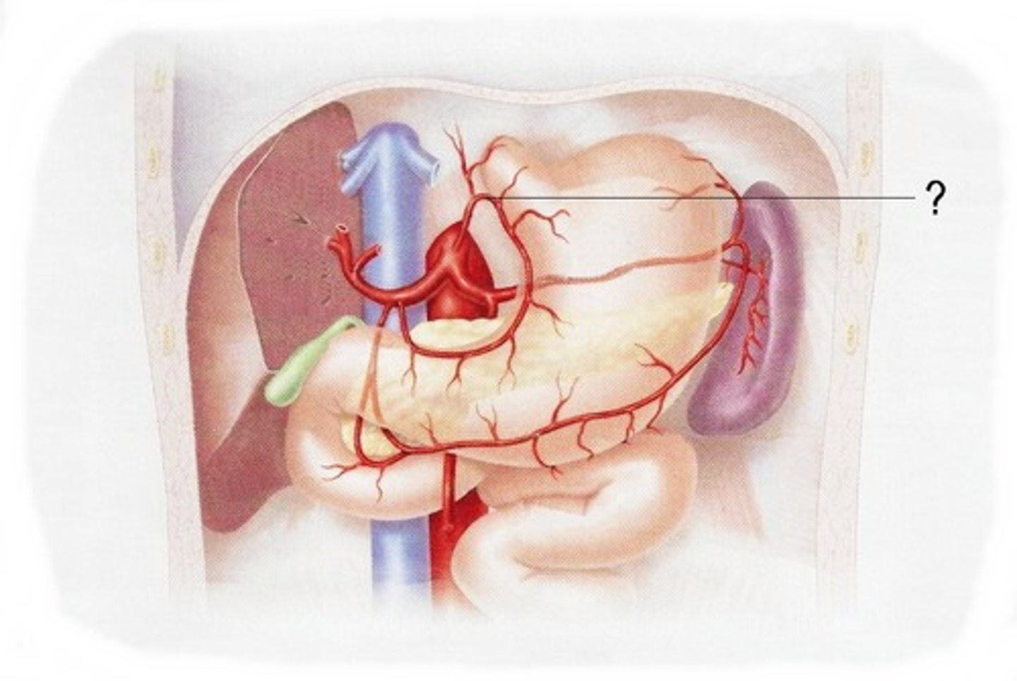
Common Hepatic Artery
arises from the celiac trunk to supply the liver
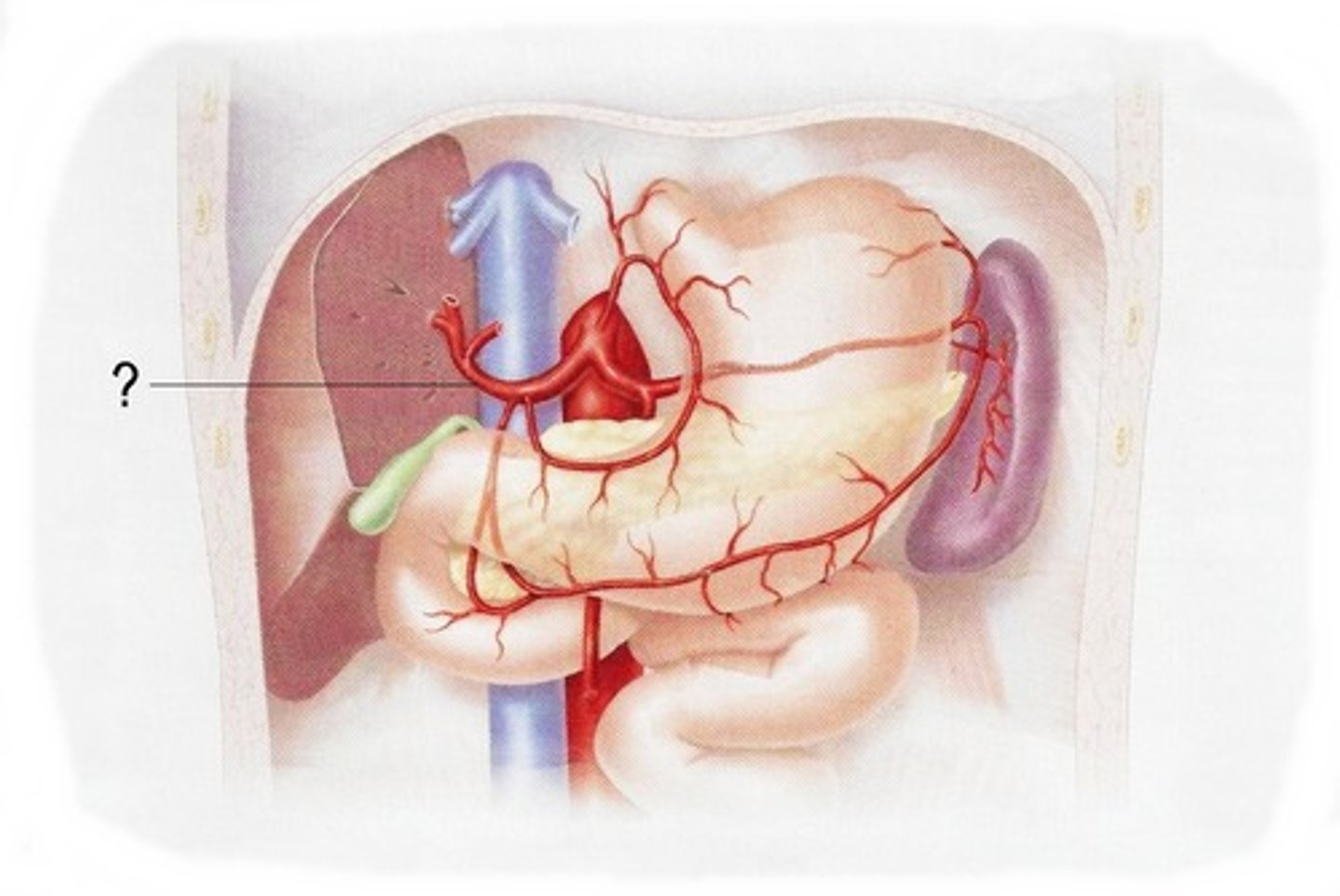
Splenic Artery
supplies the spleen
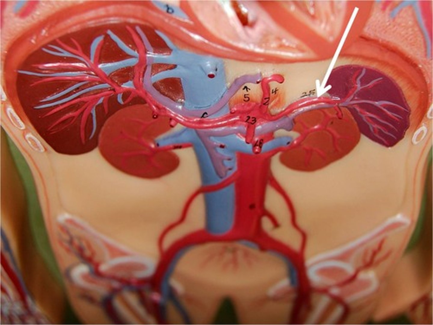
Superior Mesenteric Artery
large abdominal artery; feed large and small intestine
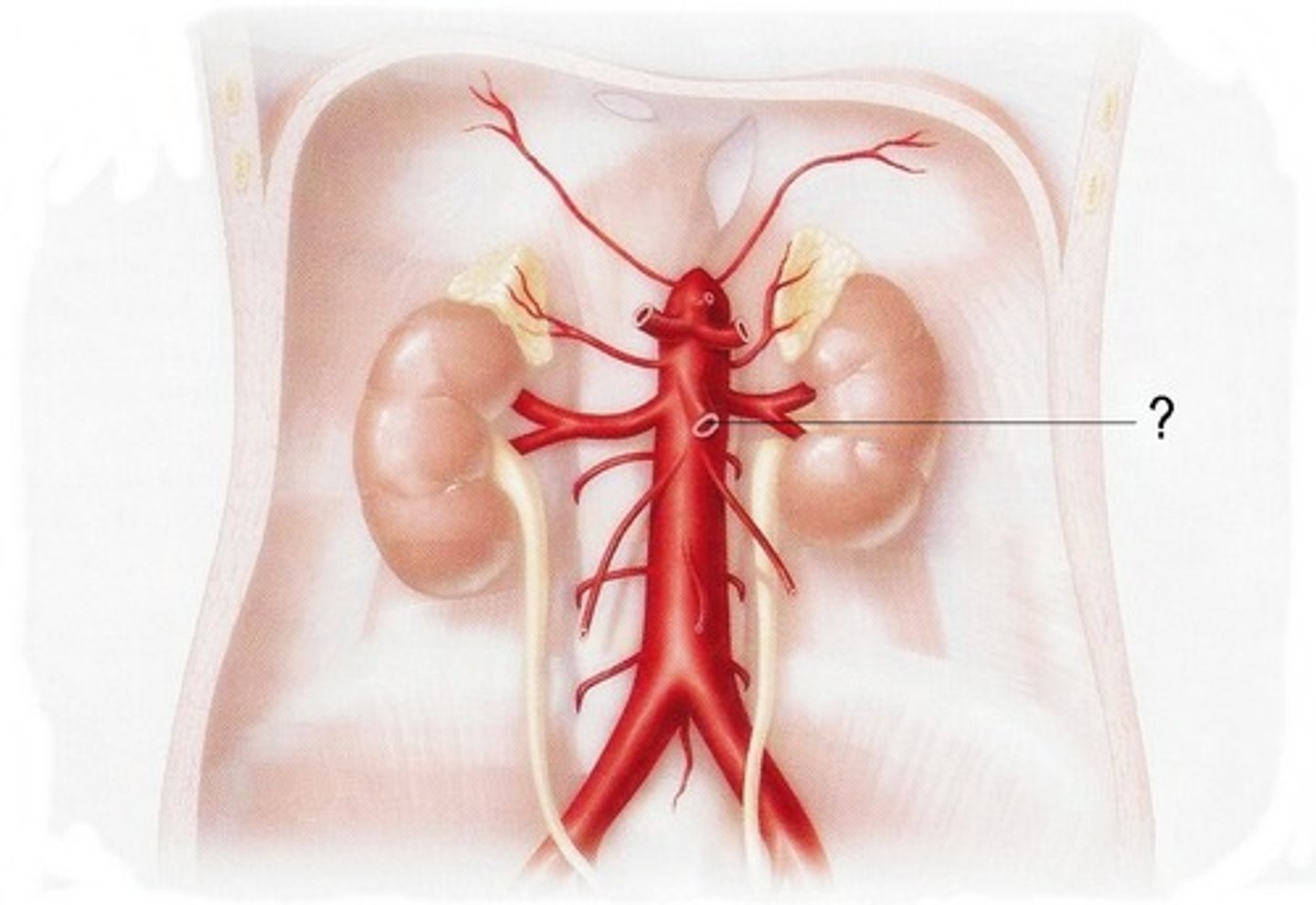
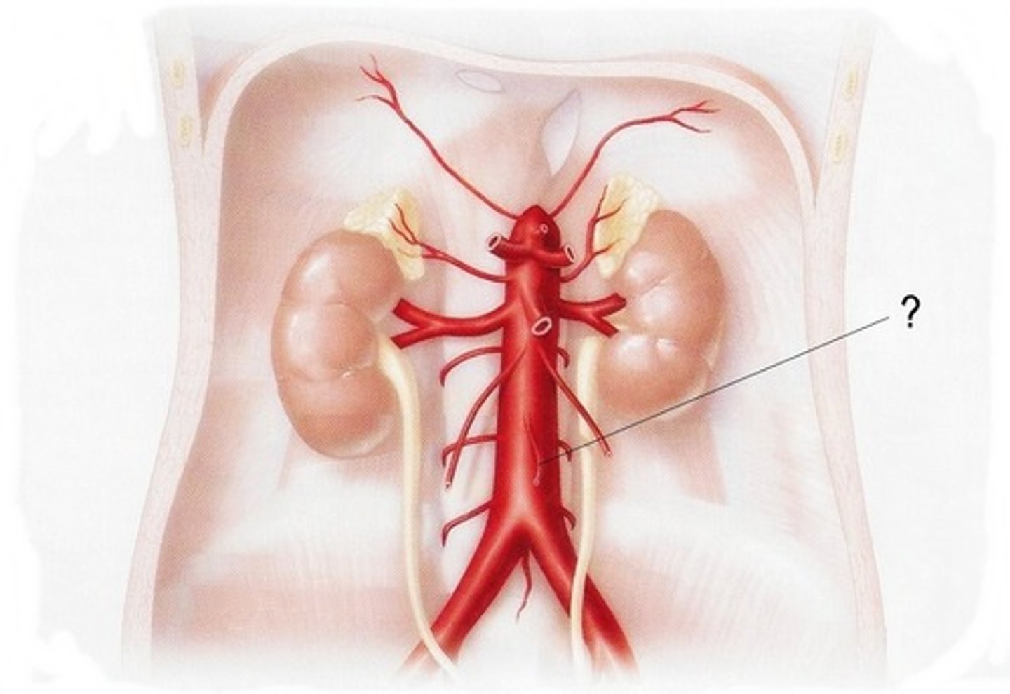
Inferior Mesenteric Artery
Supplies arterial blood to the large intestine.
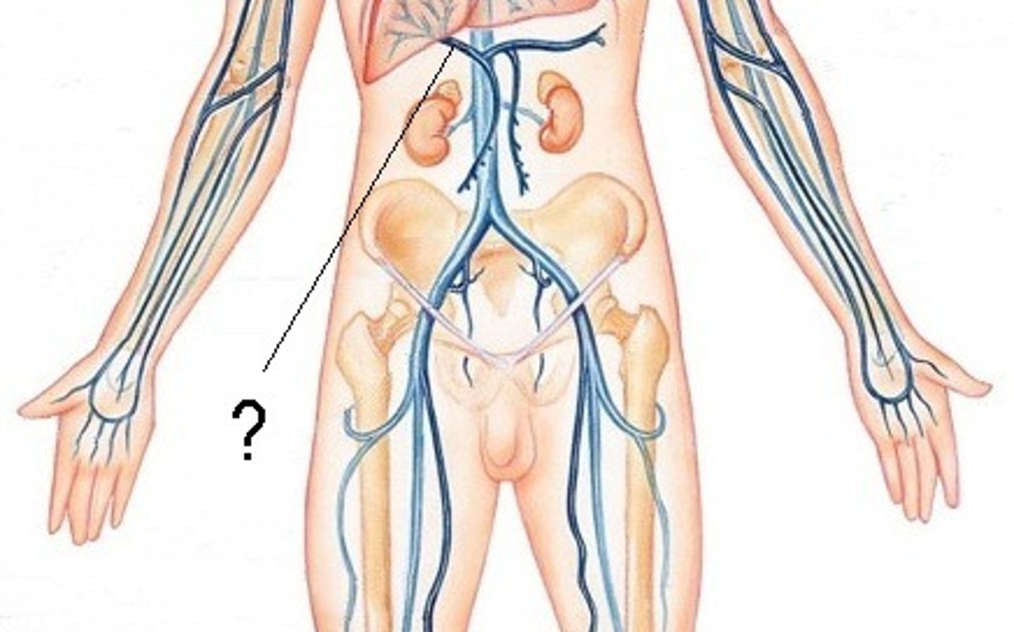
Hepatic Portal Vein
the vein that collects blood from the GI tract and conducts it to the liver
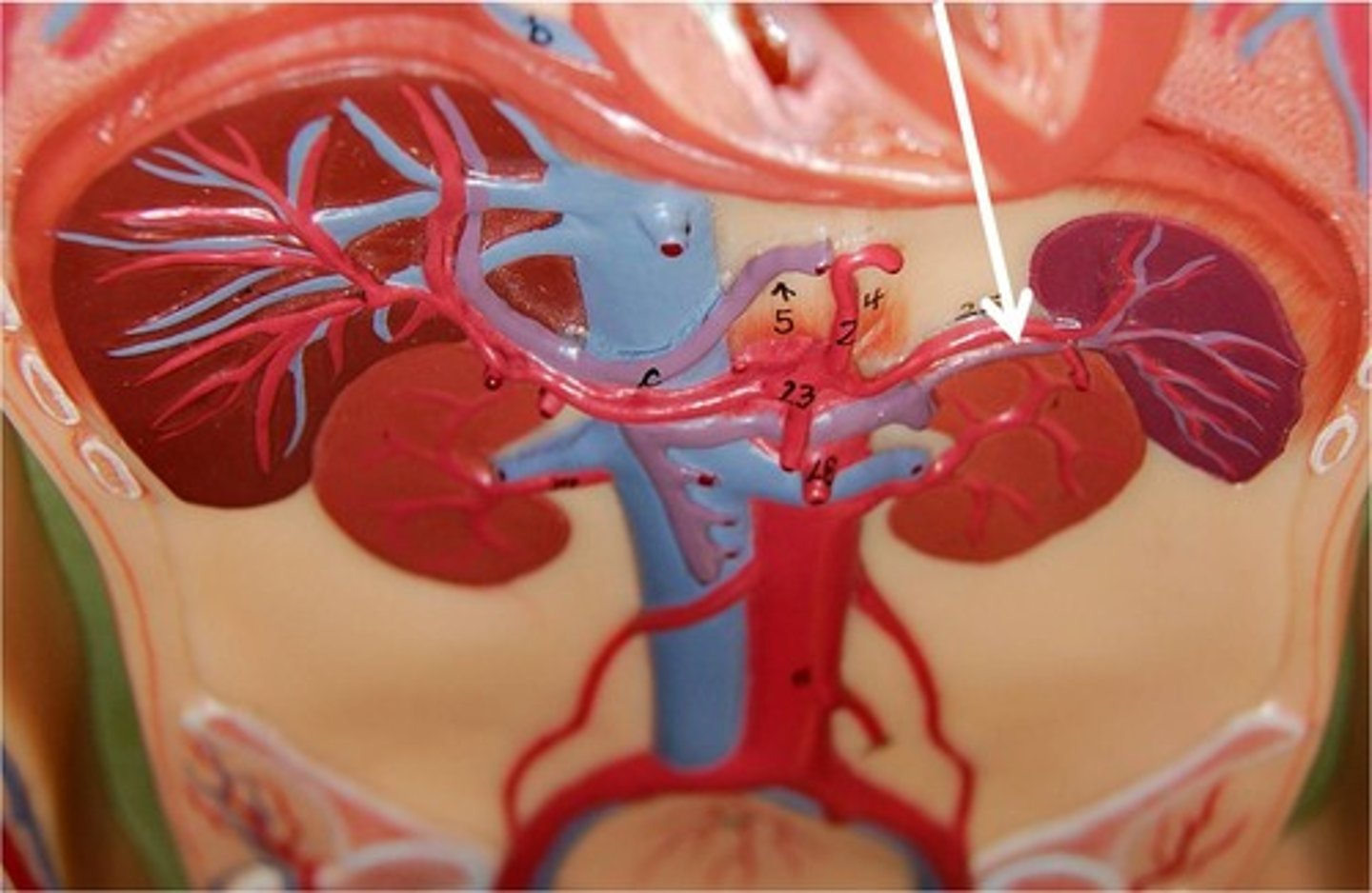
Splenic vein
drains the spleen, pancreas, and stomach
Superior Mesenteric Vein
drains small intestines and ascending colon
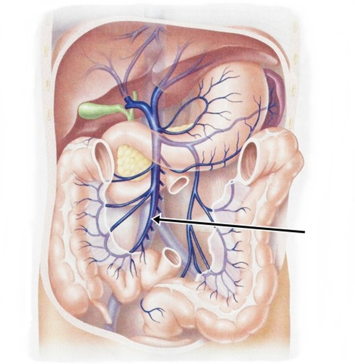
Inferior Mesenteric Vein
drains distal portion of large intestine
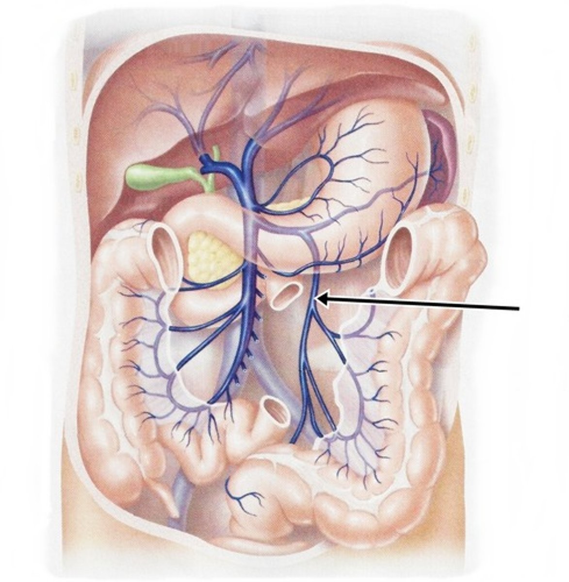
Circumvallate papillae
large papillae with taste buds- on back of tongue
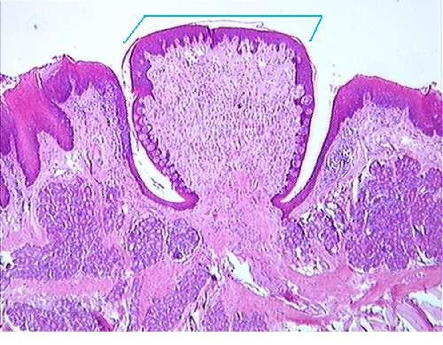
Taste Buds
sensory organs in the mouth that contain the receptors for taste
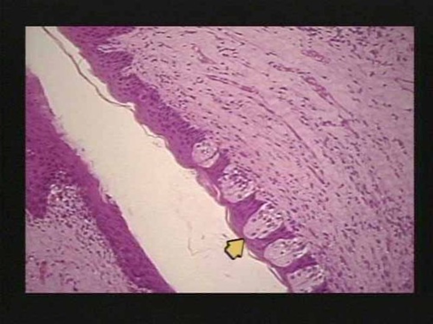
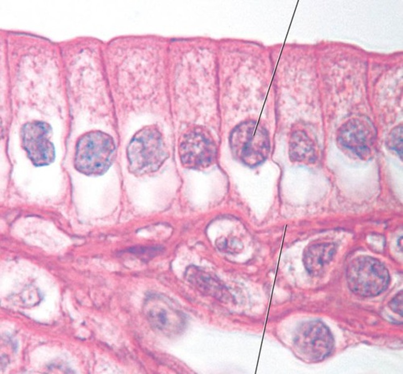
Simple Columnar Epithelium
epithelium that lines the GI tract
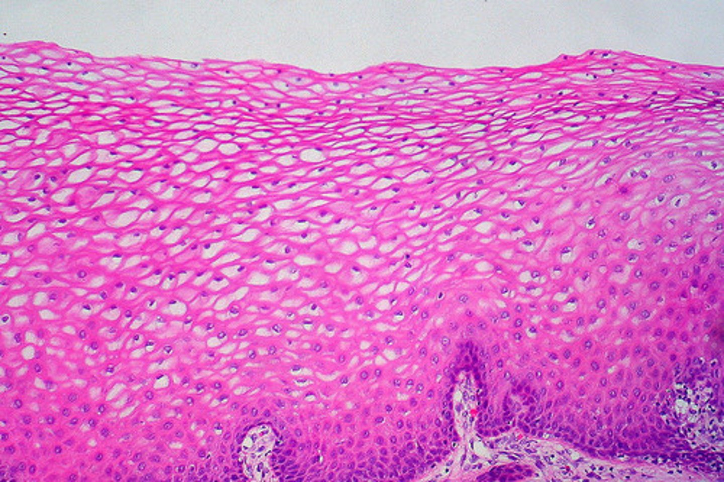
Nonkeratinized Stratified Columnar Epithelium
epithelium that lines the esophagus
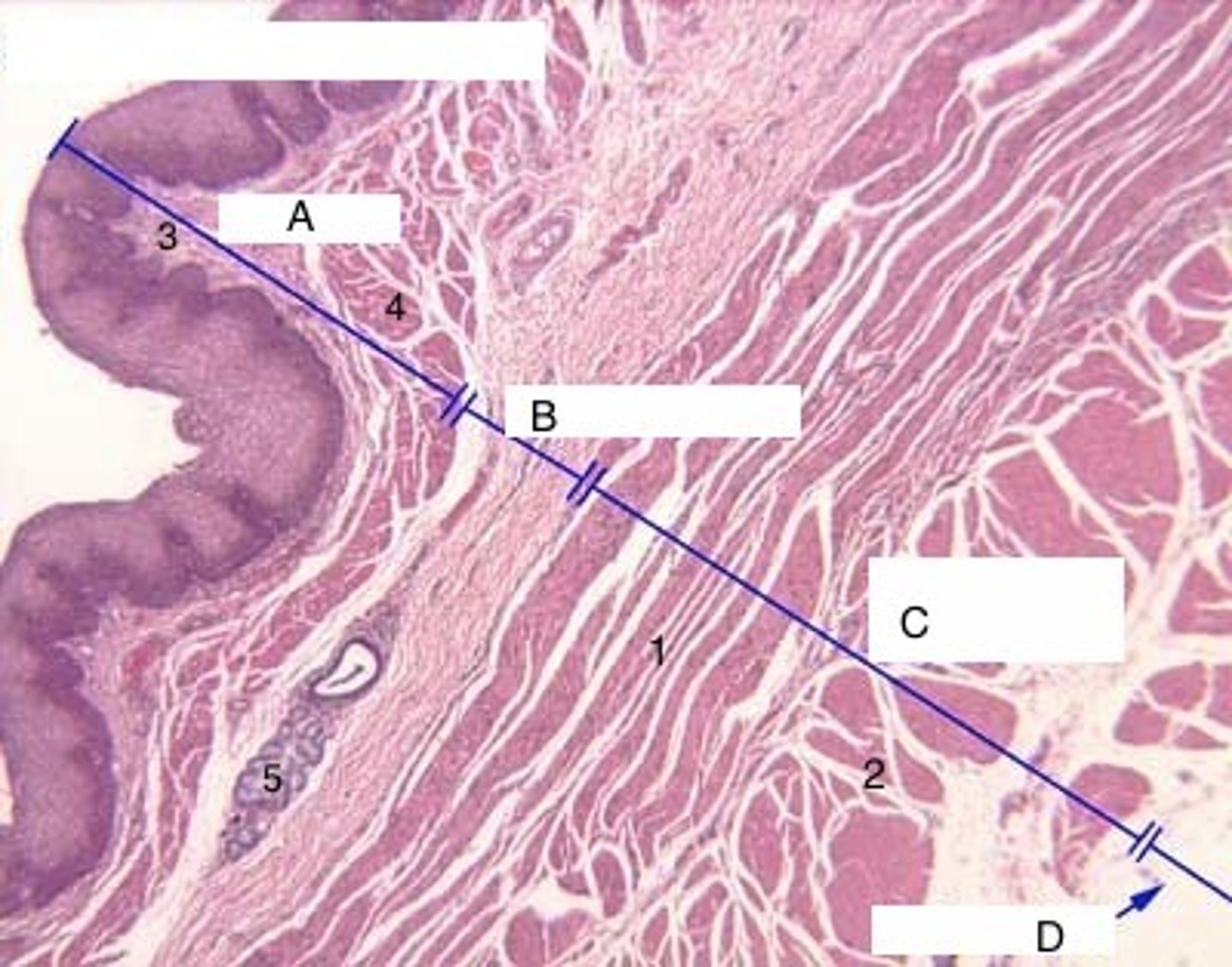
Muscularis of Esophagus
(C) superior third of the esophagus is skeletal muscle, the intermediate third is skeletal and smooth muscle, the inferior third is smooth muscle.
Mucosa of stomach
Simple columnar epithelium
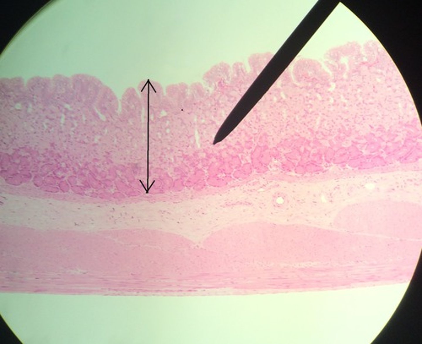
Gastric pits
Gastric glands that release secretions into the stomach
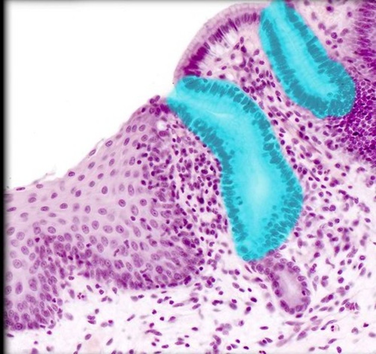
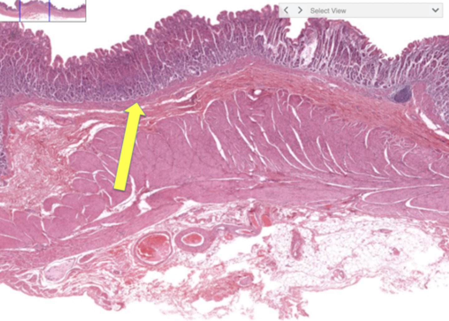
Muscularis mucosae of stomach
smooth muscle that produces local movements of mucosa in the stomach
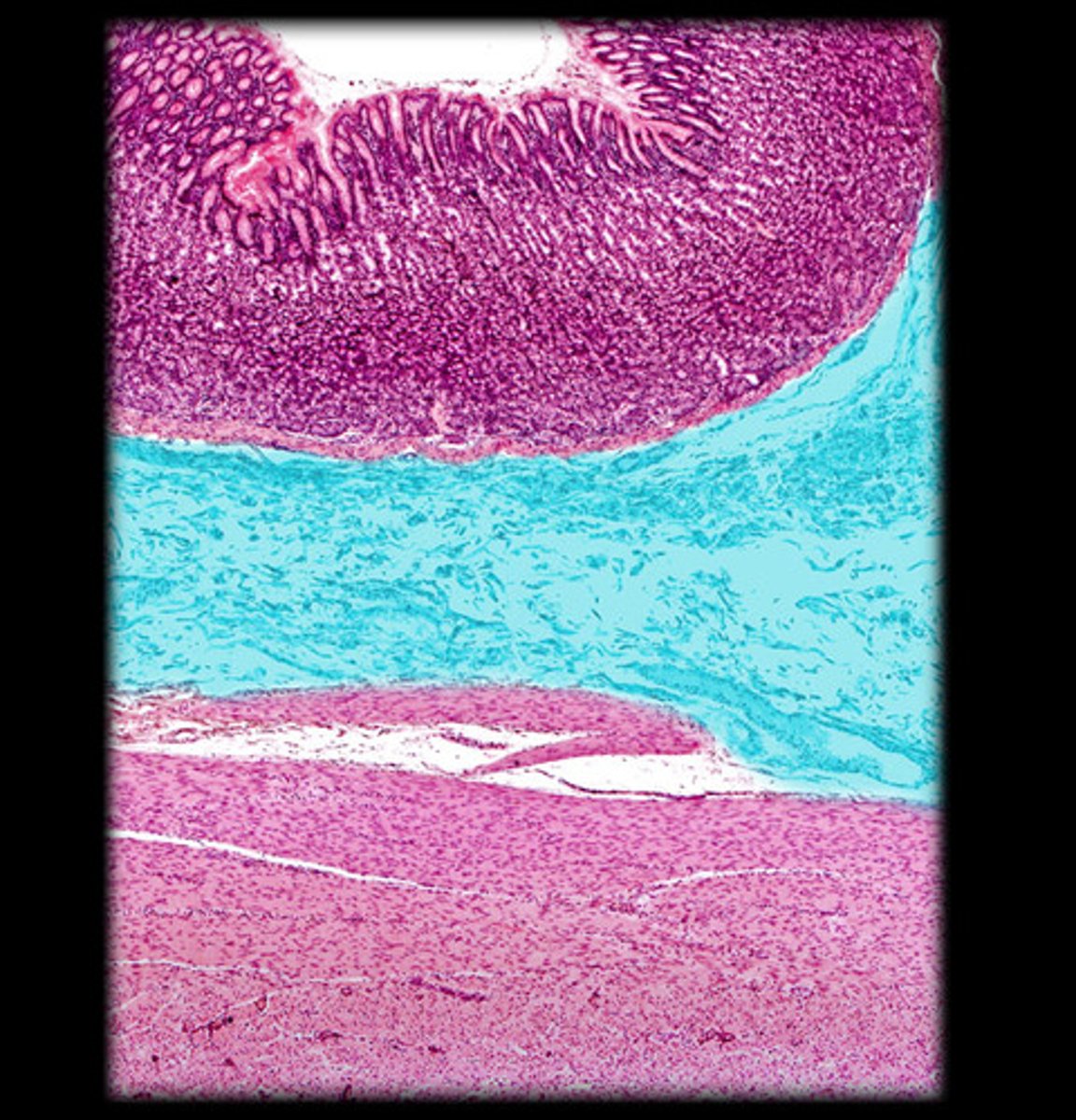
Submucosa of stomach
thicker areas of this layer are the bases of rugae
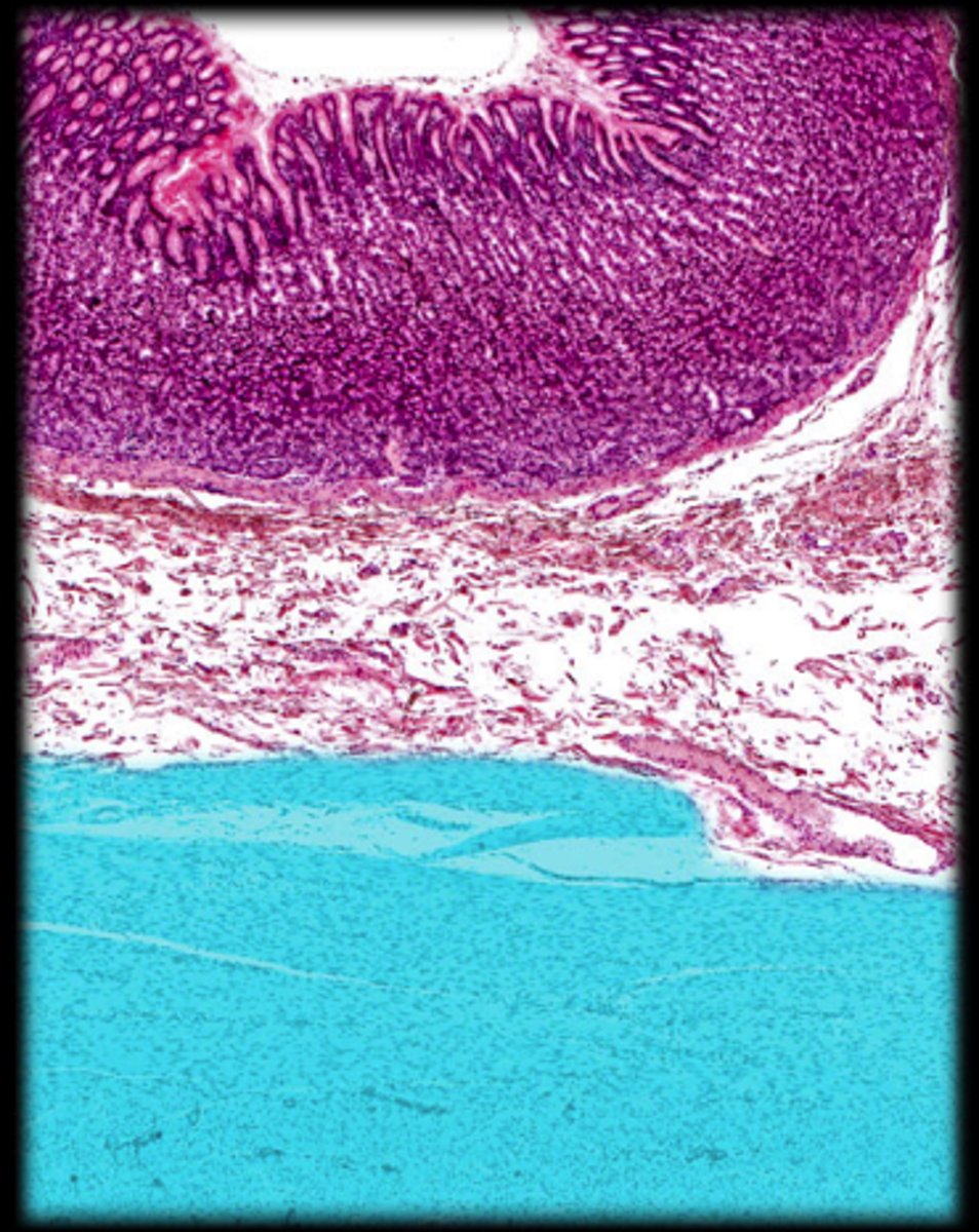
Muscularis of stomach
very thick in the stomach. 3 layers
Mucosa of Small Intestine
(Black) simple columnar epithelium
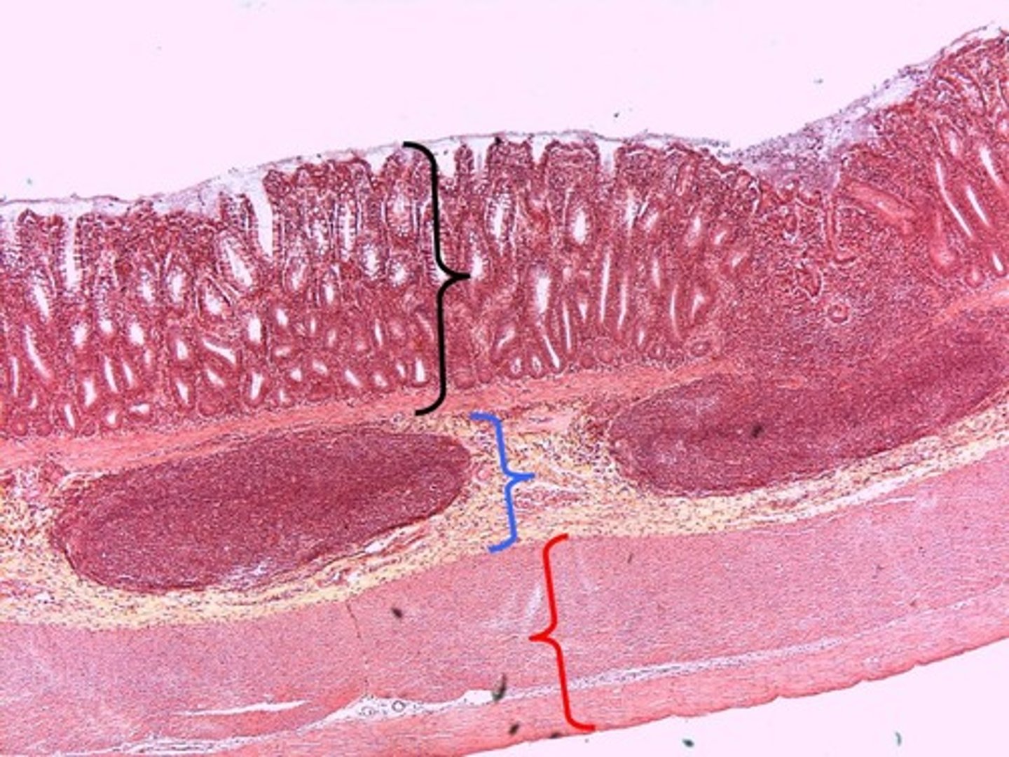
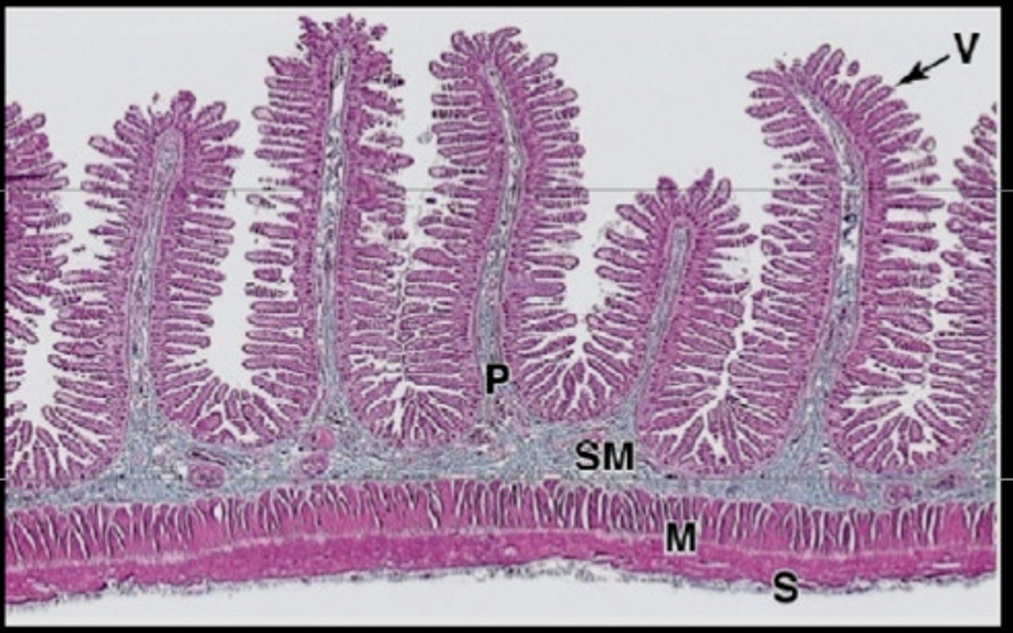
Villi
Fingerlike extensions of the intestinal mucosa that increase the surface area for absorption
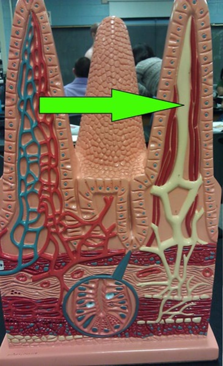
Lacteal
a lymph tubule located in the villus that absorbs fatty acids
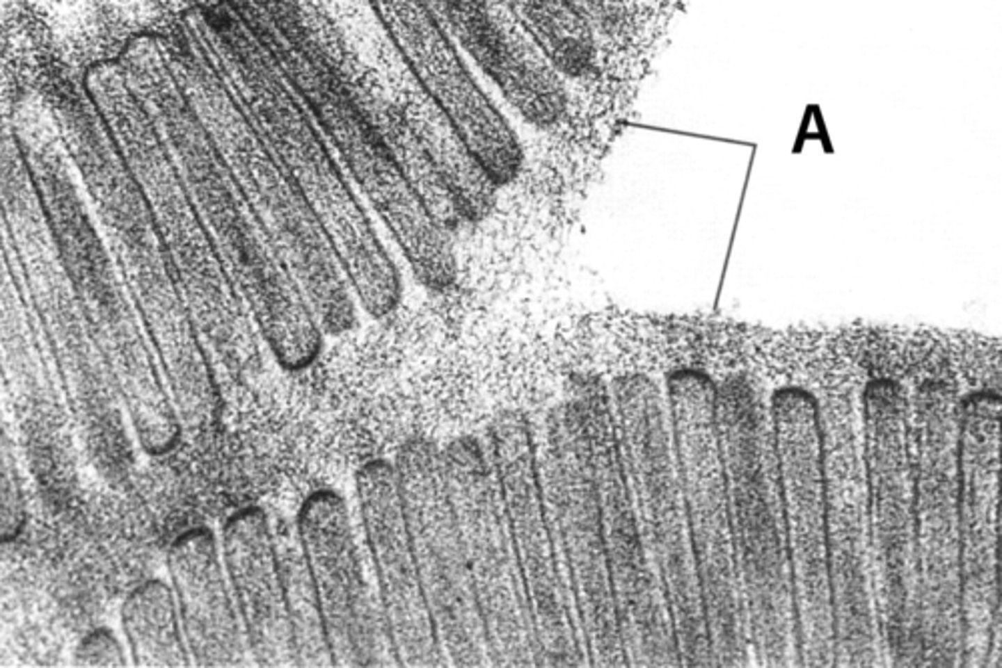
Brush Border
Surface of a cell covered with microvilli. increases surface area of a cell for absorption; in small intestine
Intestinal glands
produce an enzyme-poor "juice" containing mucus; found in the submucosa of the small intestine
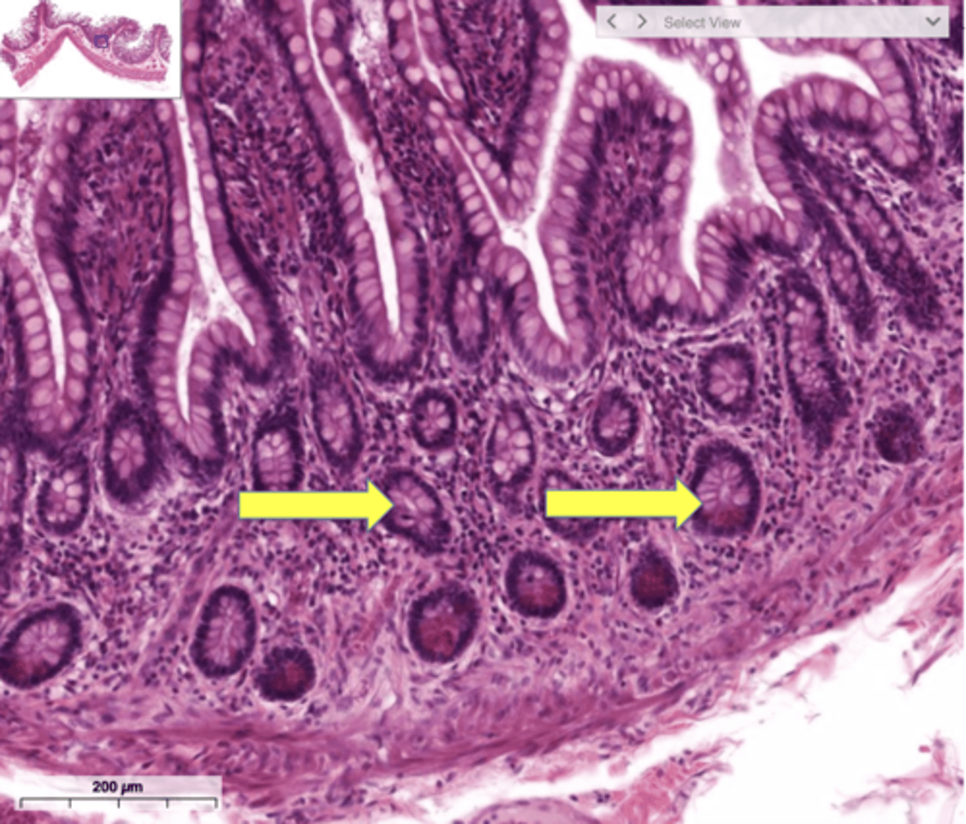
Muscularis mucosae of small intestine
smooth muscle layer forming the deep border of the mucosa in small intestine
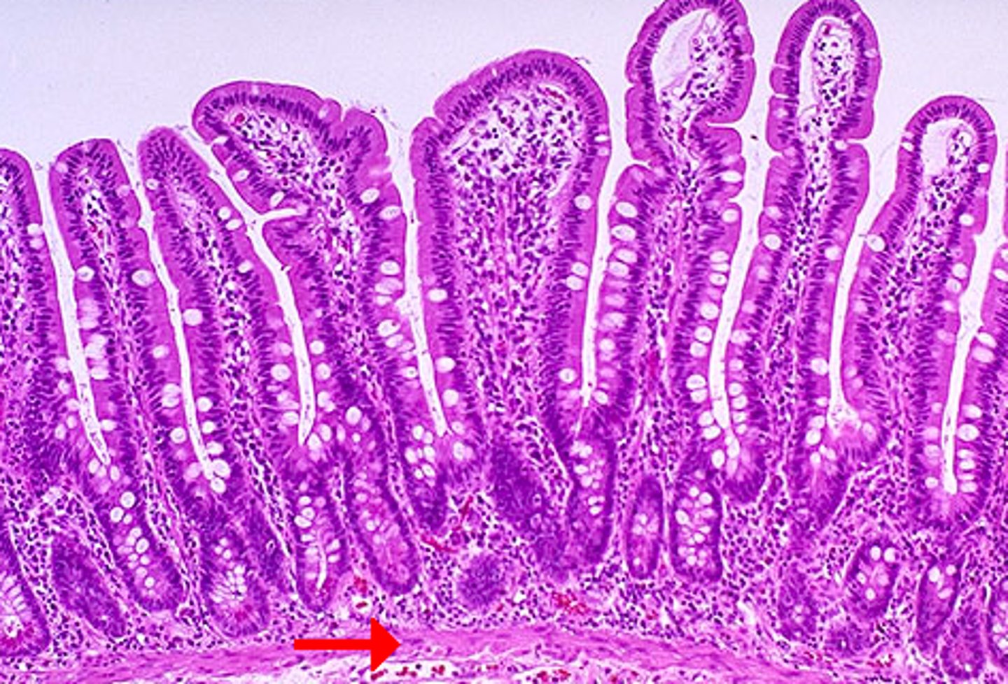
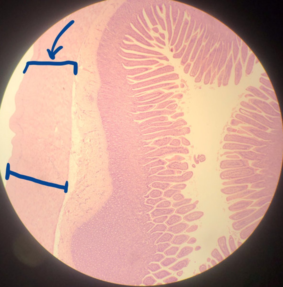
Muscularis of small intestine
2 layers of smooth muscle; outer longitudinal layer and inner circular layer
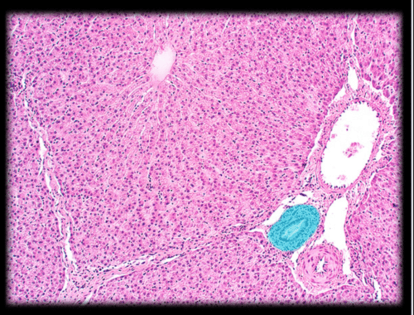
Liver histology
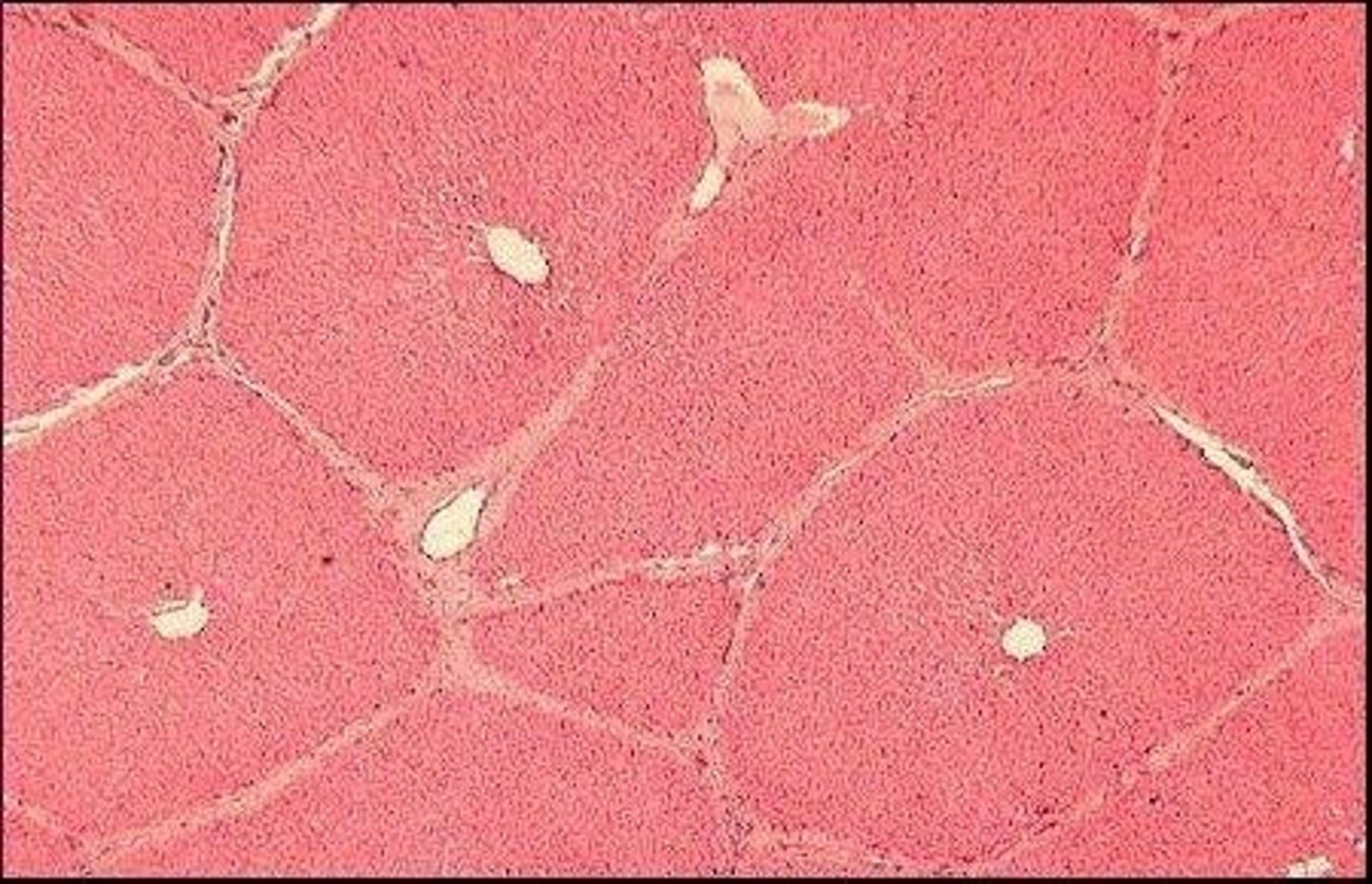
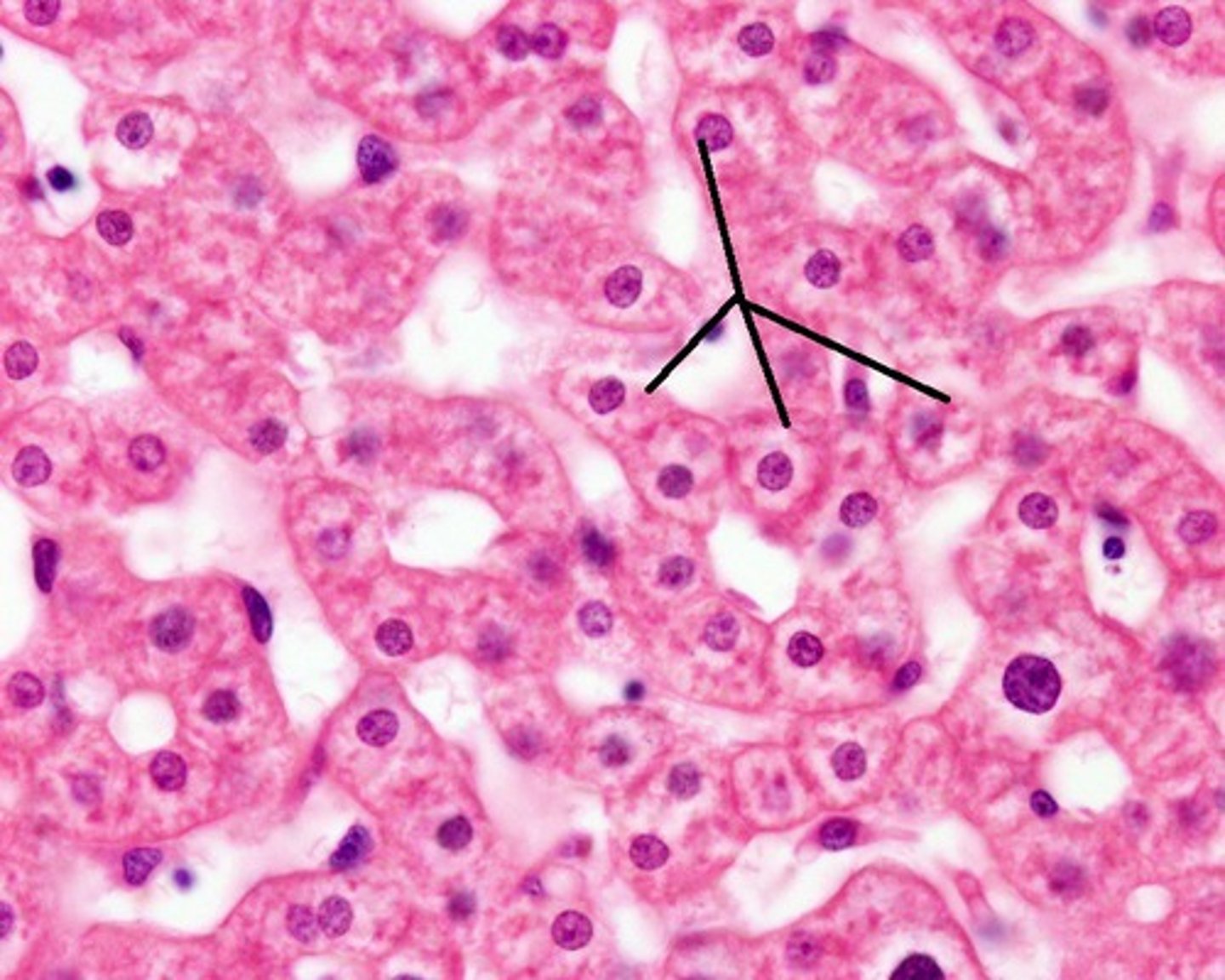
Hepatocytes
liver cells; produce bile, absorb and secrete nutrients like glucose, and secrete plasma proteins; cuboidal shape
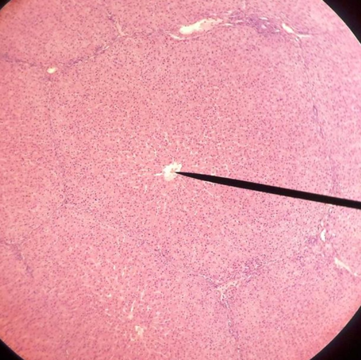
Central vein
vein that receives blood from hepatic sinusoids
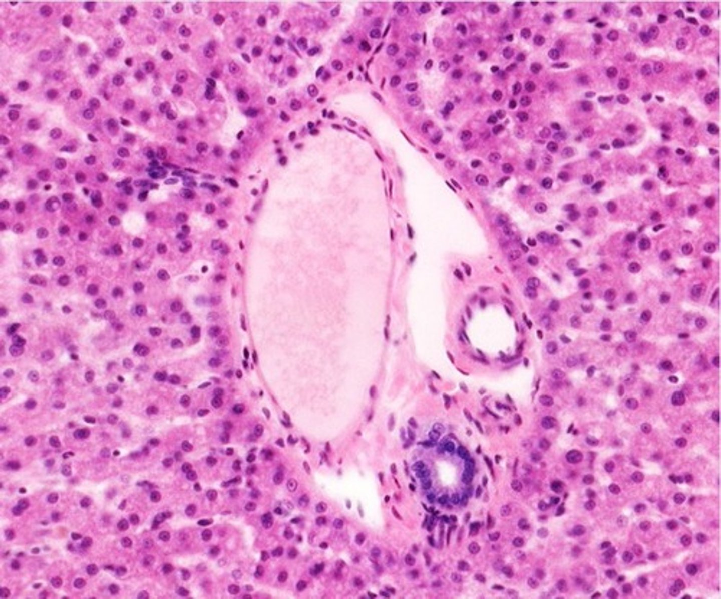
Hepatic triad
branches of hepatic artery, hepatic portal vein, and bile duct
Branch of hepatic portal vein
- blood from the digestive tract
- brings nutrients into the sinusoids
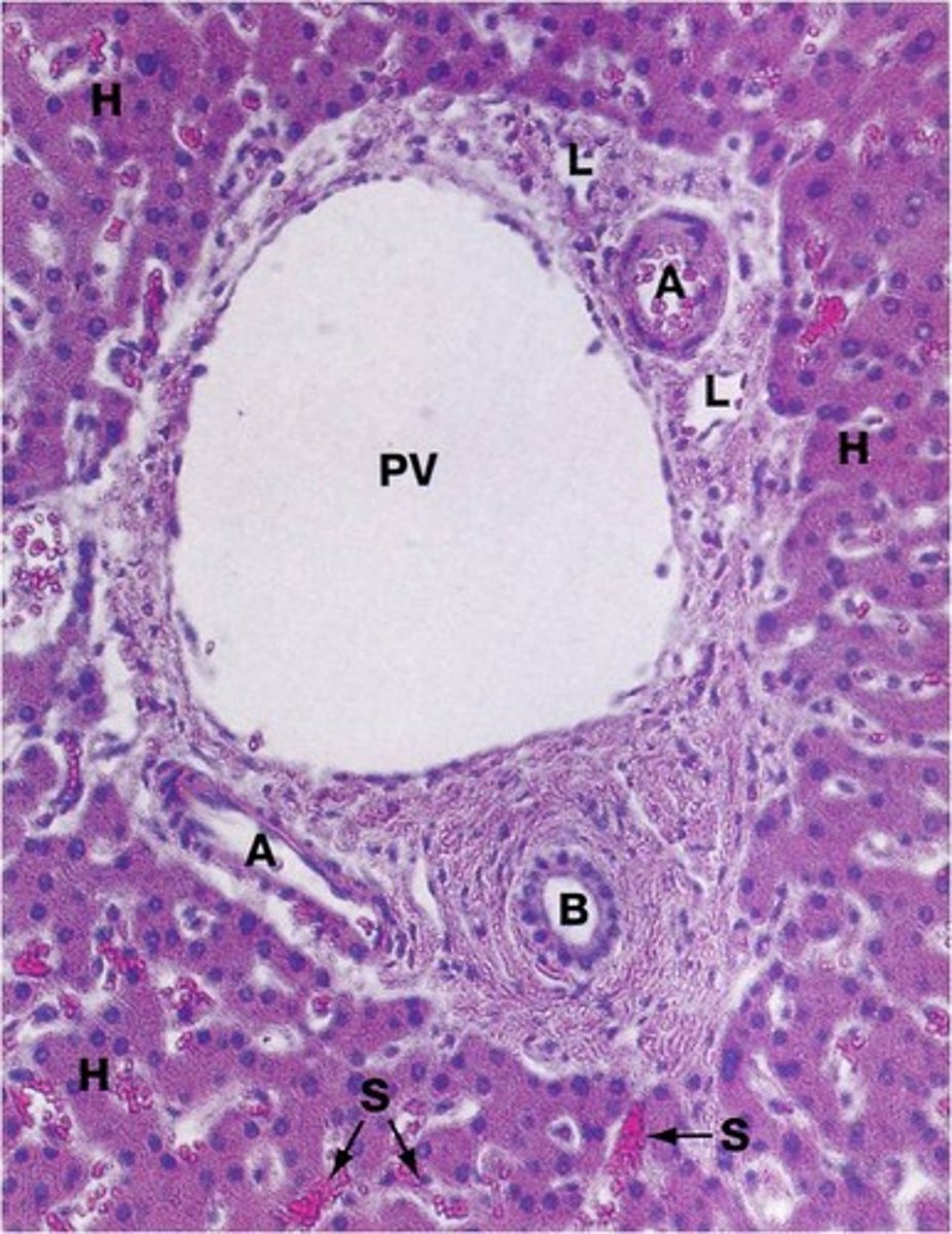
Branch of hepatic artery
fresh oxygenated blood into liver
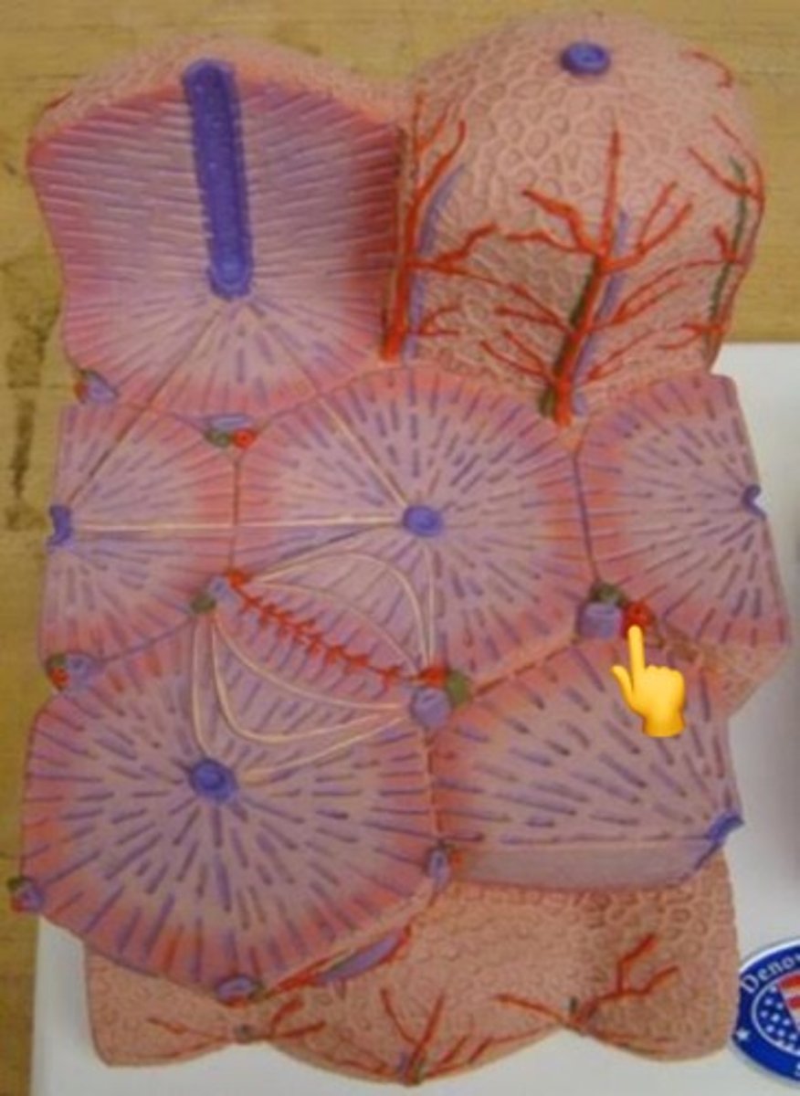
Bile duct branch
collects bile made by hepatocytes; lined by simple cuboidal epithelium
Pancreas histology
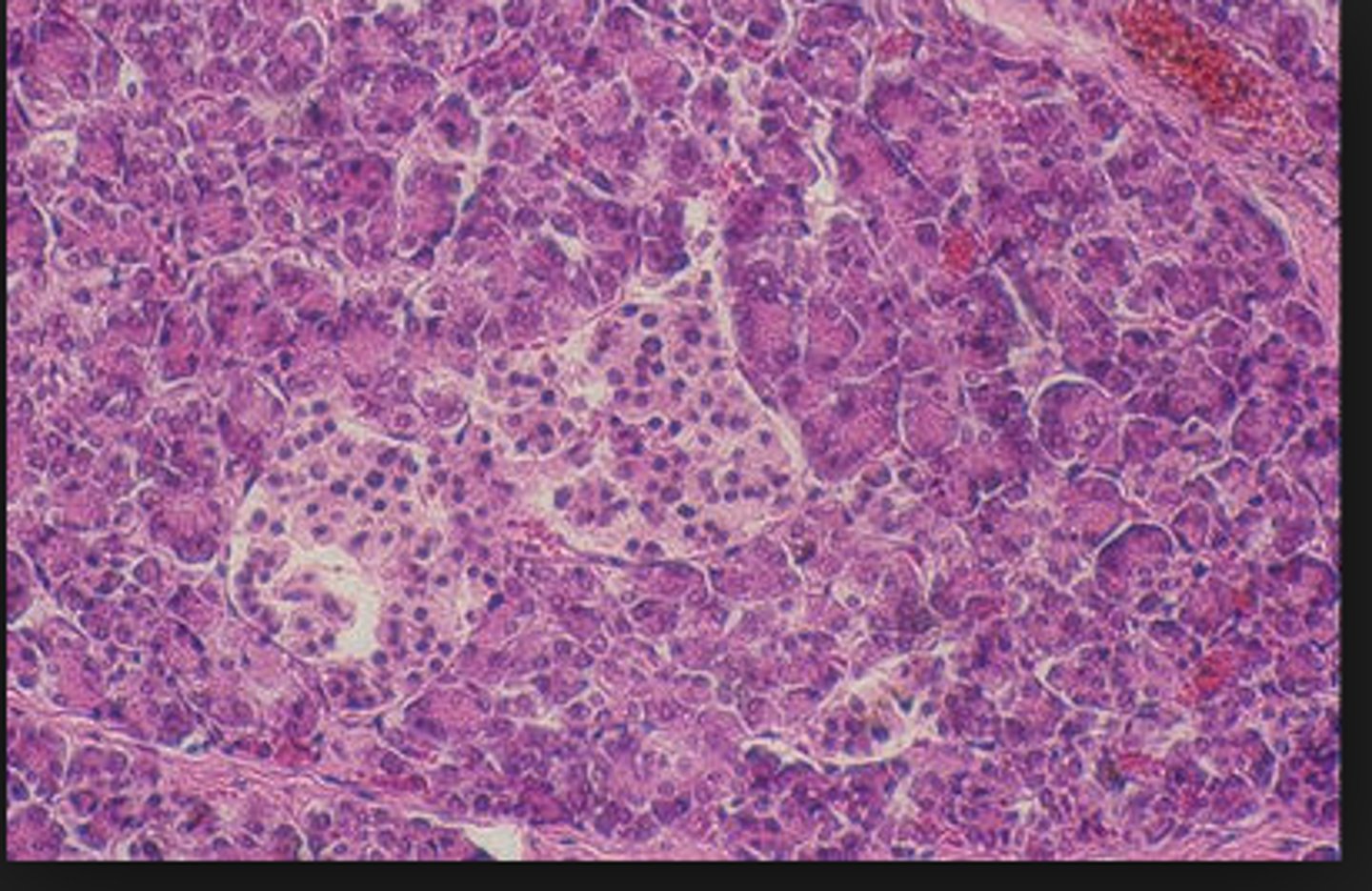
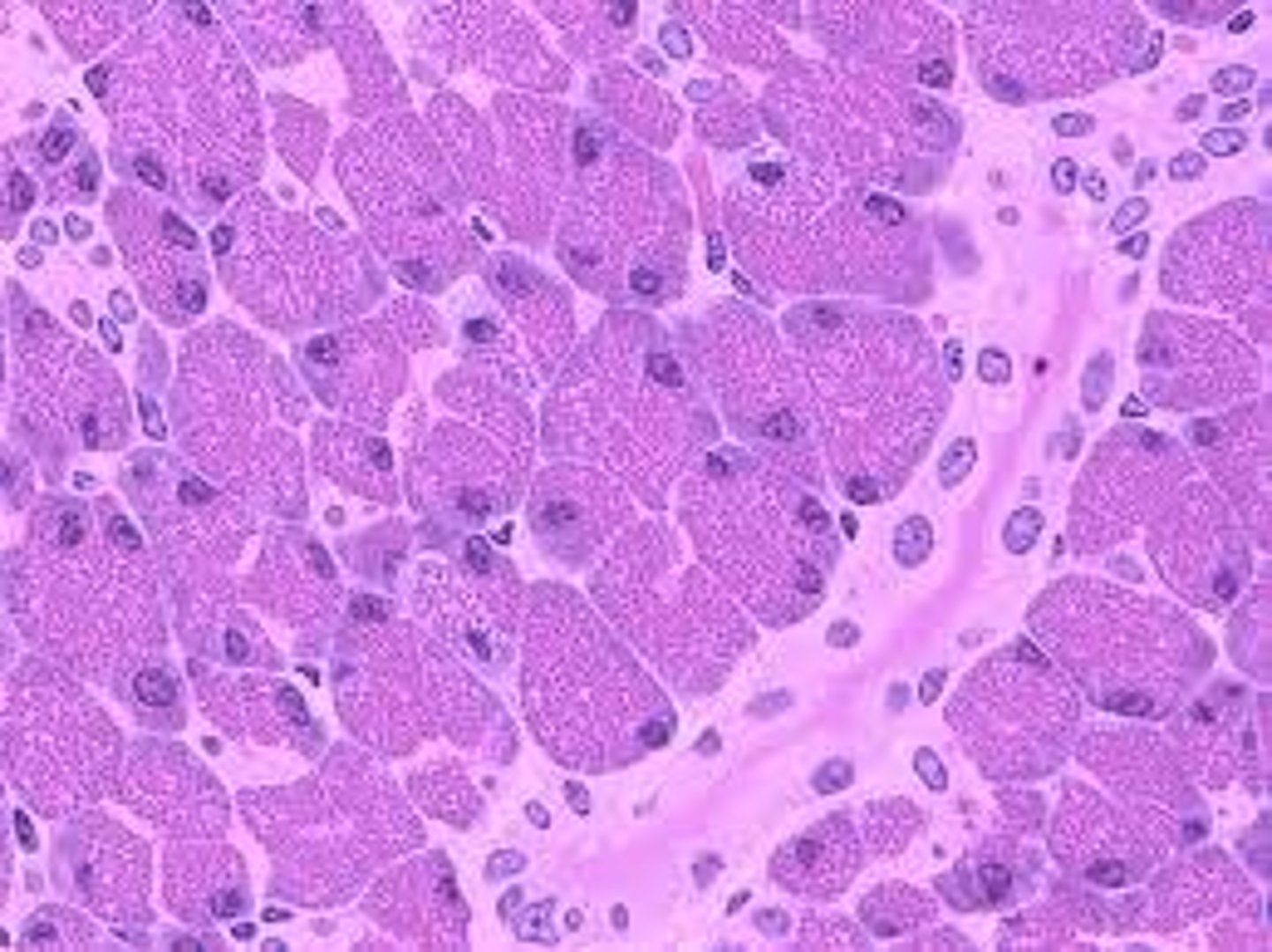
Acinar cells
enzyme-secreting cells of the pancreas; apical nuclei