Cardiovascular & respiratory system
 Call Kai
 Learn
 Practice Test
 Spaced Repetition
 Match
 Flashcards
 Knowt Play
1/69

There's no tags or description
Looks like no tags are added yet.
70 Terms
What are the main functions of the cardiovascular system?
Transport O₂ and nutrients to cells.
Remove CO₂ and metabolic waste (e.g. urea and creatinine).
Distribute hormones.
Transport immune cells for defence.
Haemostasis: stop bleeding following vessel damage.
Thermoregulation: regulate skin blood flow to control heat loss.
What is the spleen and what does it do?
It is a large flat oval organ located below the diaphragm, it’s main function is to store blood and filter it.
How often does the heart beat per minute?
70 times
How is heart contraction controlled?
Controlled by intrinsic pacemaker and autonomic nervous stimulation
What is microcirculation?
Where exchange with tissues occurs; capillaries - very thin walls (endothelial cells only); site of exchange of gases and nutrients
What does the heart of a fish consist of?
Single atrium and single ventricle
What does the heart of an amphibian contain?
Double circulatory circuit, heart - 2 atria and a single ventricle
Which side of the heart pumps blood to the systemic circulation?
Left
What does the right side of the heart pump deoxygenated blood to?
Pulmonary circulation
What is contraction called
systolic pressure
What is relaxation called
diastolic pressure
What is the pericardial sac and what does it consist of?
It is a double-walled fluid filled membrane that surrounds the heart. It consists of the epicardium (lines the inside of the heart) and parietal pericardium (outer layer).

Identify the labels on the numbered parts of the heart
Cranial vena cava
Aorta
Pulmonary veins
Pulmonary artery
Pulmonary veins
Right atria
Left atria
Coronary veins
Coronary arteries
Left veintricle
Right ventricle
Caudal vena cava
apex

identify the labels for the 3 numbers
Endocardium
Myocardium
Epicardium
What are the semilunar valves?
Aortic and pulmonary valve
What are the atrioventricular valves
Mitral (bicuspid) and tricuspid valve
When ventricles contract, how is eversion prevented?
Prevented by AV valves by action of the papillary muscles through the chordae tendinae.
What valves are inlet and outlet valves?
AV are inlet valves, semilunar are outlet.
How many cusps do semilunar valves have?
3 cusps
What are the semilunar valves?
Aortic and pulmonary
Where do the left and right coronary arteries branch from?
The base of the aorta
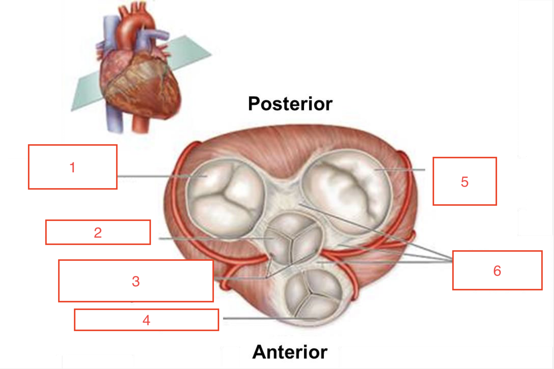
Identify the labels on the numbered parts of the heart
Right AV valve (tricuspid)
Aortic valve
Opening to coronary arteries
Pulmonary valve
Left AV valve (bicuspid)
Fibrous skeleton
What are the 5 types of blood vessels?
Elastic vessels, conduit and feed vessels, resistance vessels, exchange vessels and capacitance vessels.
What are elastic vessels?
Large arteries, accomodate stroke volume (high elastance). Convert intermittent ejection (stop-go flow) into a continuous flow.
What are conduit and feed vessels?
Medium to small arteries, conduct blood flow to organs.
What are resistance vessels?
Arterioles, terminal arteries; they control arterial blood pressure and local blood flow.
What are exchange vessels?
Capillaries, deliver nutrients to cell, lymph formation and metabolic waste removal.
What are capicitance vessels?
Venules and veins, control cardiac filling pressure and reservoir of blood.
What is the cell type of the tunica media?
Smooth muscle
What is the cell type of the tunica intima?
Endothelial cells
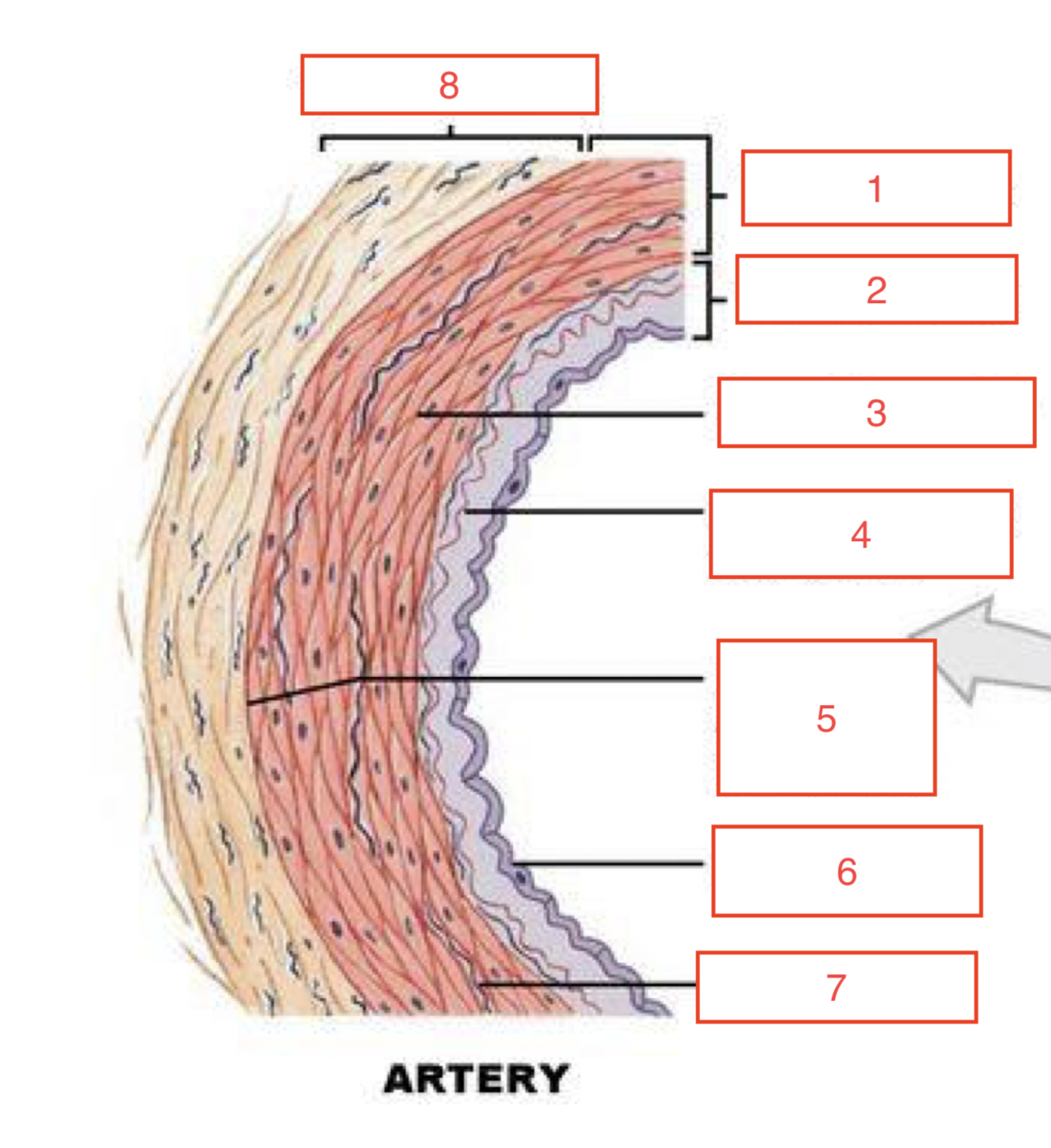
Identify the labels on the numbered parts of the artery
Tunica media
Tunica intima
Smooth muscle
Internal elastic membrane
External elastic membrane
Endothelium
Elastic fibre
Tunica externa

Fill in the table

How large are arterioles in micrometers?
40 - 300µm
How large are capillaries in micrometers?
3 - 30µm
Why does velocity slow down at capillary network?
To allow time for exchange
What is flow (velocity) in terms of blood?
Amount of blood flowing through a vessel at a given time (ml/min).
What is perfusion in terms of blood?
Flow per unit mass of tissue.
What is the relationship between blood flow and blood (hydrostatic) pressure gradient?
If the pressure gradient increases, blood flow speeds up (proportional relationship)
What is the relationship between blood flow and peripheral resistance?
inversely proportional, if resistance increases blood flow decreases.
How can resistance be changed?
By altering vessel diameter
How does fluid move across the capillary wall?
By 2 opposing forces, Oncotic pressure and hydrostatic pressure.
How is interstitual fluid formed?
By bulk flow: protein-free plasma moves across the capillary wall into the surrounding tissues.
Define oncotic and hydrostatic pressure.
Oncotic pressure: Pressure exerted by the proteins
Hydrostatic pressure: Pressure exerted by the blood
What are the four Starling forces and which direction do they move fluid?
Starling force | Effect on fluid movement |
|---|---|
Capillary hydrostatic pressure (Pc) | Pushes fluid out of capillary |
Plasma oncotic pressure (πp) | Pulls fluid into capillary |
Interstitial fluid hydrostatic pressure (Pif) | Pushes fluid into capillary |
Interstitial fluid oncotic pressure (πif) | Pulls fluid out of capillary |
Why may there be an abnormal accumulation of interstitial fluid in tissues and what is it called?
Excess filtration, defective resorption, defective lymphatic drainage. Abnormal accumulation known as oedema.
When does interstitial fluid become lymph?
When it enters lymphatic capillaries.
What is the sequence of electrical conduction through the heart?
SA node → right and left atrial cardiomyocytes → AV node → Bundle of His (left and right bundle branches) → purkinje fibre network → right and left ventricular cardiomyocytes
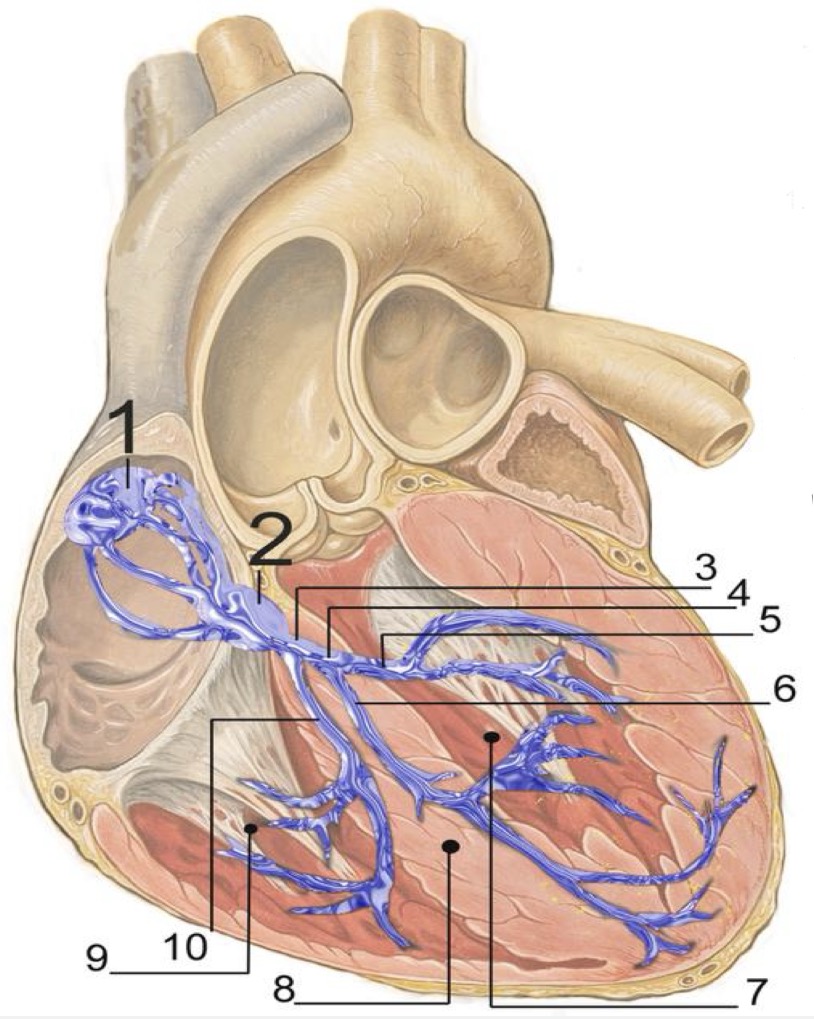
Identify the numbered labels on the heart
SA node
AV node
Bundle of His
Left bundle branch
Left posterior fascicle
Left anterior fascicle
left ventricle
ventricular septum
Right ventricle
Right bundle branch
What are the main features and components of the cardiac conduction system?
Specialised non-contractile cardiac cells, larger than standard cardiomyocytes.
Fewer contractile proteins
More glycogen and mitochondria
connected by desmosomes and gap junctions.
Immature intercalated discs.
What is the term for cardiac muscles initiating action potentials by themselves?
Intrinsic rhythmicity
What happens when Na+ channels open?
Move in, depolarise
What happens when K+ channels open?
Move out, repolarise
What happens when Ca+ channels open?
Move in, depolarise
What are pacemaker cells?
Myocardial cells of the SA and AV nodes
What do SA nodal cells do?
Depolarise and set the rate
What is synchronisation?
When the action potential is conducted down the conduction system as an electrical impulse and between cardiomyocytes via gap junctions.
What is the normal pacemaker rate of the SA node?
75 bpm
What is the normal pacemaker rate of the AV node?
50 bpm
What is the normal pacemaker rate of the Bundle and Purkinje fibres?
30 bpm
Which node depolarises fastest and what does that result in?
SA node depolarises the fastest, so it overrides all the others.
What do gap junctions provide between cardiac muscle cells?
Electrochemical link
What do desmosomes and adherens junctions provide?
Mechanical continuity
Cardiac muscle is a ________ ______ - Cells (myocytes) are electrically and mechanically coupled.
functional synctium
What are the main features of cardiac muscle?
Self-contracting
Single nucleus
Intercalated disks located between cardiac muscle cells
Gap junctions provide passage for ions and small molecules
Desmosomes and adheren junctions provide mechanical continuity
High mitochondrial density - a continuous supply of ATP needed to support contraction.
What seperates each sarcomere?
Dark Z bands
What do intercalated disks do?
Connect muscle cells, to allow unified contraction.
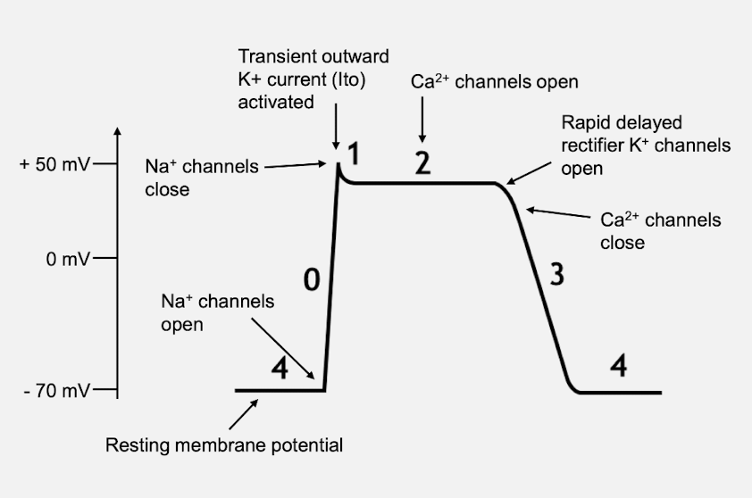
What are the 5 phases of cardiac conduction (start with phase 2), mention which ion channels are opening and closing and what the membrane potential is.
Phase 0 = Rapid depolarisation phase Na+ channels open and membrane potential becomes positive (+50 mV).
Phase 1 = Slow release K+ current activated, small drop in membrane potential triggering phase 2.
Phase 2 = Plateau phase, Ca2+ channels open, and influx of ions balances out K+ efflux component of effective refractory period when Ca2+ influx stimulates intracellular Ca2+ release initiating contraction.
(No further action potential can occur - Absolute refractory period)
Phase 3 = Repolarisation, Rapid K+ efflux returns membrane potential back to -70 mV.
Phase 4 = Resting potential
How does cardiac muscle contraction work with Ca2+?
Action potential conducted to contractile myocytes through gap junctions.
As the action potential travels between sarcomeres down the sarcolemma, it activates Ca2+ channels in the T-tubules depolarising the cell membrane.
Voltage-sensitive dihydropyridine (DHP) receptors open, leading to an influx of Ca2+ ions into the cell.
Calcium influx triggers release of calcium stored in sarcoplasmic reticulum through calcium-release channels called Ryanodine receptor 2 (RyR2).
Ca2+ binds to troponin C which moves tropomyosin covering the actin binding site.
Now the actin-binding site is free, so myosin heads can bind.
How does cardiac muscle relaxation work?
Ca2+ is moved out of the cell across the cell membrane and
Ca²⁺ is moved back into the sarcoplasmic reticulum (SR).
a) ACROSS CELL MEMBRANE: Ca2+ can be removed via facilitated transport, wherein there is a Na+/Ca2+ exchanger (3:1) taking calcium out of the cell. There is also a pump that uses ATP to pump Ca2+ across the cell membrane
b) Sarcoplasmic reticulum: SERCA2a membrane protein pump hydrolyses ATP into ADP allowing for the uptake of Ca2+ into the SR.Ca2+ removal returns troponin complex to its position on actin, ending contract as actin filaments return to initial position, relaxing the muscle.