8. Summary Case analysis and diangosis of management of nonstrab BV disorders Summary
 Call Kai
 Learn
 Practice Test
 Spaced Repetition
 Match
 Flashcards
 Knowt Play
1/50

There's no tags or description
Looks like no tags are added yet.
51 Terms
What is the diagnostic pattern for convergence insufficiency (CI)?
The patient must have:
Greater exophoria at near than distance, plus at least one of:
Receded NPC: break ≥6 cm
Reduced near PFV: BO break ≤15Δ or failure of Sheard’s criterion
Reduced vergence facility: ≤10 cpm, with difficulty clearing BO
How does convergence deficit differ from convergence insufficiency?
Both demonstrate deficient convergence findings:
Receded NPC: ≥6 cm break
Reduced PFV: ≤15Δ BO break or Sheard’s criterion not met
Vergence facility ≤10 cpm, with difficulty clearing BO
Difficulty with binocular plus lenses may be present
However, convergence deficit does not require greater exophoria at near than distance, whereas CI does.
What findings define divergence insufficiency (DI)?
The patient must have:
Esophoria greater at distance than near
At least one sign of inadequate divergence compensation:
Reduced distance NFV
Failure of Sheard’s criterion at distance
What is the diagnostic pattern for basic exophoria?
The patient must have exophoria at distance and near that is approximately equal:
At least 4Δ exophoria at distance and near
Distance-near difference no greater than 3Δ
Plus two supporting findings:
Receded NPC: ≥6 cm
Reduced PFV: ≤15Δ BO break or Sheard’s criterion not met at near and distance
Difficulty with binocular plus lenses may also occur.
What is the diagnostic pattern for basic esophoria?
The patient must have:
At least 3Δ esophoria at distance and near, with a difference no greater than 2Δ
At least one supporting NFV finding:
Near BI break <8Δ
Failure of Sheard’s criterion at near and distance
Difficulty with binocular minus lenses may be present.
What findings define fusional vergence dysfunction?
There is poor function in both vergence directions, despite no defining phoria pattern. Either may establish the diagnosis:
Reduced vergence amplitudes:
BO break ≤15Δ or Sheard’s criterion not met
BI break <8Δ or Sheard’s criterion not met
Vergence facility ≤10 cpm, with difficulty clearing both BO and BI
Difficulty with binocular plus lenses may also occur.
What is the diagnostic pattern for convergence excess (CE)?
The patient must have:
Near esophoria >3Δ, typically greater at near than distance
At least one sign of reduced divergence at near:
BI break <8Δ or failure of Sheard’s criterion
Vergence facility ≤10 cpm, with difficulty clearing BI
Difficulty with binocular minus lenses commonly supports the diagnosis.
What is the diagnostic pattern for divergence excess (DE)?
The characteristic findings are:
Exophoria greater at distance than near
Inadequate distance PFV relative to the deviation
Failure of Sheard’s criterion at distance
An intermittent exotropia may occur at distance.
What symptoms are characteristic of convergence excess?
Symptoms occur primarily with sustained near work:
Asthenopia or headaches
Ocular burning or tearing
Intermittent blur or diplopia
Words appearing to move
Loss of concentration
Sleepiness while reading
Reduced comprehension over time
Slow reading
Symptoms worsening later in the day
Symptoms are often chronic and longstanding. A CISS score around 16 may help differentiate symptomatic CE in young adults.
What clinical findings support convergence excess?
Common findings include:
Greater esophoria at near than distance
Reduced near NFV for the magnitude of the phoria
Failure of Sheard’s criterion
High accommodative lag
Low PRA
Reduced binocular accommodative facility, especially difficulty with minus
Eso fixation disparity at near
Generally good stereopsis
Possible intermittent suppression
Possible vertical deviation
A high lag can conceal part of the esodeviation because the patient is accommodating less and therefore producing less accommodative convergence.
Why must accommodation be carefully controlled when measuring the near phoria of a patient with suspected CE?
Underaccommodation produces less accommodative convergence and can make the measured esophoria appear smaller than it truly is.
To reveal the deviation:
Use a small, detailed target
Encourage the patient to maintain clarity
Compare the phoria with accommodative testing and MEM findings
How can untreated convergence excess affect binocularity over time?
Persistent CE may produce:
Eso fixation disparity
Intermittent suppression
Reduced stereopsis
Increasing inability to compensate for the eso deviation
Possible decompensation into strabismus
This is why treatment is appropriate even when the patient maintains relatively good stereopsis initially.
What functional disorders should be differentiated from convergence excess?
Differentiate CE from:
Basic esophoria
Divergence insufficiency
Accommodative disorders
Uncorrected or latent hyperopia
A cycloplegic refraction is important when hyperopia may be contributing.
What findings suggest that a new esodeviation is pathological rather than ordinary convergence excess?
Red flags include:
Acute onset
Unilateral findings
Neurological symptoms
Significant medical problems
Recent medication or drug exposure
Signs of ocular inflammation
Potential causes include:
Spasm of accommodation or convergence
Iritis or other local inflammation
Sympathetic pathway dysfunction
CNS disease
Medication effects, including miotics such as pilocarpine
What is the first management step for convergence excess?
Begin with accurate refractive management:
Obtain a cycloplegic refraction when appropriate.
Correct clinically significant refractive error.
Reevaluate the distance and near phorias after correction.
Determine whether additional near plus, vision therapy, or prism is needed.
CE generally has an excellent prognosis when properly treated.
How are near plus lenses selected for convergence excess?
Near plus reduces the accommodative demand and therefore reduces accommodative convergence.
Selection should consider:
Magnitude of the near esophoria
Change in phoria through plus
Gradient AC/A ratio
Working distance
Symptoms and functional demands
The prescription may be provided as a:
Bifocal
Progressive or near addition
Separate reading prescription
What is the role of vision therapy in convergence excess?
Vision therapy primarily develops:
Negative fusional vergence or divergence
Vergence facility
Flexibility between convergence and divergence
Accommodation-vergence integration
A typical program may require approximately:
12 to 16 sessions per 10Δ to 15Δ change in divergence ability
Approximately 12 to 24 sessions, depending on severity and response
When might prism be added for convergence excess?
A patient with clinically significant esophoria at both distance and near, with greater eso at near, may also require base-out prism if the deviation cannot be adequately managed with lenses and therapy alone.
Prism direction follows the deviation:
Eso → BO relieving prism
Exo → BI relieving prism
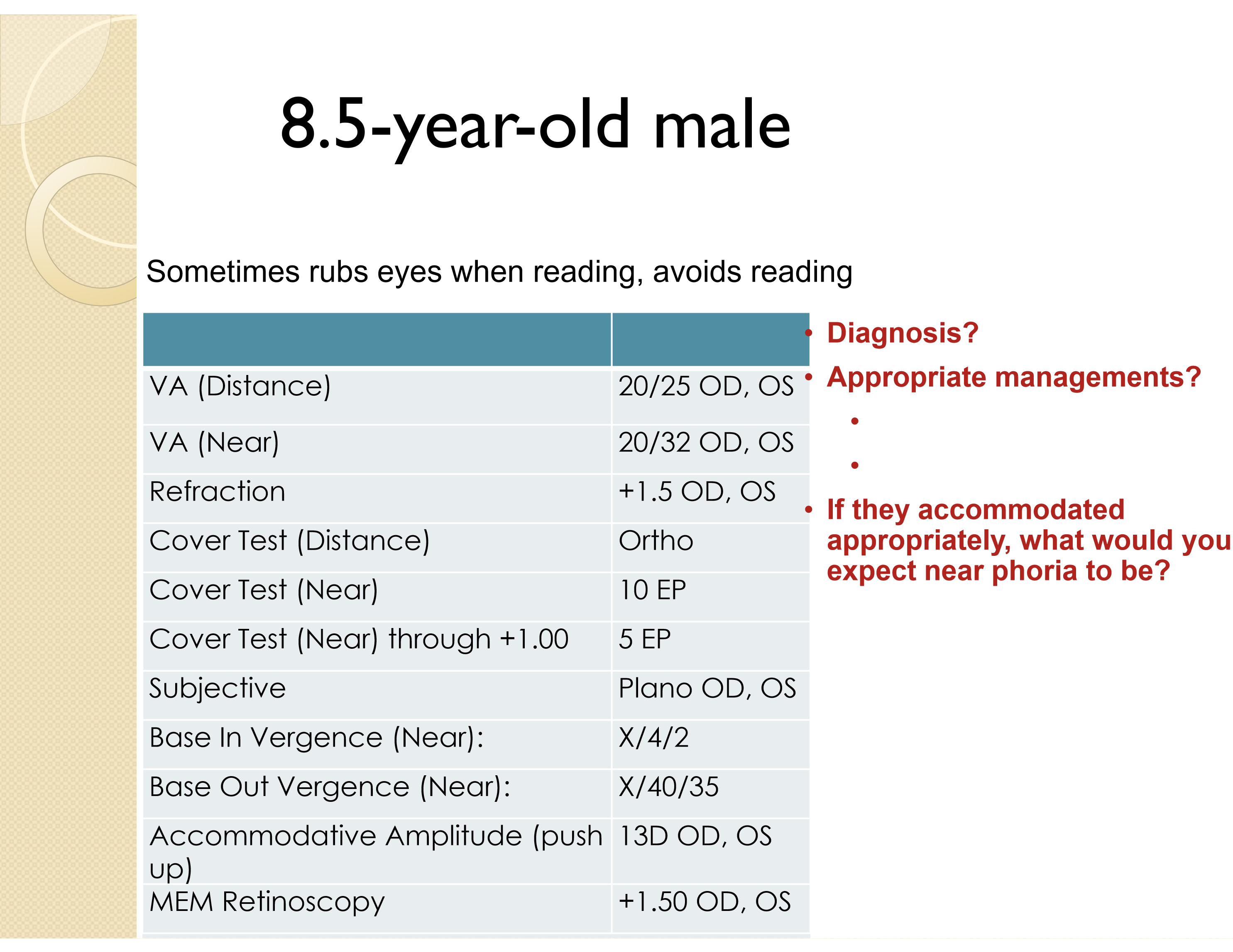
An 8.5-year-old has 10Δ near esophoria, orthophoria at distance, BI vergence of X/4/2, BO vergence of X/40/35, and a high MEM of +1.50 D. What is the primary binocular diagnosis?
Convergence excess.
Evidence:
Esophoria is greater at near than distance
Near BI break is only 4Δ, showing deficient NFV
BO is very large, consistent with excessive convergence ability
High MEM lag may be masking an even larger near esophoria
Reading avoidance and eye rubbing are consistent with a symptomatic near disorder
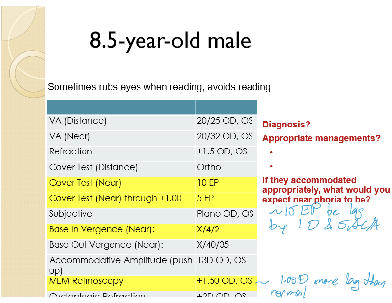
In the CE case, why is the measured 10Δ near esophoria probably an underestimate?
MEM is +1.50 D, approximately 1.00 D more lag than expected. This means the child is underaccommodating and generating less accommodative convergence.
The cover test changed from:
10Δ EP normally
5Δ EP through +1.00 D
This suggests a gradient AC/A of approximately 5:1. If the child accommodated about 1.00 D more appropriately, the near phoria would be expected to increase to approximately 15Δ EP.
What management should be considered for the child with CE, a high accommodative lag, and uncorrected hyperopia?
Management should include:
Confirm the refractive finding with cycloplegia
Address the clinically significant hyperopia
Reassess accommodation and near alignment after correction
Consider an appropriate near addition based on the gradient AC/A and response
Consider vision therapy to improve NFV and accommodation-vergence integration
Monitor for suppression or progression toward strabismus
What is the typical presentation of divergence excess?
DE usually develops in childhood and is characterized by:
Exodeviation greater at distance than near
Frequent intermittent exotropia at distance
Average deviation larger at distance than near
Worsening control with:
Inattention
Fatigue
Illness
Daydreaming
Parents may be more aware of the deviation than the child.
What symptoms are typical of divergence excess?
Common reports include:
One eye turning outward intermittently
Asthenopia
Photophobia
Possible diplopia
Closing one eye in bright light
Occasional near discomfort, although near symptoms may be absent
Patients with suppression at distance often do not report diplopia.
What examination findings support divergence excess?
Findings include:
Greater exodeviation at distance than near
Exotropia occurring more frequently at distance
Comitant deviation
Normal NPC
Inadequate PFV relative to the exodeviation
Possible suppression or anomalous correspondence at distance
Typically normal stereopsis at near
Often an eso fixation disparity at near
Possible inferior oblique overaction
Usually no primary vertical deviation
Why can a patient with divergence excess have numerically normal or large PFV and still be unable to compensate?
Fusional vergence must be interpreted relative to the size of the phoria, not solely by comparison with population norms.
A patient with a large distance exophoria may have an apparently normal BO range but still fail:
Sheard’s criterion
Sustained control
Real-world binocular demands
Clinical rule: Compare compensating vergence with the deviation.
What AC/A findings may occur in divergence excess?
DE commonly demonstrates:
High stimulus AC/A
Normal to slightly high response AC/A, approximately 3:1 to 9:1
This distinction matters because the near deviation may appear smaller due to accommodative convergence or other fusion-related mechanisms.
How are true and simulated divergence excess differentiated?
Dissociate the patient to uncover a hidden near deviation using:
30 minutes to 3 hours of monocular occlusion, with no fusion before the post-occlusion cover test
A +3.00 D lens test OU at near
If the near exodeviation increases and becomes similar to the distance deviation, the patient has simulated DE, not true DE.
Concept: Simulated DE is often caused by tenacious proximal fusion or an altered near accommodative-convergence response.
What disorders are included in the differential diagnosis of divergence excess?
Differentiate DE from:
Basic exophoria
Simulated divergence excess
Convergence insufficiency
The most important distinctions are:
Distance-near relationship
Response to occlusion or +3.00 D testing
NPC
Near PFV and near symptoms
What is the primary goal of vision therapy for divergence excess?
Therapy aims to improve:
Awareness and control of the distance exodeviation
Positive fusional vergence
Vergence facility
Accommodation-vergence integration
Ability to distinguish physiological from pathological diplopia, when normal retinal correspondence is present
A typical course may be approximately 24 to 36 sessions, or 6 to 8 sessions per 10Δ to 15Δ change.
What factors predict a poorer vision-therapy prognosis in divergence excess?
Negative prognostic factors include:
Large horizontal deviation
Large vertical deviation
Noncomitancy
Abnormal sensory correspondence
Poor ability to recognize diplopia or misalignment
What options besides vision therapy may be used for divergence excess?
Additional options include:
Added minus lenses
Therapeutic lenses
Full-time spectacle wear when indicated
Surgical referral for large, poorly controlled, or refractory deviations
Mechanism of added minus: Minus stimulates accommodation, increasing accommodative convergence and helping reduce the exodeviation.

An 8-year-old has 16Δ distance exophoria with occasional intermittent exotropia, 5Δ near exophoria, normal NPC, and distance BO of X/20/10. What is the primary diagnosis?
Divergence excess with intermittent exotropia at distance.
Evidence:
Distance exo exceeds near exo by 11Δ
Intermittent exotropia is observed at distance
NPC is normal, arguing against CI
Distance BO break of 20Δ is inadequate for 16Δ exophoria because Sheard’s criterion would require approximately 32Δ of compensating PFV

Why is this DE patient symptomatic even though the distance BO break is 20Δ?
A 20Δ BO break may look numerically reasonable, but it is inadequate relative to the patient’s 16Δ distance exophoria.
Using Sheard’s criterion:
Compensating reserve should be at least twice the phoria
Required PFV ≈ 2 × 16Δ = 32Δ
Measured PFV break = 20Δ
Therefore, the patient lacks enough PFV to compensate comfortably.
What secondary finding should be considered in the DE case if binocular accommodative facility is only 5 cpm?
The low binocular accommodative facility suggests a possible accompanying accommodative dysfunction.
However, a specific accommodative diagnosis requires knowing:
Whether difficulty occurred with plus, minus, or both
Monocular facility
Accommodative amplitude
MEM
NRA and PRA
Do not diagnose accommodative infacility from the cpm value alone without identifying the lens side causing difficulty.
What symptoms suggest a clinically significant vertical phoria?
Common symptoms include:
Headache and asthenopia
Blur
Diplopia
Frequent loss of place while reading
Skipping or repeating lines
Motion sickness
Difficulty concentrating during sustained tasks
Fatigue or sleepiness during prolonged visual work
Even a small vertical deviation may be symptomatic because vertical fusional reserves are limited.
What signs may accompany a vertical phoria?
Common signs include:
Compensatory head tilt
Reduced horizontal positive and negative fusional ranges
Abnormally low or exceptionally large vertical fusion ranges, depending on chronicity
Possible association with reading or learning difficulties
A longstanding deviation may develop unusually large vertical fusion ranges as compensation.
What ocular or mechanical factors can cause a vertical phoria?
Possible causes include:
Hyperorbital asymmetry
Abnormal extraocular muscle insertion
Vertical rectus anomalies
Oblique muscle dysfunction
Ocular surgery, including lens extraction
Unilateral or bilateral pseudophakia
Acquired deviations require comparison with prior alignment and surgical history.
Which causes of vertical diplopia require investigation for neurological or neuromuscular disease?
Important etiologies include:
Skew deviation
CN III palsy
CN IV palsy
Ocular myasthenia
Inferior oblique overaction
Superior oblique overaction or dysfunction
Vertical rectus abnormalities
Acute vertical diplopia, incomitancy, ptosis, pupil abnormalities, or neurological symptoms should not be assumed to be a benign vertical phoria.
How is a symptomatic vertical phoria managed?
Treatment options include:
Vertical prism, especially when the symptomatic deviation is approximately 0.75Δ or greater
Some clinicians consider treatment beginning around 0.50Δ
Prism based on the associated phoria or X-intercept of the vertical fixation-disparity curve
Vertical fusional vergence therapy in selected patients
Treatment or referral for the underlying cause when the deviation is acquired or pathological
What symptom pattern supports a functional accommodative or binocular vision disorder?
Functional disorders typically produce symptoms that are:
Chronic or longstanding
Associated with specific visual tasks
Worse with prolonged visual activity
Worse later in the day
Relieved by stopping the provoking task
Symptoms that do not follow this pattern should raise concern for an organic cause, unreliable reporting, or another diagnosis.
What history questions help rule out an organic cause of new binocular or accommodative symptoms?
Ask about:
Exact onset and progression
Recent illness
New medications
Appetite, sleep, or weight changes
Dizziness or balance problems
Fainting
New or worsening headaches
Headaches that awaken the patient
Recent trauma
Muscle weakness
Numbness or tingling
What minimum examination should be performed when an organic cause of binocular symptoms is possible?
External examination
Pupils, including APD testing
Versions
Distance and near cover testing
Cover testing in at least two positions of gaze
Evaluation for a vertical deviation
Color vision
Ophthalmoscopy
Confrontation visual fields or formal field testing when indicated
The goal is to identify incomitancy, neurological asymmetry, optic nerve dysfunction, or ocular disease.
What is the basic sequence for analyzing accommodative and binocular findings?
Determine whether an accommodative abnormality is present.
Determine whether a significant lateral phoria exists at distance or near.
If a phoria is present, ask whether fusional reserves adequately compensate.
Evaluate the distance-near pattern.
Evaluate the AC/A relationship.
Integrate vergence, accommodation, fixation disparity, stereopsis, and symptoms.
Rule out organic disease before labeling the condition functional.
If no significant lateral phoria is present, what disorders should still be considered?
Accommodative dysfunction
Fusional vergence dysfunction
Oculomotor dysfunction
Hyperphoria
Fixation disparity
Aniseikonia
Latent hyperopia
Cyclophoria
What should be reconsidered when the initial findings do not produce a clear diagnosis?
Reassess:
Accuracy and repeatability of test results
Objective observations versus subjective responses
Whether difficulty occurs with one specific task or with all tasks
Effect of fatigue and time of day
Possibility of organic disease
Comprehension of test instructions
Possibility of noncredible or exaggerated responses
How should the clinician interpret apparently conflicting BV test results?
Findings should be understood as an interacting system rather than isolated pass/fail values.
Consider how:
Accommodation changes accommodative convergence
Phoria magnitude determines the fusional reserve required
Suppression may eliminate diplopia
Chronic deviations may produce unusually large compensating ranges
A high accommodative lag may conceal esophoria
A numerically normal vergence range may still fail Sheard’s criterion
How should a binocular vision diagnosis be explained to the patient or parent?
The explanation should:
Use nontechnical language
Connect examination findings to the patient’s symptoms
Describe common symptoms of the condition
Explain why measurable signs may exist without current symptoms
Demonstrate the problem or treatment effect when possible
The patient should understand not only the diagnostic label, but how the disorder affects visual function.
What must be established before beginning treatment for a BV disorder?
Establish a complete diagnosis, including:
Refractive status
Accommodative function
Phoria pattern
Compensating vergence ability
AC/A relationship when relevant
Sensory status
Presence or absence of pathology
Treatment should target the mechanism causing the symptoms, not a single abnormal test value.
What should be included when presenting management options for a binocular vision disorder?
Discuss:
Available treatment options
Advantages and disadvantages of each option
What the treatment involves
Expected duration
Expected success rate
Patient responsibilities and keys to success
Alternatives if the initial plan is unsuccessful
The goal is to help the patient or parent make an informed treatment decision.
What should be included in reports to parents or other professionals?
Reports should:
Use language appropriate for the recipient
Avoid unexplained abbreviations
Summarize relevant findings
State the diagnosis and functional implications
Describe the treatment plan
Provide useful recommendations
Be sent promptly
Include acknowledgment or thanks when appropriate
How can the major horizontal binocular vision disorders be rapidly differentiated by their phoria pattern?
Near exo > distance exo: Convergence insufficiency
Distance eso > near eso: Divergence insufficiency
Similar exo at distance and near: Basic exophoria
Similar eso at distance and near: Basic esophoria
Near eso > distance eso: Convergence excess
Distance exo > near exo: Divergence excess
No defining phoria pattern + poor BI and BO: Fusional vergence dysfunction
Then confirm the diagnosis by determining whether the patient’s compensating fusional vergence is inadequate.