Week 9 ,, Protozoa Part 2
 Call Kai
 Learn
 Practice Test
 Spaced Repetition
 Match
 Flashcards
 Knowt Play
1/65

There's no tags or description
Looks like no tags are added yet.
66 Terms
Cardiovascular System Infection
Malaria, Babebiosis, and Trypanosomiasis
Skin and Mucous Membrane Infection
Leishmania
Central Nervous System Infection
Toxoplasmosis
Malaria Causal Agent
Plasmodium spp.
Malaria Vector
Anopheles gambiae (mosquitoes)
Plasmodium falciparum
Most deadly and most common in hot, tropical regions
Plasmodium vivax
Most frequent worldwide ,, Common in cool tropical areas
Plasmodium malariae
Less common ,, Most widely distributed
Plasmodium ovale
Rare
Plasmodium spp. Definitive Host
Anopheles gambiae (mosquitoes)
Plasmodium spp. Intermediate Host
Humans
Plasmodium spp. Diagnostic Stage
Immature/mature trophozoites, schizont, and gametocytes
Plasmodium spp. Infective Stage
Ruptured oocyst releases sporozoites and the vector takes a blood meal (injection of sporozoites)
Plasmodium spp. Clinical Features
May be rapidly fatal if untreated ,, Fever and chills, myalgias, weakness, vomiting, and diarrhea ,, Splenomegaly, anemia, thrombocytopenia, hypoglycemia, pulmonary or renal dysfunction, and neurological changes
Plasmodium spp. Laboratory Diagnosis
Microscopic identification in Giemsa-stained blood smears is the method most frequently used to demonstrate an active infection
Female Gametocytes of P. falciparum
Deep blue cytoplasm
Male Gametocytes of P. falciparum
Redish cytoplasm

Identify the Protozoa
Plasmodium spp. ring-form trophozoites

Identify the Protozoa
Plasmodium spp. schizones

Identify the Protozoa
Plasmodium spp. gametocytes

Identify the Plasmodium Species 1
P. falciparum

Identify the Plasmodium Species 2
P. malariae

Identify the Plasmodium Species 3
P. vivax

Identify the Plasmodium Species 4
P. ovale
Babebiosis Causative Agent
Babesia spp.
Babesia microti Reservior
Rodents
Babesia microti Geographical Distribution
Worldwide ,, Most frequently identified in the US
Babesia divergens Reservior
Cattle
Babesia divergens Geographical Distribution
Most frequently identified in Europe
Babesia spp. Vector
Borrelia burgdorferi (ticks)
Babesia spp. Infective Stage
Ruptured oocyst releases sporozoites and the vector takes a blood meal (injection of sporozoites)
Babesia spp. Diagnostic Stage
Trophozoites
Babesia spp. Clinical Features
Mostly asymptomatic ,, B. divergens infection is more severe than B. microti ,, Fever, chills, sweating, myalgias, fatigue, hepatosplenomegaly, and hemolytic anemia ,, More severe in patients who are immunosuppressed, splenectomized, and/or elderly
Babesia spp. Laboratory Diagnsosis
Identification of the parasite inside RBC is the method most frequently used to demonstrate infection ,, Ring-form can be vacuolated ,, Intra-erythrocytic forms ,, Clumps of extracellular forms are indicative of parasite ,, Tetrad
Trypanosomiasis Causative Agent
Trypanosoma brucei and Trypanosoma cruzi
African Trypanosomiasis
Trypanosoma brucei
American Trypanosomiasis
Trypanosoma cruzi
What is African Trypanosomiasis also called?
Sleeping sickness
Trypanosoma brucei Vector
Tsetse flies
Trypanosoma brucei Infective Stage
Vector takes a blood meal and injects metacyclic trypomastigotes into the host
Trypanosoma brucei Diagnostic Stage
Trypomastigotes in the blood
Trypanosoma brucei Clincial Features
1. Development of trypanosomal chancre at the site of inoculation ,, 2. Hemolymphatic stage (fever, pruritus, lymphadenopathy) ,, 3. Meningoencephalitic stage (invasion of the central nervous system with headaches, somnolence, abnormal behavior, and leads to the loss of consciousness and coma)
Trypanosoma brucei Laboratory Diagnosis
Microscopic identification of chancre fluid, lymph node aspirates ,, blood and bone marrow, and the cerebrospinal fluid ,, Trypomastigotes are the only stage found in patients
What is American Trypanosomiasis also known as?
Chagas’ disease
Trypanosoma cruzi Vector
Triatomine bugs
Trypanosoma cruzi Infective Stage
Vector takes a blood meal and passes metacyclic trypomastigotes in feces ,, Trypomastigotes enter the bite wound or mucosal membrane
Trypanosoma cruzi Diagnostic Stage
Intracellular amastigotes transform into tyrpomastigotes that then burst out of the cell and enter the bloodstream
Trypanosoma cruzi Clincial Features
Romaña's sign (periocular swelling) ,, Chronic form can manifest as cardiomyopathy and pathologies of the digestive tract and weight loss ,, Complications of chronic Chagas disease can be fatal
Trypanosoma cruzi Laboratory Diagnosis
Microscopic examination of motile parasites in fresh anticoagulated blood (or its buffy coat) or of parasites in thin and thick blood smears stained with Giemsa

Identify the Protozoa
Trypanosoma spp.
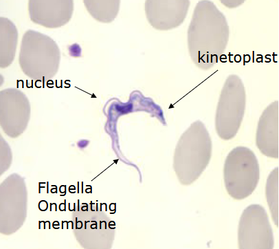
Identify the Protozoa
Trypanosoma spp.

Identify the Protozoa
Trypanosoma spp.
Leishmaniasis spp. Vector
Sand flies
Leishmaniasis spp. Diagnostic Stage
Promastigotes transform into amastigotes inside macrophages ,, Amastigotes multiply in cells of various tissues
Leishmaniasis spp. Infective Stage
Vector takes a blood meal and injects promastigotes into the skin
Leishmaniasis spp. Laboratory Diagnosis
Microscopic examination of Giemsa-stianed slides of the relevant tissue is the method most frequently used to detect the parasite

Identify the Protozoa
Leishmaniasis spp.
Cutaneous Leishmaniasis
One or more cutaneous lesions that can change shape (volcano, with a raised edge
and central crater) ,, Swollen glands near the sores
Visceral Leishmaniasis
Fever, weight loss, an enlarged spleen and liver, and swollen glands ,, Anemia, neutropenia, and thrombocytopenia ,, Sometimes post kala-azar dermal leishmaniasis
Toxoplasmosis Causative Agent
Toxoplasma gondii
Toxoplasma gondii Definitive Hosts
Cats
Toxoplasma gondii Infective Stage
Fecal oocysts and tissue cysts
Toxoplasma gondii Diagnostic Stage
CSF serum
Toxoplasma gondii Laboratory Diagnosis
Observation of parasites in patient specimens, such as bronchoalveolar lavage material
from immunocompromised patients, or lymph node biopsy ,, Serologic testing is the routine method of diagnosis
Congenital Toxoplasmosis
Severity vary with the trimester during which infection was acquired ,, Patients are often
asymptomatic until the second or third decade of life, when lesions develop in the eye ,, Most infants will develop ocular infection

Identify the Protozoa
Toxoplasma gondii tachyzoites