6.1.13 Recognises ocular manifestations of systemic disease
 Call Kai
 Learn
 Practice Test
 Spaced Repetition
 Match
 Flashcards
 Knowt Play
1/21

There's no tags or description
Looks like no tags are added yet.
22 Terms
What is high blood pressure?
Normal blood pressure reading = between 90/60mmHg and 120/80mmHg
Retinal signs of hypertension
Arterial narrowing (changes in A/V ratio)
Arteriovenous crossing changes
Flame haemorrhages
Copper/silver wiring
Retinal oedema
If SEVERE: cotton wool spots, hard exudates/ macular star, optic nerve head swelling
What are the signs of malignant hypertension, and what are the symptoms?
Signs of malignant hypertension = over 180/120mmHg
Hypertensive retinopathy due to chronic hypertension is usually asymptomatic.
Malignant or accelerated hypertension are usually symptomatic:
Intermittent blurring
Visual field defects
Headache
Flushed and red appearance of face
Nausea and vomiting
Sudden painless loss of vision due to vessel occlusion
Sings of end-organ damage
High blood pressure
Systemic risk factors:
Poorly controlled BP
Diabetes Mellitus
Cardiovascular disease
Obesity
Smoking
Alcohol consumption
Poor diet
What is the Keith-Wagener-Barker classification?
Grade 1: mild, generalised constriction of retinal arterioles
Grade 2: definite focal narrowing of arterioles and AV nicking
Grade 3: grd 2 with flame-shaped haemorrhages, cotton wool spots and hard exudates
Grade 4: grd 3 with papilloedema and/or signs of retinal oedema
How is hypertension managed according to the Keith-Wagener-Barker classification?
Grade 1 & 2 - refer if undiagnosed, hypertension control advice
Grade 3 - routine refer for GP intervention for stricter hypertension control
Grade 4 - Emergency referral to HES
What are the systemic risk factors for vein occlusions?
Hypertension - arteriosclerosis compresses adjacent veins
High cholesterol - promotes fatty deposits and vessel occlusion.
Giant cell arteritis - arterial inflammation compressing venous outflow
In younger patients, investigate hypercoagulable (abnormality of blood coagulation that increases the risk of thrombosis) or inflammatory causes
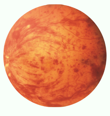
What is a Central Retinal Vein Occlusion (CRVO),
Signs
Symptoms
Blockage of the main retinal vein, causing blood and fluid to leak into the retina.
Signs:
“Blood and thunder” fundus,
diffuse retinal haemorrhages,
dilated tortuous veins,
CWS,
macular oedema,
optic disc swelling,
reduced VA
Symptoms:
Sudden, painless vision loss
What are the actions required and clinical management for CRVO?
Actions required:
OCT for macular oedema,
Fundus photography
Clinical Management:
Urgent referral to ophthalmology (same day if suspect ischaemia)
What is a Branch Retinal Vein Occlusion (BRVO), and what are its signs and symptoms?
Blockage of a smaller branch of the retinal vein resulting in localised retinal swelling and bleeding.
Signs:
Localised flame/blot haemorrhages along one retinal quadrant,
Venous dilation,
CWS,
possible macular oedema
Symptoms:
Sudden, painless visual field defect, may be asymptomatic if peripheral

What are the actions required and clinical management for BRVO?
Actions required:
OCT for macular oedema,
fundus photography
Clinical Management:
Non-urgent referral to ophthalmology (1-2 weeks unless macula)
What are the causes of artery occlusions in patients under and over 50 years?
<50 years - Cardiac emboli, hypercoagulable states, vasculitis, migraine, drug-induced vasospasm
>50 years - Atherosclerosis, carotid disease, cardiac emboli, hypertension, diabetes, giant cell arteritis
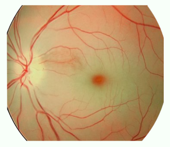
What is CRAO?
Signs
Symptoms
Sudden blockage of the central retinal artery, cutting off blood supply to the retina.
Signs:
Pale retina,
cherry red spot at macular,
attenuated arterioles (white/yellow spots),
“boxcarring” of blood columns (discontinuous columns of red blood cells)
Symptoms:
Sudden, painless vision loss, often NLP or just hand movements
CRAO
Actions required
Clinical Management
Actions required:
Ocular emergency! Ocular massage, IOP-lowering drops
Clinical Management:
Immediately same-day referral to A&E or stroke unit
What is BRAO?
Signs
Symptoms
Blockage of a branch of the central retinal artery leading to sectoral lack of blood flow.
Signs:
Localised retinal pallor,
embolus may be visible,
attenuated arterioles
Symptoms:
Sudden painless sectoral vision loss

BRAO
Actions required
Clinical management
Actions required:
Discuss stroke risk to patient
Clinical Management:
Urgent referral to A&E or stroke unit
What conditions are associated with AAION?
Giant Cell Arteritis - Results in luminal narrowing or occlusion → ischaemia of the optic nerve head.
Behçet's disease - a rare, chronic condition causing inflammation in blood vessels (vasculitis) throughout the body.
What conditions are associated with NAION?
Giant Cell Arteritis - Results in luminal narrowing or occlusion → ischaemia of the optic nerve head.
Atherosclerosis
What is AAION?
Signs
Symptoms
Inflammation of the arteries (usually GCA) leading to reduced blood flow to the optic nerve.
Signs:
Pale swollen optic disc, chalky white disc,
RAPD,
CWS,
altitudinal defect - particularly lower half,
thick and pulsating temporal artery
Symptoms:
Sudden severe vision loss,
headache,
jaw claudication,
scalp tenderness

AAION
Actions Required
Clinical Management
Actions required:
Medical emergency! Stress importance of fast treatment
Clinical Management:
Immediate same-day referral to hospital A&E
What is NAION?
Signs
Symptoms
Non-inflammatory condition where poor blood flow damages the optic nerve.
Signs:
Optic disc swelling (often segmental),
small cup-to-disc ratio,
flame haemorrhages,
altitudinal defect - particularly lower half
Symptoms:
Sudden painless monocular vision loss,
often upon waking

NAION
Actions required
Clinical Management
Actions required:
Medical emergency! Stress importance of fast treatment
Clinical Management:
Immediate same-day referral to hospital A&E