Chapter 13: Special Senses - The Eye and Ear
 Call Kai
 Learn
 Practice Test
 Spaced Repetition
 Match
 Flashcards
 Knowt Play
1/60

There's no tags or description
Looks like no tags are added yet.
61 Terms
Optometry
Professional practice that provides care for the eyes and associated structures
Doctor of Optometry or Optometrist (OD)
Provides primary care for the eyes including examining eyes for disease, treating diseases, assessing visual acuity, and prescribing corrective lenses
Optician
Grinds and fits prescription lenses and contacts as prescribed by a physician or optometrist
Audiology
Professional practice that provides care for communication disorders arising from hearing loss
Doctor of Audiology or Audiologist (AuD)
Diagnoses and treats hearing impairment and associated communication disorders
Eye
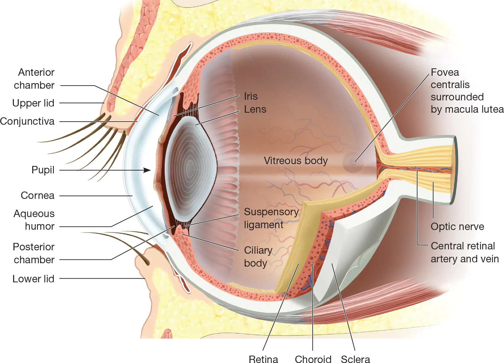
Contains the sensory receptor cells for vision
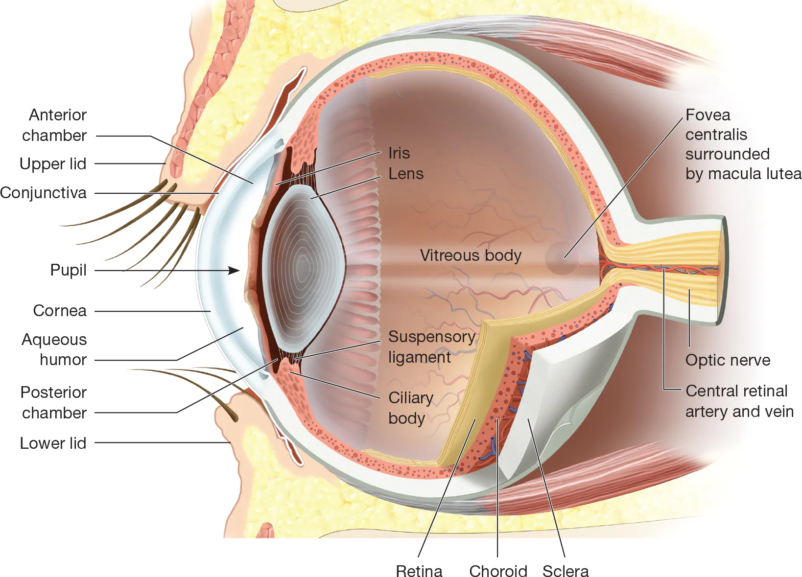
choroid
conjunctiva
eye muscles
eyeball
eyelids
lacrimal apparatus
retina
Primary structures that comprise the eye:

Concentrated
The five special senses are those that have sensory receptors _______ in a specific organ
Vision (Eye)
Hearing (Cochlea of the ear)
Equilibrium (Inner ear)
Smell (Nose)
Taste (Tongue)
What are the five special senses?
Ophthalmology (Ophth)
Study of the eye; Branch of medicine specializing in condition of eye; Physician is an example
Eyeball
Organ of sight that transmits an external image by way of the nervous system—the optic nerve—to the brain; Eye by itself, without any appendages such as eye muscles or tear ducts
Optic Nerve
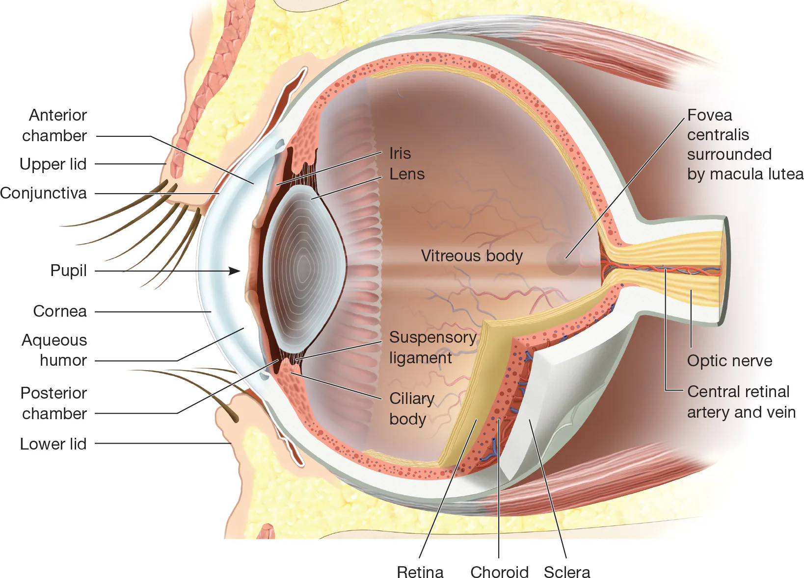
Second cranial nerve that carries impulses from retina to brain

The brain then ______ sensory impulses from the retina into an image with computer-like accuracy.
Translates
Eye Muscles
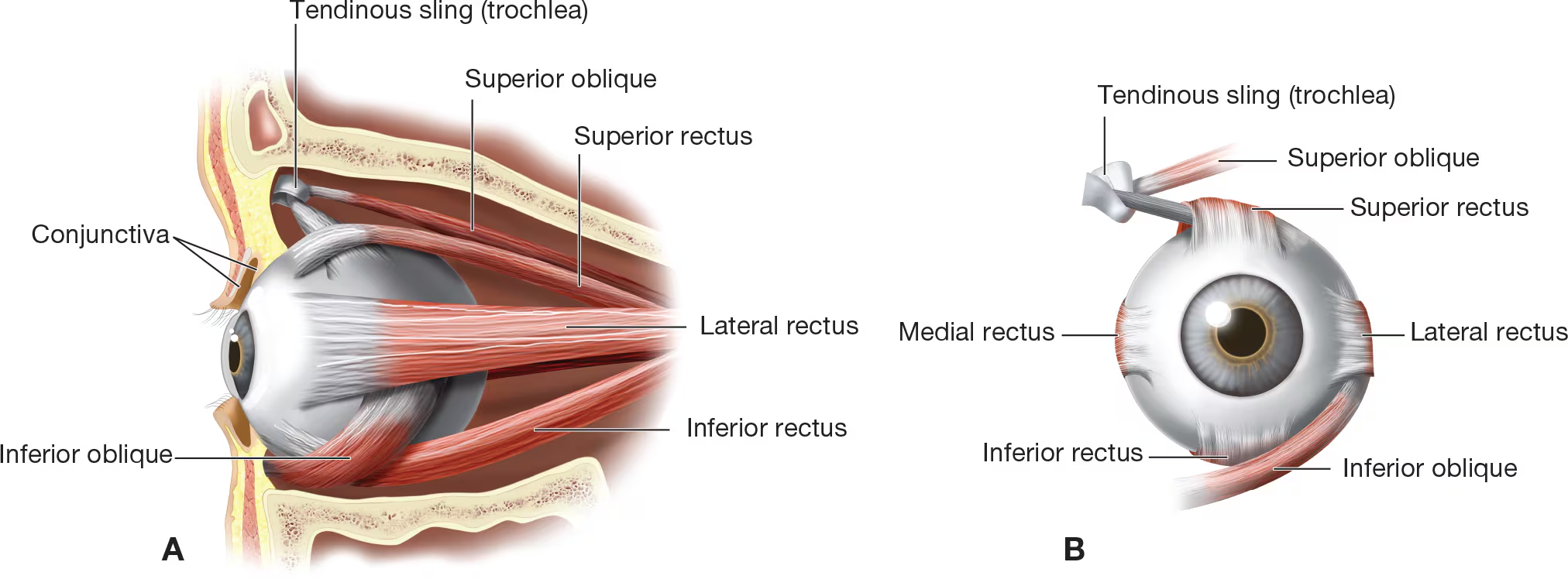
Six muscles that connect eyeball to orbit cavity; Allow for rotation of eyeball and change in the direction of each eye’s sightline; Provide support for the eyeball in the socket
Eyelids
Upper and lower folds of skin that provide protection from foreign particles, injury from sun and intense light, and trauma; Both upper and lower edges have cilia; Sebaceous glands are located in eyelids

Conjunctiva
Protective mucous membrane lining on underside of each eyelid and across anterior surface of each eyeball

Lacrimal Apparatus
Consists of lacrimal gland, lacrimal canals, and nasolacrimal duct
Orbit
Opening in skull formed by portions of frontal, zygomatic, maxillary, ethmoid, sphenoid, lacrimal, and palatine bones; Houses and protects eyeball
sclera
choroid
retina
The eyeball is composed of three layers:

Sclera
Tough protective outer layer of eyeball; Commonly referred to as “white of eye”; Light rays enter the eyeball through the anterior structures of the sclera; First layer of the eye

Choroid
Second or middle layer of eyeball; Provides blood supply for eye

Retina
Third and innermost layer of eye; Contains visual receptors called rods and cones that receive light impulses and transmit them to brain via optic nerve

Focused
Light rays enter the eyeball through the anterior structures of the sclera and are ______ onto the sensory receptor cells of the retina where they are converted to electrical signals that travel to the brain via the optic nerve.
Cornea
Portion of sclera that is clear and transparent and allows light to enter interior of eye; Plays role in bending (refacting) light rays

Refracts
Bending of light rays as they enter eye
iris
pupil
ciliary body
The anterior portion of the choroid layer consists of:

Iris
Colored portion of eye; Contains smooth muscle; Can dilate or constrict to change size of pupil and control amount of light entering interior of eye
Pupil
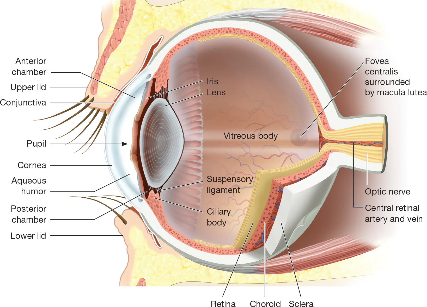
Hole in center of iris; Allows light rays to enter the eyeball; Size is changed by iris dilating or constricting
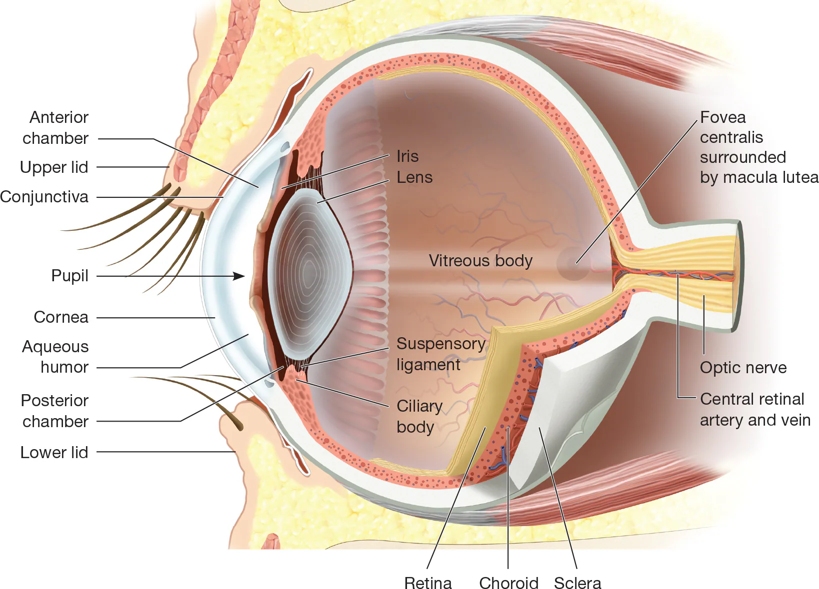
Ciliary Body
Intraocular eye muscles that change shape of the lens; Ring of smooth muscle
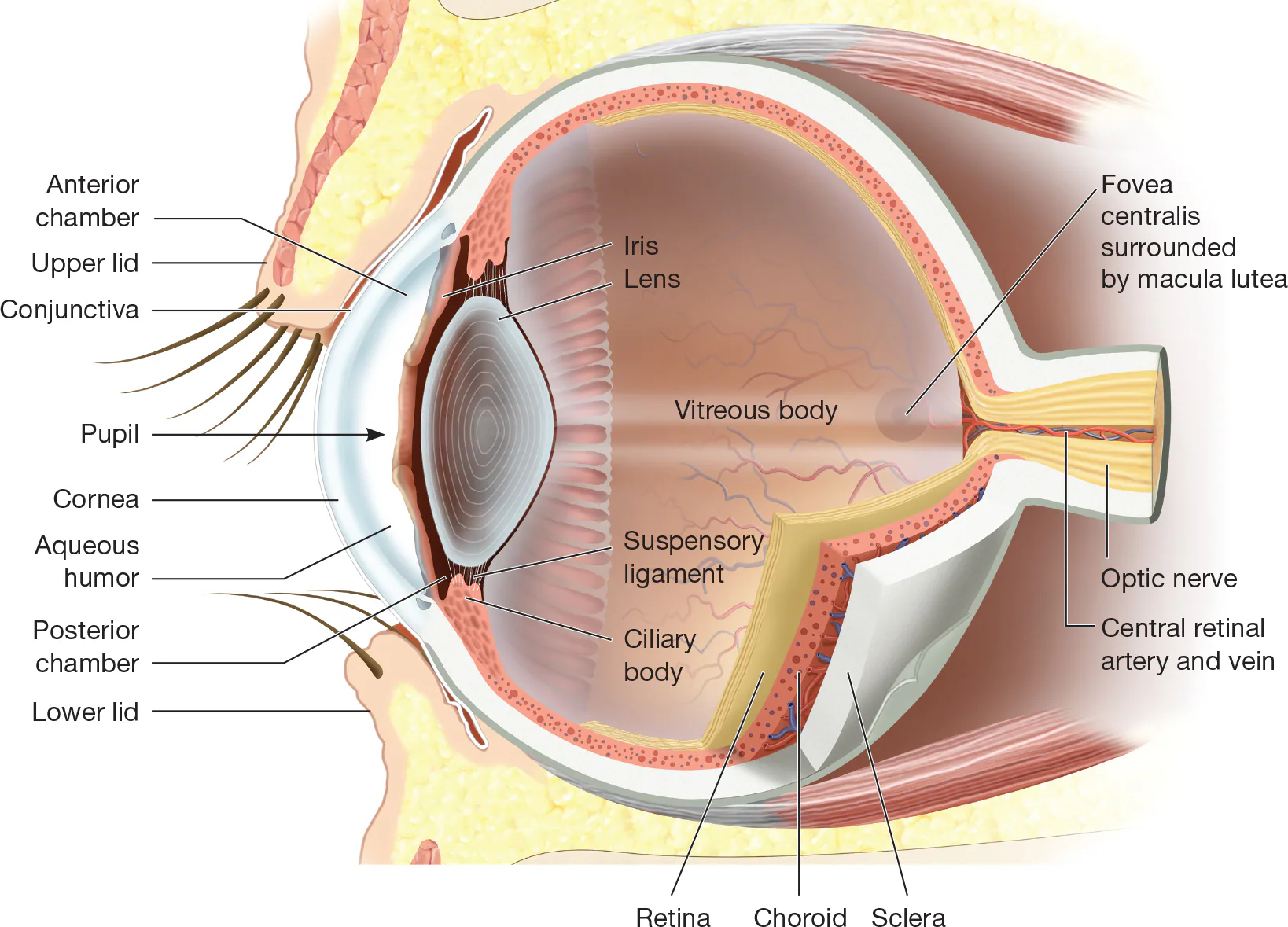
Lens
Transparent structure behind pupil and iris; Functions to bend light rays so they land on retina; Not apart of the choroid layer; Attached the ciliary body by suspensory ligaments
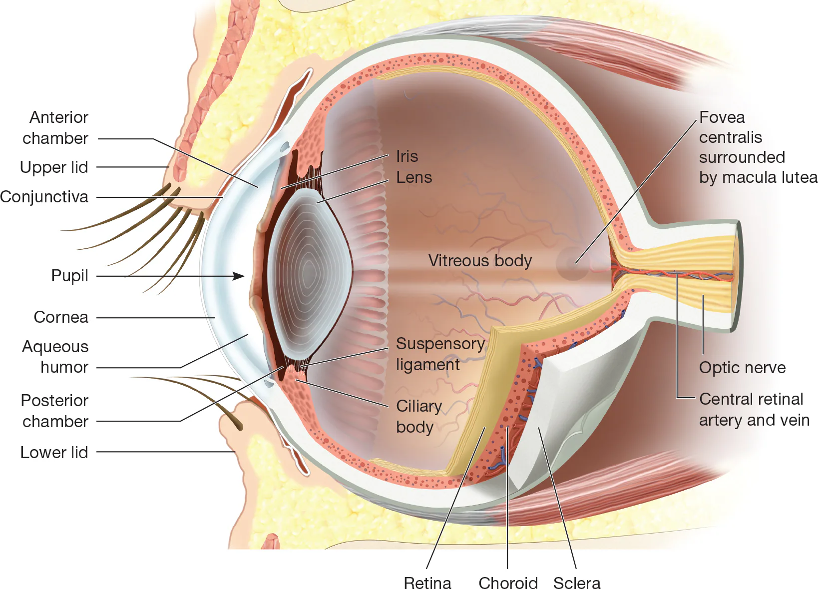
Suspensory Ligaments
Many thin ligaments
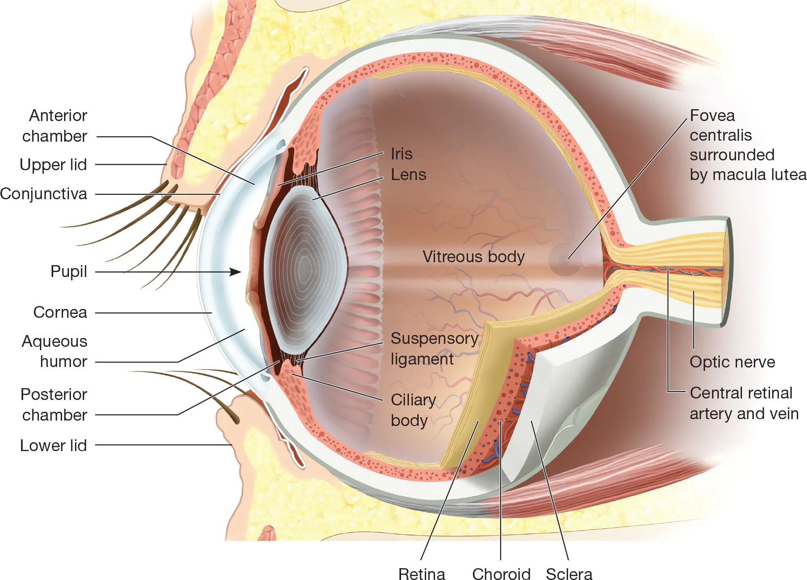
Accommodation
Process of ciliary body contracts or relaxes to pull on the edge of the lens, changing shape of lens so it can focus incoming light onto retina
Rods
Sensory receptors of retina that are active in dim light and do not perceive color; Help to see gray tones
Cones
Sensory receptors of retina that are active in bright light and see in color; Responsible for color vision
Macula Lutea
Area of retina onto which straight-ahead images are projected and come into focus; Also referred to as “yellow spot”
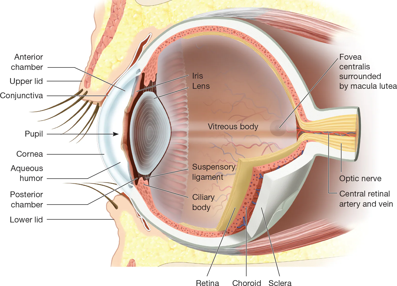
Fovea Centralis
Area of retina that has sharpest vision; Located in the center of the macula lutea; Means central pit; Contains a high concentration of sensory receptor cells making it the point of clearest vision

Optic Disk
Area of retina associated with optic nerve; Also called blind spot; The point where the retinal blood vessels enter and exit the eyeball and where the optic nerve leaves the eyeball

Retinal blood vessels
Blood vessels that supply oxygen to rods and cones of retina

Sensory Receptor
There are no _________ cells in the optic disk and therefore it causes a blind spot in each eye’s field of vision.
Blind Spot
Because the _________ of each eye is set to the side, one eye is able to cover for the missing information from the other. For this reason, a person is not generally aware of their existence.
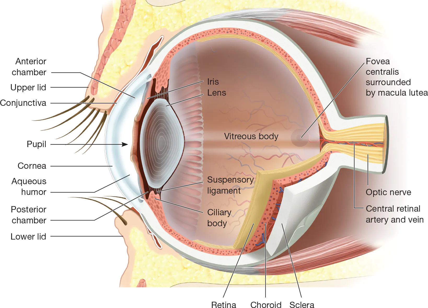
Aqueous Humor
Watery fluid filling spaces between cornea and lens

Vitreous Humor
Transparent jellylike substance inside eyeball; Between the lens and retina; semisolid gel
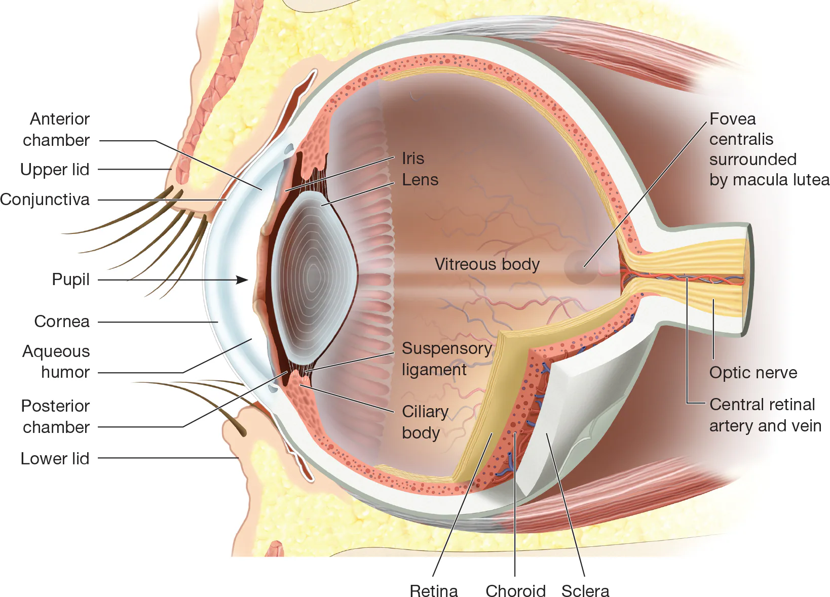
Strabismus
Children may be born with a weakness in some of the eye muscles and may require treatments such as eye exercises or even surgery to correct this problem; Commonly referred to as crossed eyes

Rectus Muscles
Means straight muscle; Four of the eye muscles are this type of muscle; Pull the eye up, down, left, or right, in a straight line
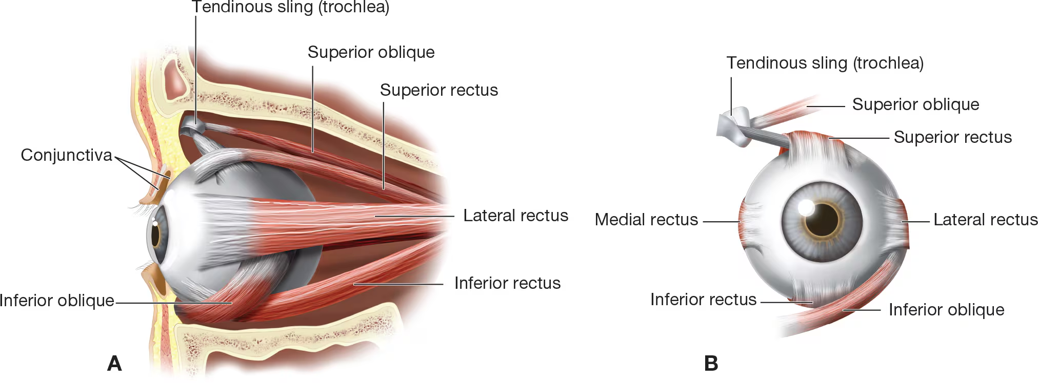
Oblique Muscles
Means “slanted” muscle; Two eye muscles are this type of muscle; On an angle and produce diagonal eye movement

Cilia
Term for eyelashes that protect eye from foreign particles or for nasal hairs that help filter dust and bacteria out of inhaled air
Sebaceous Glands
Produce substance called sebum that lubricates skin surfaces and eyeball; also called oil glands
Mucous Membrane
Membrane that lines body passages that open directly to exterior of body, such as conjunctiva of the eye; Secretes thick substance, or mucus; Forms a continuous covering on the underside of each eyelid and across the anterior surface of each eyeball; Serves as protection for the eye by sealing off the eyeball in the socket
Lacrimal Gland
Located under the outer upper corner of each eyelid; Produces and washes anterior surface of eye with fluid called tears


Tears
Fluid that washes and lubricates anterior surface of eyeball
Lacrimal Canals
Located in inner corner of eye socket; Collect tears and drain them into the nasolacrimal duct

Nasolacrimal Duct
Collects tears from inner corner of eye socket (lacrimal canals) and drains them into nasal cavity

Nasal Cavity
Large cavity just behind external nose that receives outside air; Covered with mucous membrane to cleanse air; Nasal septum divides nasal cavity into left and right halves

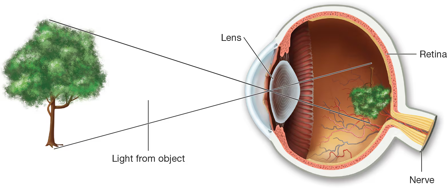
Light Rays
When _____ strike the eye, they first pass through the cornea, pupil, aqueous humor, lens, and vitreous humor. They then strike the retina and stimulate the rods and cones.

Upside-Down
When the light rays hit the retina, an _______ image is sent along nerve impulses to the optic nerve. The optic nerve transmits these impulses to the brain, where the _______ image is translated into the right-side-up image being looked at
Coordination of the external eye muscles so that both eyes move together
The correct amount of light admitted by the pupil
The correct focus of light on the retina by the lens
The optic nerve transmitting sensory images to the brain
Vision requires proper functioning of four mechanisms:
Cataract
Development of opaque or cloudy lens, resulting in diminished vision; Most common causes are aging, eye trauma, or radiation (especially sunlight) exposure but may be present at birth; Treatment is usually surgical removal of effected lens and replacement with prosthetic lens
Glaucoma
Increase in intraocular pressure; if untreated, may result in atrophy (wasting-away) of optic nerve and blindness; treated with medication and surgery; Increased risk in person over age 60, those of African ancestry, people who have sustained serious eye injury, or anyone with a family history diabetes of the disease
Legally Blind
Describes person who has significant visual disability; Usually defined as having visual acuity of 20/200 that cannot be improved with corrective lenses or having visual field of less than 20 degress
Chalazion
Small, firm mass formed when an oil gland deep in the eyelid becomes blocked, it may or may not become infected; DIfferent from a s