UNIT 10. SEROUS FLUID
 Call Kai
 Learn
 Practice Test
 Spaced Repetition
 Match
 Flashcards
 Knowt Play
1/97

There's no tags or description
Looks like no tags are added yet.
98 Terms
parietal membrane - lines cavities
visceral membrane - lines organs
Pleural, Pericardial, Peritoneal Cavities are lined by 2 membranes, namely ?
Serous Fluid
__ is the body fluid responsible for lubrication in the serous membrane
Pleural Cavity - lodges the lungs
Pericardial Cavity - lodges the heart
Peritoneal/Visceral Cavity - lodges the visceral organs
ENUMERATE
Body cavities
serous space
The space between the visceral and parietal membranes is called the ___.
lymphatic capillaries
The difference in pressure between the parietal and visceral capillaries creates a small excess of fluid.
This excess fluid is reabsorbed by __ within the membranes.
T
T or F
Both membranes contain blood vessels — specifically capillaries — that allow the exchange of fluids.
serous space
The ultrafiltrate of plasma naturally moves into the ___
endothelial cells
not the same
Since capillaries have ___ that act as filters, the fluid that enters the serous space comes from the blood, but its composition is ___ as that of blood because some blood components cannot pass through the endothelial barrier.
oncotic
same
The __ pressure between or in both sides of the parietal and visceral membrane are the _.
ultrafiltrate of plasma
where does serous fluid come from
Oncotic pressure
__ is the force yield by your protein in pulling back the water back to the circulation/blood vessel.
hydrostatic pressure
__ is the pressure yielded because of a natural phenomenon caused by gravity.
Hydrostatic
__ pressure is responsible for the filtration process.
Abnormal effusion
__ is the excess accumulation of fluid in the serous space.
EFFUSION
By the action of oncotic pressure the pressure that pulls back the excess fluid into the circulation and by hydrostatic pressure the pressure that brings in fluid in the cavity the volume of serous fluid remains to be constant unless there is __
Effusion
__ is the abnormal accumulation of fluid within a body cavity, usually due to inflammation, increased capillary permeability, or an imbalance between hydrostatic and oncotic pressures.
Transudate
__ are effusions that form because of a systemic disorder that disrupts the balance in the regulation of fluid filtration and reabsorption
Transudate
An effusion produced because of metabolic disruptions in the production and reabsorption of serous fluid.
Occurs when there is no inflammation or infection, but rather a disturbance in pressure balance within the capillaries.
Exudate
An effusion produced because of inflammation or infection.
This is not a metabolic problem, but rather a pathological response where fluid escapes due to increased permeability or tissue damage
congestive heart failure
hypoproteinemia associated with nephrotic syndrome.
Transudate are effusions that form because of a systemic disorder that disrupts the balance in the regulation of fluid filtration and reabsorption—such as the changes in hydrostatic pressure created by ___ or ___
Exudates
___ are produced by conditions that directly involve the membranes of the particular cavity, including inflammations/infections and malignancies.
congestive heart failure
salt & fluid retention
Causes of Transudates due to Increased hydrostatic pressure
nephrotic syndrome
hepatic cirrhosis
malnutrition protein-losing enteropathy
hypoproteinemia
Causes of Transudates due to Decreased oncotic pressure
protein wasting
Nephrotic Syndrome is Characterized by __.
80%
total protein, albumin, and globulin (TPAG)
In cirrhosis, about __ of hepatocytes are damaged, resulting in decreased _
Edema
__ is also a common manifestation in patients with cirrhosis or kidney problems.
microbial infections
membrane inflammation
malignancy
Causes of Exudates due to Increased Capillary permeability
malignant tumors
lymphomas
infection/inflammation (W. bancrofti or B. malayi)
thoracic duct injury
Causes of Exudates due to Lymphatic obstruction
>100 ml
Main Needle Aspiration: __ is collected
needle aspiration
Fluids for laboratory examination are collected by __ from the respective cavities
Pleural = Thoracentesis
Pericardial = Pericardiocentesis
Peritoneal = Paracentesis
NEEDLE ASPIRATION
Pleural = ___
Pericardial = ___
Peritoneal = ___
EDTA
Sterile Heparinized
SPS
Heparin
[SESH]
ENUMERATE
Anticoagulants used in Body fluid aspiration
EDTA
Anticoagulants used in Body fluid aspiration
Cell counts & Differential counts
Sterile Heparinized & SPS
Anticoagulants used in Body fluid aspiration
Microbiology & Cytology
Plain tubes / Heparin
Anticoagulants used in Body fluid aspiration
Chemistry
transported on ice or
maintained in anaerobic conditions
pH requirement for body fluids aspiration
< 10 ml
Normal volume of Pleural fluid
10-50 ml
Normal volume of Pericardial fluid
< 100 ml
Normal volume of Peritoneal fluid
PLEURAL FLUID TRANSUDATE
Pleural fluid cholesterol = < 45-60 mg/dL
Pleural fluid: serum cholesterol ratio = < 0.3
Pleural fluid: bilirubin ratio = < 0.6
PLEURAL FLUID TRANSUDATE
Pleural fluid cholesterol =
Pleural fluid: serum cholesterol ratio =
Pleural fluid: bilirubin ratio =
PLEURAL FLUID TRANSUDATE
Pleural fluid cholesterol = > 45-60 mg/dL
Pleural fluid: serum cholesterol ratio = > 0.3
Pleural fluid: bilirubin ratio = > 0.6
PLEURAL FLUID EXUDATE
Pleural fluid cholesterol =
Pleural fluid: serum cholesterol ratio =
Pleural fluid: bilirubin ratio =
recollect for sediment analysis
wtd if malignant cells are found on CSF?
Clear & pale yellow
Normal appearance of Pleural fluid
Microbial infection (Tuberculosis)
immune disorder (Rheumatoid Arthritis)
Turbid white appearance of Pleural fluid
transudate
Transudate or Exudate?

Exudate

fibrinogen
Specimen clotting in exudates is due to the presence of __
Transudate = Clear, water-like
Exudate = Cloudy, pus-like
SPX APPERANCE
Transudate =
Exudate =
WBC
Cloudy, pus-like appearance of exudate is due to increased ___
Hematology - EDTA
Microbiology - SPS
Chemistry - Heparin/Plain
Order of Department priority for serous fluid analysis
cytology
Histopathology
If the hematology section finds malignant cells during differential count, a __ exam is requested, then to the _ section for further examination.
Differential Count (Wright Stain, Cytocentrifuged Specimen)
Examines not only WBCs but also normal and malignant cells
The purpose is to examine not only WBCs, but also other nucleated cells present in the specimen.
Manual (Neubauer Counting Chamber)
Count all nucleated cells, inclusion tissue cells, and debris
including degenerated or depressed cells.
Electronic Counters
Used to prevent debris interference during cell counting
Hemothorax (Traumatic injury)
Hemorrhagic Exudate (Malignancy)
Traumatic Tap
ENUMERATE
Causes of blood pleural fluid
Hemothorax (Traumatic injury)
PF Hct > 50% of WB Hct
effusion from blood due to injury
Hemorrhagic Exudate (Malignancy)
PF Hct < WB Hct
blood less than pleural fluid
Traumatic Tap
Cause of streaked & uneven pleural fluid
Rupture of amoebic liver abscess (E. histolytica)
cause of brown pleural fluid
Aspergillus infection
cause of black pleural fluid
Malignant mesothelioma (presence of hyaluronic acid)
cause of viscous pleural fluid
Hemothorax
Caused by traumatic injury—a blood vessel is damaged, allowing blood to enter the pleural fluid.
Uneven blood distribution across collected specimen tubes
1st tube: Dark red (high blood content)
2nd tube: Pink (less blood)
3rd tube: Light-colored (further diluted)
HEMOTHORAX
1st tube:
2nd tube:
3rd tube:
Hemorrhagic
Caused by active bleeding within the pleural cavity.
Even blood distribution across all tubes — all remain red regardless of how many are collected
Chylous Effusion
Caused by damage to the thoracic duct
Triglyceride level: High
Cholesterol level: Low
Pseudochylous Effusion
Caused by chronic inflammation or lymphatic obstruction.
Triglyceride level: Low
Cholesterol level: High
Chylous effusion

Pseudochylous effusion

macrophage

Lymphocytes
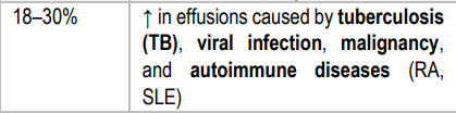
Neutrophils

Eosinophils

Mesothelial Cells
Membranes lining the serous cavities consist of a single layer of these cells.
Normal cells: small or large, round, with round nuclei and uniform dark purple cytoplasm
Reactive Mesothelial Cells
Found in clusters, multinucleated, with eccentric nuclei, resembling malignant cells.
tuberculosis
Lack of mesothelial cells with plasma cells indicates ___.
Malignant Cells
Show nuclear and cytoplasmic irregularities and hyperchromatic nucleoli.
pneumonia
malignancy
Malignant cells in pleural fluid is increased in __ and _.
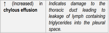
Triglyceride
Glucose
↓ (Decreased) in tuberculosis, rheumatoid inflammation, and infections
Lactate
↑ (Increased) in bacterial infections
pneumonia
need for tube drainage
< pH 7.2 would suggest ?
gastric fluid issues
esophageal rupture
≤ pH 6.0 would suggest ?
malignancy
≥ pH 7.4 (alkaline) may be related to __
acidosis
advanced infection
pH 0.30 lower than blood pH indicates ?
ADA (Adenosine Deaminase)
> 40 U/L
High __ levels may indicate malignancy or tuberculosis.
Pleural Fluid & Salivary Amylase
Tested for assessment of pancreatitis, Esophageal rupture and malignancy

>1.1 = Transudate
<1.1 = Exudate
Serum-Ascites Albumin Gradient (SAAG)
>1.1 =
<1.1 =
PERITONEAL LAVAGE
Sensitive test to detect intra-abdominal bleeding in blunt trauma cases
RBC count >100,000/mL
parameter used as an indication of Blunt trauma injuries
bile
Green/Dark-brown Peritoneal fluid indicates the presence of __
bacterial peritonitis or cirrhosis
>500 cells/µL in peritoneal fluid indicates
Ascitic exudates
show malignant cells, Lipophages, Toxoplasma gondii
Psammoma bodies
seen in benign conditions & w/ Ovarian & Thyroid malignancies
GIT malignancy, colorectal cancer
CEA in peritoneal fluid
ovaries, fallopian tubes, endometrium cancers
(+) CA125, (-) CEA
Pancreatitis, GIT perforation
Increased amylase & lipase in peritoneal fluid
GIT perforation
Increased ALP in peritoneal fluid