Visceral Pathology Lab Midterm
 Call Kai
 Learn
 Practice Test
 Spaced Repetition
 Match
 Flashcards
 Knowt Play
1/284

There's no tags or description
Looks like no tags are added yet.
285 Terms
Concentric Appearance with Thicker Wall
What is the Normal Morphology of the Left Ventricle?

Cresenteric Appearance with Thinner Wall
What is the Normal Morphology of the Right Ventricle?

300g; Fist Sized
What is the Normal Size and Weight of a Healthy Heart?
Right Side
Which Side of the Heart "Drives" the Other?
Right Side has enough force to push deoxygenated blood to lungs and back to Left Atrium
How does the Right Side "Drive" the Left Side of the Heart?
Concentric Hypertrophy of Left Ventricle
What is being shown?
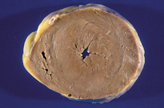
Pressure Overload
What causes the Hypertrophy shown here?

In Parallel
How are Sarcomeres being added here?
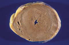
Left Ventricle
What Chamber is predominantly affected by Concentric Hypertrophy?
Chronic Hypertension & Valvular Stenosis
What conditions predispose one to what is shown?
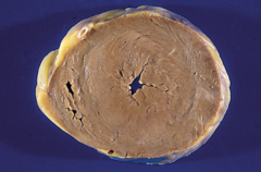
Cytoplasmic Hypertrophy with Enlarged Nuclei (Boxcar Nuclei)
What Histological Change is shown?

Occurs as a result of overload and corresponding hypertrophy
Why does what is being shown occur?

Concentric Hypertrophy
What is Shown by A?

Eccentric Hypertrophy
What is Shown by B?

Volume Overload
What causes the Hypertrophy shown in B?

In Series
How are Sarcomeres added in B?

Ischemia
What is a Potential Complication of Myocardial Hypertrophy?
Pulmonary Edema within Alveoli
What is being shown?
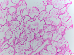
Left Sided Heart Failure
What is a common cause of this?
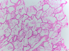
Left Ventricular Failure
What is a Specific Cardiogenic Cause of this?

Hemosiderin-Laden Macrophages
What Cells would one Expect to see if Magnified?

Neck Vein Distension
What is being shown?
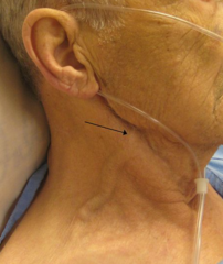
Right Sided Heart Failure
What causes this?

Elevated Jugular Venous Pressure, Hepato-Abdomino-Jugular Reflex, Ascites, Bilateral Pitting Edema, Hepato/Splenomegaly
What other signs may accompany what is being shown?

Pitting Edema
What is being shown here?

RSHF, Renal Failure, Nephrotic Syndrome, Kwashiorkor
What are potential causes of this?

Depth, or Time of Resorption
How is this assessed?

Ventricular Septal Defect
What is being shown?
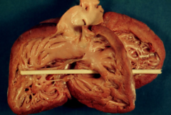
Ample Trabeculae Carnae
How do we know this is VSD?

Holosystolic Murmur
What finding would be present with auscultation here?
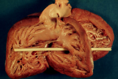
Volume Overload
What type of overload would be present here?

Eccentric Hypertrophy
What type of hypertrophy would be present here?

Atrial Septal Defect
What is being shown?
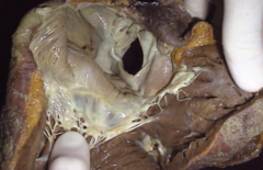
Reversal of Shunting to R->L due to Increased Pulmonary Vascular Resistance
What is Eisenmenger's Complex?
Mitral Stenosis & Ostium Secundum
What is Lutembacher's Syndrome
Patent Ductus Arteriosus
What is being shown?

Arch of Aorta & Pulmonary Trunk
What abnormal communication is visualized?

Congenital Bicuspid Aortic Valve (rather than Tricuspid Valve)
What is being shown?
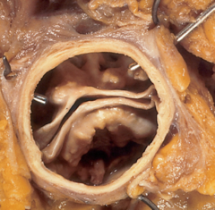
Congenital Valvular Aortic Stenosis
What is this condition called?

Stenosis
What impact does this have on the lumen?

Pressure Overload
What type of overload would be present?

Senile Aortic Stenosis, Coarctation of Aorta, Rheumatic Aortic Stenosis
What other conditions may decrease diameter of this lumen?

Exertional Dyspnea, Angina Pectoralis, Syncope
What are the major clinical features of this condition?

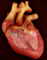
Tetrology of Fallot
What is being show?

Pulmonary Valve Stenosis, Right Ventricular Hypertrophy, Ventricular Septal Defect, Over-Riding Aorta
What anomalies compose the disorder shown?

Right-to-Left
What kind of shunt is present?
Cyanotic
How does a patient with this condition present at birth?
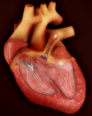
Boot-Shaped Heart
What characteristic term is given to this radiograph?

Right Ventricular Hypertrophy associated with Tetrology of Fallot Rotates Heart
What causes this abnormality?
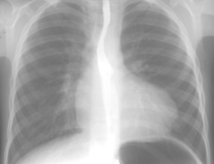
Coarctation of Aorta
What is being shown?

Hypertension resulting in Headache & Nosebleeds
What Outward Signs are present in the Upper Body?

Hypotension leading to Vascular Claudication
What Outward Signs are present in the Lower Body?
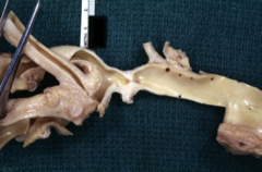
Coarctation of Aorta
What is being shown?

Rib Notching
What other Radiographic Features could be expected from this?

Upper Posterior Intercostal Arteries
What arteries are responsible for rib notching?
Angina which Radiates down Arm, Patient >35 Years Old
How does a Myocardial Infarct Present?
Myocardial Infarction
What is shown here?

Lack of Perfusion into the Area
What causes the area indicated by the arrows to have a pale appearance?
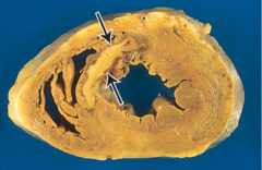
Zone of Hyperemia
What is the darker area surrounding the pale area?

Inflammatory Response has been Initiated
What does the darer area indicate?

Eosinophilia; Within 24 Hours Neutrophils will Proliferate
What Histological findings would be present?

Coagulative Necrosis
What Morphological Pattern of Necrosis is present here?
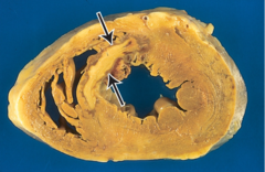
Multiple Healed Myocardial Infarctions
What is shown here?

Collagen Rich Connective Tissue
What are the Silvery-White areas?

First Neutrophils Appear
What occurs at Day 1 of Healing of a Myocardial Infarct?
Granulation Tissue Forms
What occurs at Week 1 of Healing of a Myocardial Infarct
Scar Tissue Formation
What occurs at Month 1 of Healing of a Myocardial Infarct
Different Complications arise at Different Times
Why are time periods associated with Healing of a Myocardial Infarct important?
Myocardial Infarction
What is shown here?
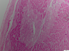
Living Myocardium surrounding a Patent Blood Vessel
What is the lighter area show?
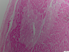
Coagulative Necrosis
What does the surrounding darker area show?

Eosinophilic
What term is given to the colour change present?

Neutrophils
What is found between spaces of dead myocytes?

24 Hours Post Infarction
When was this histological preparation prepared?
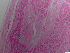
1 Week Post Infarction
When was this histological preparation prepared?

Granulation Tissue
What Type of Tissue is Present here?

Granulation Tissue is very Weak and likely to Rupture
What is a Serious Complication that may arise at this point?

Rupture of Granulation Tissue
What is shown here?

Healed Myocardial Infarction
What is shown here?

Connective Tissue
What is found at the bottom right?

Streptococcus bacteria
What is the Etiology of Rheumatic Heart Disease?
All Layers
What Layers are of the Heart are Affected in Rheumatic Heart Disease?
Acute Rheumatic Heart Disease
What is shown here?

Aschoff Bodies
What are Granulomas found in this condition called?
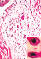
Anitchkow Cells
What are Giant Cells found in Granulomas present in this condition called?

Caterpillar Shaped Nuclei
What characteristic do the Nuclei of Giant Cells found in Granulomas present in this condition have?
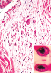
Mitral & Aortic Valve
What Valves of the Heart are commonly affected by Chronic Rheumatic Fever?
Stenosis and/or Valvulitis of Mitral & Aortic Valve
What are the Consequences of Chronic Rheumatic Fever on Valves of the Heart?
Chronic Rheumatic Valvulitis
What is shown here?

Vegetative Growths
What is shown by the arrow?

Stiff Non-Compliant Valves & Stenosis
What are consequences of this?

Rheumatic Mitral Stenosis
What is shown here?

Fish Mouth Appearance
What is this characteristic appearance termed?

Fusion of Commissures & Chordae Tendineae
What causes this appearance?

Mitral Valve Prolapse
What is shown here?
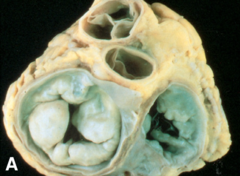
Familial Distribution, Inherited Collagen Disease
What Predisposed one to this condition?

Mid-Systolic Click with Murmur
What can be expected during Auscultation in this condition?

Endocarditis
Having this pathology predisposes one to _____

Bacterial Endocarditis of Mitral Valve
What is shown here?

Sustained Inflammation at Valve due to Prolapse
How does this condition predispose valves to Vegetations?
