Lab B4
 Call Kai
 Learn
 Practice Test
 Spaced Repetition
 Match
 Flashcards
 Knowt Play
1/45

There's no tags or description
Looks like no tags are added yet.
46 Terms
What are the major functions of connective tissue?
Binding, support/protection, immune protection, movement, storage, and transport
What are the two major components of connective tissue?
Cellular portion and matrix.
Is the matrix cellular or acellular?
Acellular
What are the two parts of the connective-tissue matrix?
Ground substance and fibrous component.
What are the three major fiber types in connective tissue?
Collagen, elastic, and reticular fibers.
What consistency can ground substance have?
It ranges from liquid to solid, often a semisolid gel.
What is a primary tissue-determining cell?
A cell that defines the primary structure/function of a specific tissue.
Example: Osteocytes define osseous tissue
What supporting cells may be present in multiple connective tissues?
Fibroblasts, macrophages, neutrophils, plasma cells, mast cells, and adipocytes.
How is areolar connective tissue described?
A “packaging” tissue
What does areolar connective tissue contain abundantly?
Extracellular fibers
What cellular feature is scattered throughout areolar tissue?
Nuclei of various cell types
Where is areolar connective tissue commonly found?
Beneath epithelia, including subcutaneous tissue.
What fibers should you be able to recognize in areolar CT?
Collagen fibers and reticular fibers.
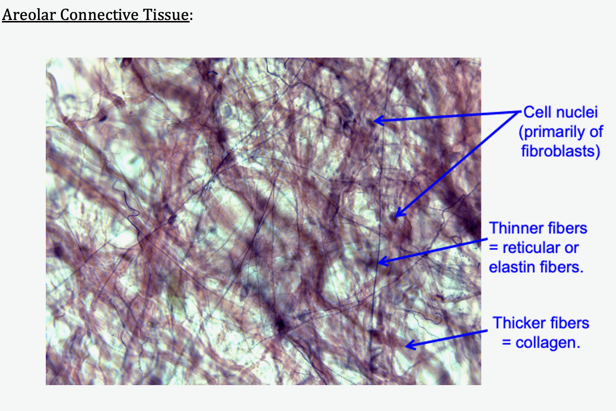
What microscopic appearance should suggest areolar CT?
Loose fibers with scattered cell nuclei.
Where is reticular connective tissue found?
Immune-system tissues such as lymph nodes and spleen.
What is the primary cell type listed for reticular connective tissue?
Lymphocytes.
What fibers make up most of reticular connective tissue's fibrous component?
Reticular fibers
What does the silver stain make especially visible in reticular tissue?
Reticular fibers, which appear dark
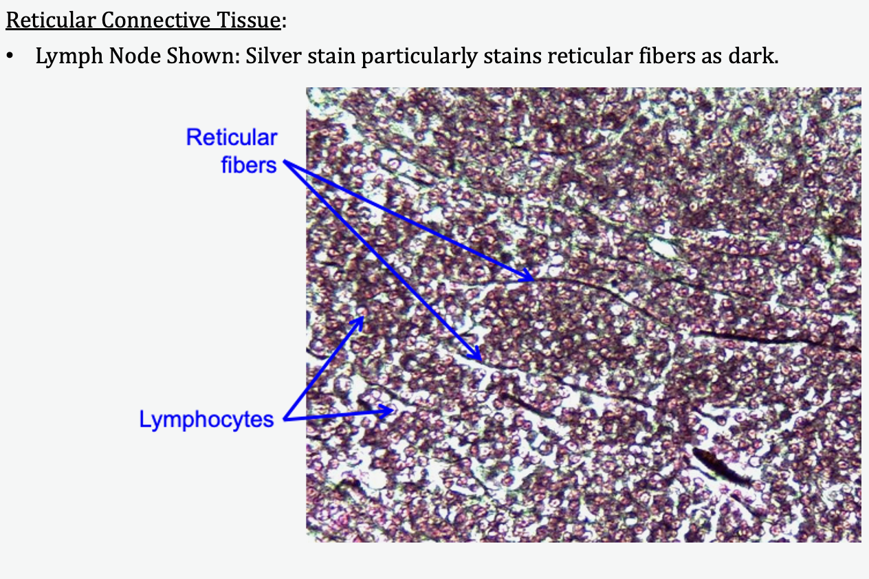
What microscopic combination suggests reticular CT?
A network of reticular fibers with many lymphocytes.
What is the characteristic cell of adipose connective tissue?
Adipocyte
What occupies most of an adipocyte?
A large triglyceride-filled lipid inclusion
Where is the nucleus located in an adipocyte?
Pushed toward the cell's periphery
Where is adipose connective tissue commonly found?
Subcutaneous tissue, breast tissue, and around organs.
What vascular structures are often visible in adipose tissue?
Capillaries and other blood vessels
Are adipocytes found only in adipose tissue?
No. Scattered adipocytes can occur in other tissues.
What microscopic appearance suggests adipose tissue?
Large empty-looking adipocytes with nuclei pushed to the edges
What is the defining cell type of cartilage?
Chondrocyte
Where are chondrocytes located?
Within lacunae.
What is a lacuna?
A space in the cartilage matrix containing a chondrocyte.
What is the primary fiber in all three cartilage types?
Collagen
What happens to the collagen fibers in hyaline cartilage under the microscope?
They are present but are not individually visible.
Where is hyaline cartilage found?
Articular cartilage, trachea/bronchi, larynx, and costal cartilages.
Does hyaline cartilage always have a perichondrium?
No. It sometimes has one.
What tissue makes up the perichondrium?
Dense irregular connective tissue
Is the perichondrium itself cartilage?
No

What microscopic appearance suggests hyaline cartilage?
Smooth/glassy matrix with chondrocytes in lacunae and no obvious individual fibers.
What fiber distinguishes elastic cartilage from the other cartilage types?
Elastin fibers
What property do elastin fibers give elastic cartilage?
Elasticity
Where is elastic cartilage found?
External ear and epiglottis.
Does elastic cartilage have a perichondrium?
Yes

What microscopic feature strongly suggests elastic cartilage?
Visible elastin fibers around chondrocytes in lacunae
Where is fibrocartilage found?
Pubic symphysis, intervertebral discs, and menisci.
How are chondrocytes often arranged in fibrocartilage?
In linear rows.
What mechanical property is emphasized for fibrocartilage?
It is strong and resists compression well.
Does fibrocartilage have a perichondrium?
No, never.
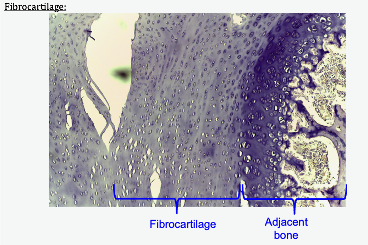
What microscopic feature strongly suggests fibrocartilage?
Chondrocytes arranged in rows among prominent fibers.