Anatomy Exam 3 Brig
 Call Kai
 Learn
 Practice Test
 Spaced Repetition
 Match
 Flashcards
 Knowt Play
1/610

There's no tags or description
Looks like no tags are added yet.
611 Terms
where does 90% of movement in the cervical spine occur
between C1 and C2
spinous process of axis
Last felt before the skull because the atlas does not have a spinous process
Vertebra prominens of C7 vertebra
Spinous process is much longer and is palpable at the base of the flexed neck
Hyoid bone
U-shaped
Directly inferior to mandible at C3 level
Laryngeal prominence
Adam's apple
-superior prominence of thyroid cartilage
-anterior to C4
Cricoid cartilage
Ring-shaped hyaline cartilage directly inferior to thyroid cartilage
-C6 vertebra
location of cricoid cartilage
C6
Bottom of the cricoid cartilage is at the bottom of the laryngopharynx
what muscle divides the neck into anterior and posterior triangles
SCM
transverse process of the atlas (C1)
Inferior to mastoid process and posterior angle of mandible where they intersect
borders of trapezius muscle
muscular ridge from external occipital protuberance to acromion process and lateral portion of the clavicle
tendon of sternal head of SCM muscle
either side of suprasternal notch
lesser supraclavicular fossa
between the 2 SCM tendons, overlies inferior portion of internal jugular
supraclavicular fossa
overlies subclavian artery
clinical significance of cervical fascia
helps limit spread of infection to one compartment
4 layers of deep cervical fascia
investing fascia
pretracheal fascia
prevertebral fascia
alar fascia + carotid sheath
Superficial cervical fascia
platysma is embedded
investing fascia
encircles the neck deep to superficial fascia
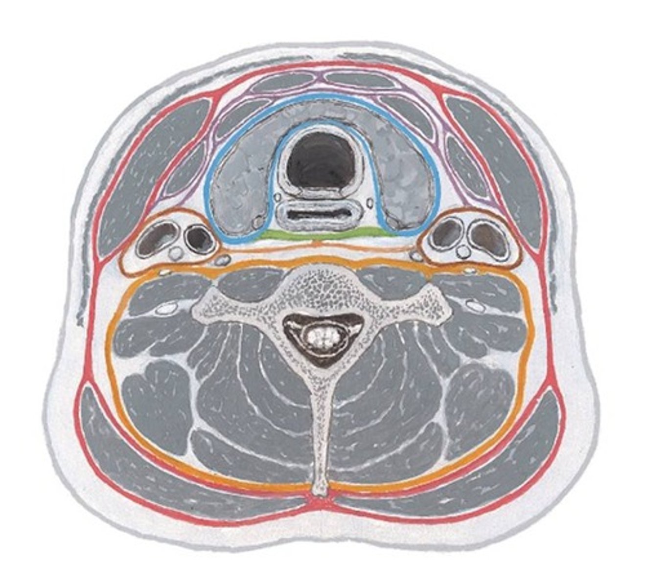
superior attachments of investing fascia
occipital bone
mastoid process
zygomatic arches
inferior border of mandible
hyoid bone
interior attachments of investing fascia
manubrium
clavicle
acromin and spine of scapula
ligamentum nuchae
Splitting of investing fascia
splits to encase SCM and trapezius mm.
Pretracheal fascia
Muscular and visceral portions
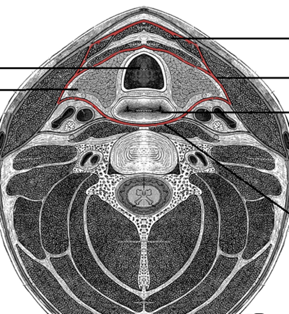
muscular portion of pretracheal fascia
only in anterior portion of the neck
-encircles infrahyoid and suprahyoid muscles
visceral portion of pretracheal fascia
larynx, pharynx, trachea, esophagus, thyroid gland, parathyroid glands, recurrent laryngeal nn., associated vessels
=visceral compartment of neck
visceral pretracheal fascia extends from...
base of skull and hyoid to fibrous pericardium creating a column
what does visceral pretracheal fascia fust
carotid sheaths laterally
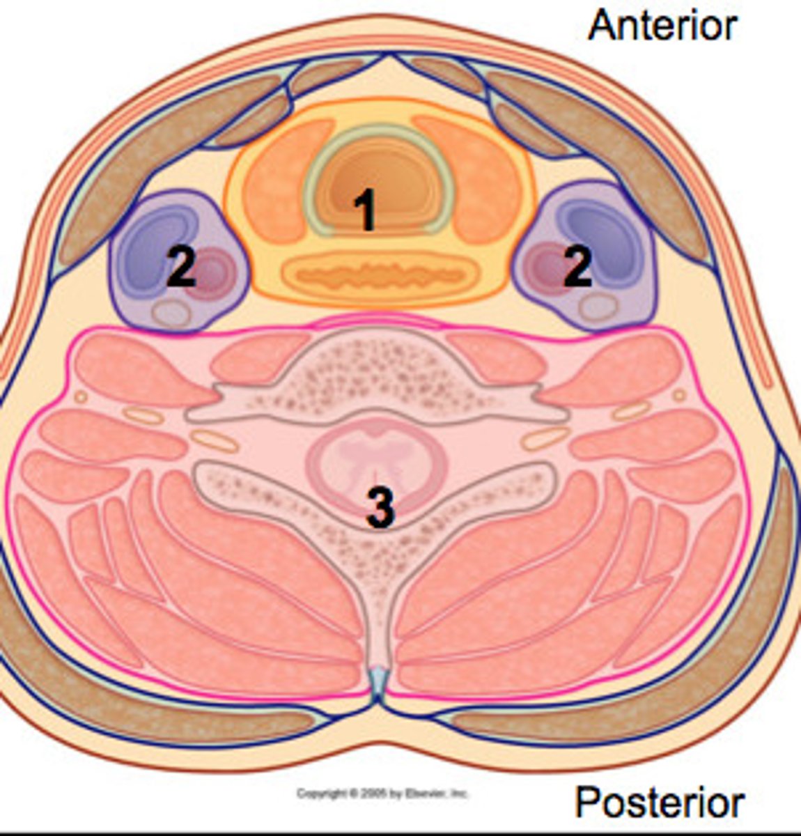
Prevertebral fascia
Encircles prevertebral muscles, vertebral column, and intrinsic muscles of the neck
also, dorsal and ventral rami of cervical spinal nerves
vertebral compartment
made of prevertebral fascia
largest compartment of the neck
inferior blending of prevertebral fascia
blends with endothoracic fascia and encases cervical sympathetic trunk
laterally forms axillary sheath to enclose brachial plexus and axillary artery
carotid sheaths
b/l tubes of fascia that hold common carotid, internal carotid, internal jugular vein, and vagus nerve
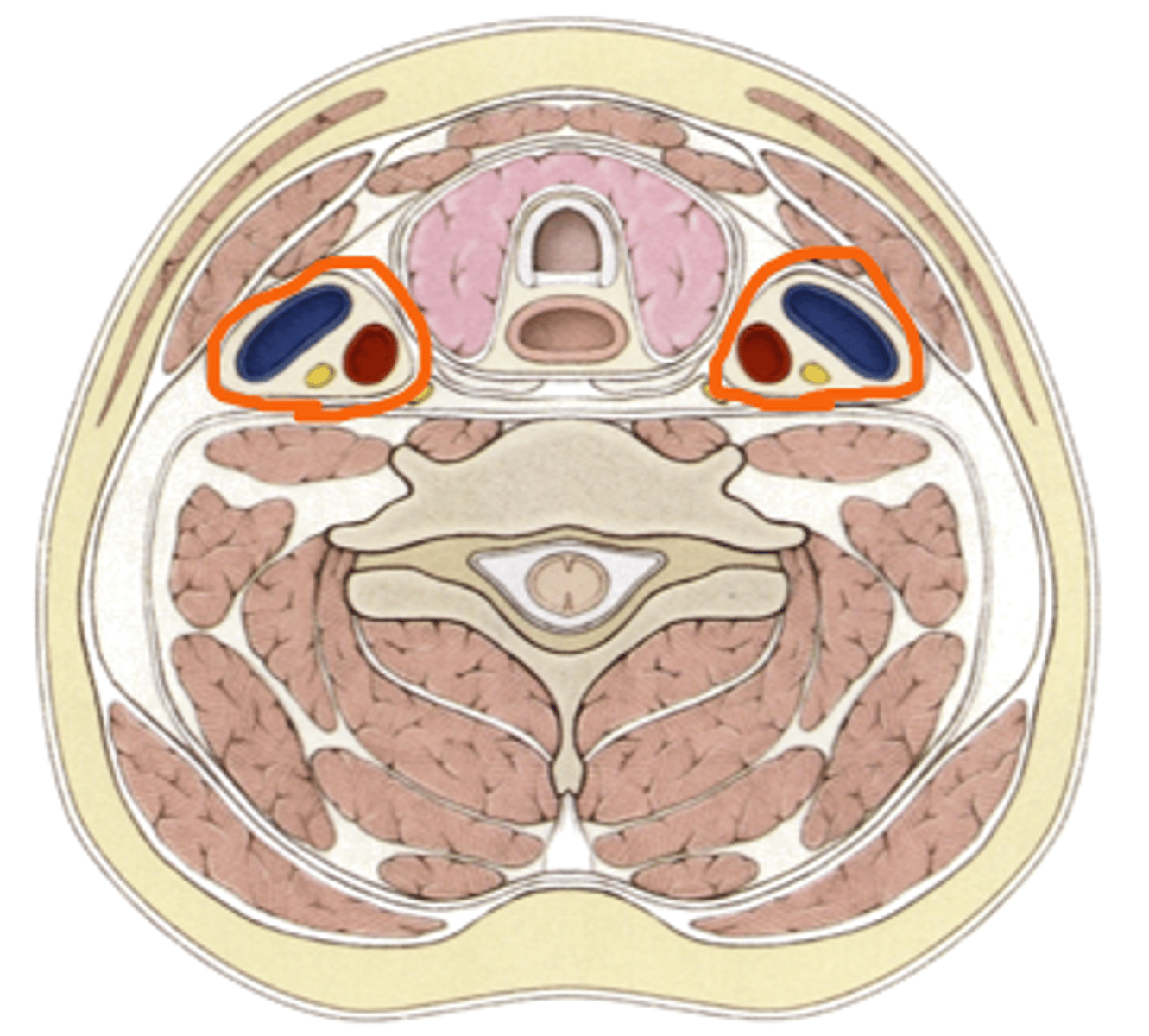
blending of carotid sheaths
investing layer anterolaterally
pretracheal fascia anteromedially
alar fascia medially
prevertebral fascia posteriorly
alar fascia
connects 2 carotid sheaths and subdivides neck into visceral and vertebral compartments
retropharyngeal space
potential space
between pretracheal fascia and alar fascia
skull to superior mediastinum
function of retropharyngeal space
permits movement of contents in visceral compartment (food swallowing)
alar (danger) space
Potential space between alar fascia and prevertebral fascia
infection here can spread to diaphragm
narrow band of thyroid tissue that joins the 2 lobes
isthmus
pyramidal thyroid lobe
on isthmus, is random and poses no risk to patient
follicle cell composition
simple cuboidal cells - follicular cells
function of T3 and T4
increase ATP production
increase transcription and translation of cells
=increased cellular metabolism
Grave's disease
antibodies to TSH receptors cause over-secretion of T3 and T4
-hyperplasia of follicular cells = diffusely enlarged thyroid gland
Hashimoto's
Anti-T3
Anti-T4
Anti-TSH
Anti-thyroid antibodies
=Undersecretion of thyroid hormones and gradual thyroid destruction
borders of neck rectangle
Posterior - trapezius m
Anterior - midline of body or sternohyoid m
Inferior - clavicle
Superior - mandible
anterior triangle connects head to..
thorax
posterior triangle connects head to..
upper limb
Inferior belly of the omohyoid
divides posterior triangle into 2 triangles:
1. superior occipital triangle
2. inferior supraclavicular triangle
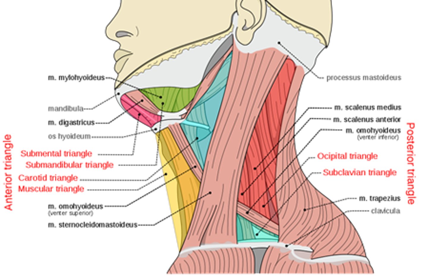
Superior belly of the omohyoid
divides inferior anterior triangle in 2:
1. carotid triangle
2. muscular triangle
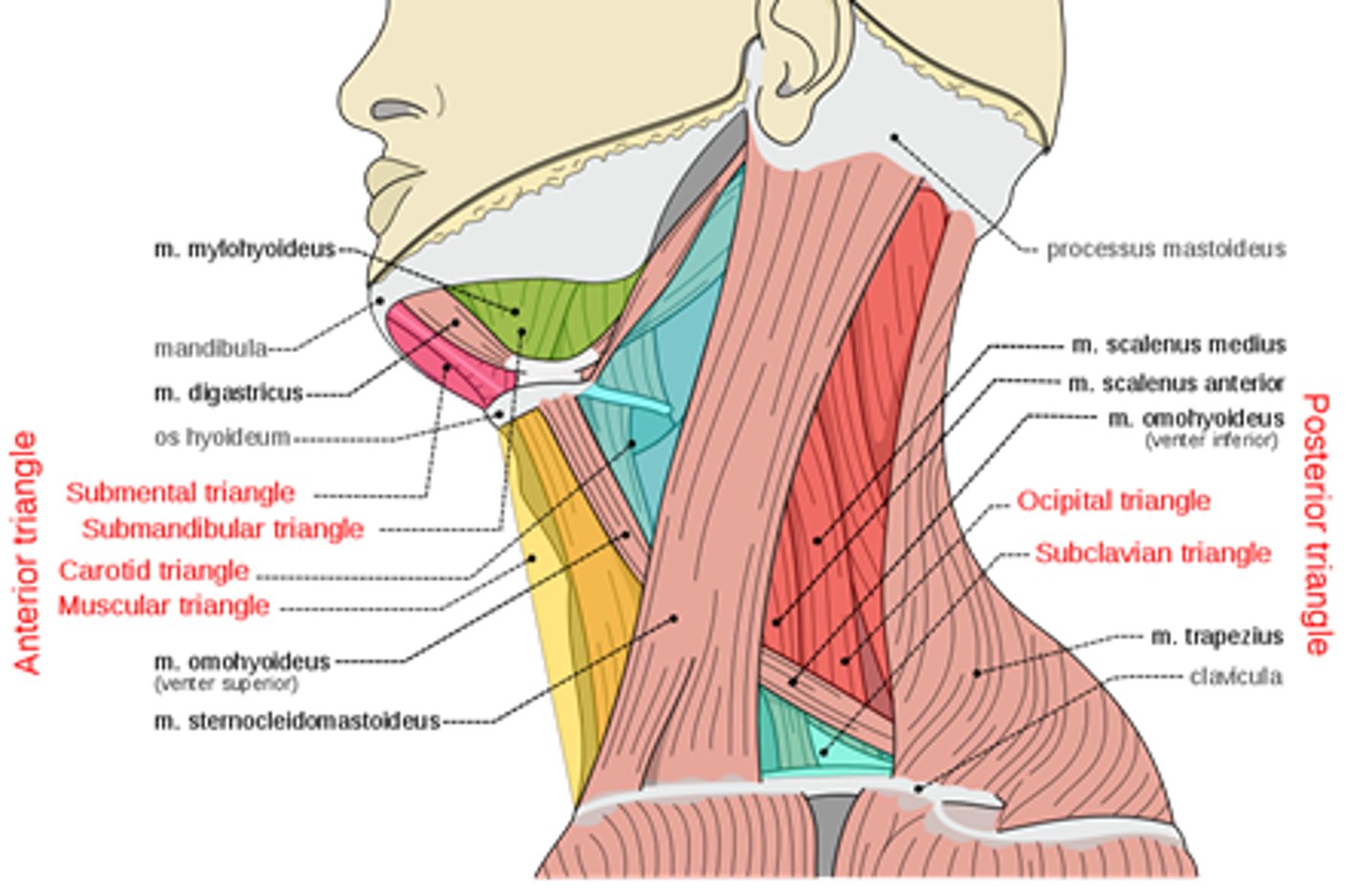
Digastric muscle
divides superior and anterior parts of the triangle into 2:
1. submandibular triangle
2. submental triangle
borders of the occipital triangle
anteriorly - SCM
posteriorly - trapezius
inferiorly - inferior belly of the omohyoid
contents of the occipital triangle
external jugular vein
accessory nerve
supraclavicular nerves
levator scapular muscle
anterior, middle, posterior scalene muscles
supraclavicular triangle borders
superior - inferior belly of the omohyoid muscle
anterior - SCM
inferior - medial portion of clavicle
contents of supraclavicular triangle
part of brachial plexus
subclavian artery
suprascapular artery
borders of the carotid triangle
superiorly - posterior belly of digastric muscle
posteriorly - SCM
inferiorly - superior belly of omohyoid muscle
contents of carotid triangle
Common carotid (internal and external)
internal jugular vein
hypoglossal nerve
vagus nerve
submandibular triangle borders
superior - inferior border of mandible
anterior - anterior belly of digastric
posterior - posterior belly of digastric
contents of submandibular triangle
submandibular gland
facial artery and vein
stylohyoid muscle
mylohyoid muscle and nerve
what 2 muscles form floor of mouth
stylohyoid and mylohyoid
borders of submental triangle
superior - anterior belly of digastric muscle
anterior - median plane of neck
inferior - hyoid bone
submental triangle contents
anterior jugular vein
mylohyoid muscle
borders of the muscular triangle
superior - superior belly of omohyoid
anterior - median plane (medial sternothyroid and sternohyoid)
inferior - SCM
contents of muscular triangle
sternothyroid muscle
sternohyoid muscle
3 superficial neck muscles
platysma
SCM
trapezius
platysma origin
pectoral fascia
platysma insertion
inferior border of mandible and local muscles
platysma action
tense neck skin
platysma innervation
cervical branch of facial nerve
SCM origin of sternal head
anterior surface of manubrium
SCM origin of clavicular head
medial 1/3 of clavicle
SCM insertion
mastoid process of temporal bone
innervation of SCM
accessory nerve
function of SCM
lateral rotation and flexion
b/l = cervical flexion
5 muscles of the posterior neck triangle
Splenius capitis
Levator scapulae
Posterior scalene
Middle scalene
Anterior scalene
splenius capitis origin
ligamentum nuchae
spinous processes of C7-T3
splenius capitis muscle insertion
mastoid process and lateral superior nuchal line
splenius capitis muscle innervation
dorsal rami of C2 and C3
splenius capitis muscle action
lateral flexion and rotation
b/l = cervical extension
levator scapulae origin
transverse processes of C1-C4
levator scapulae insertion
superior angle of scapula
levator scapulae innervation
dorsal scapular nerve
levator scapulae action
elevate scapula
rotate glenohumoral joint inferiorly
posterior scalene muscle origin
transverse processes of C4-C6
posterior scalene insertion
external surface of second rib
posterior scalene innervation
ventral rami of C7-C8 spinal nerves
posterior scalene action
lateral flexion
elevate second rib
middle scalene origin
Transverse processes of C2-C7
middle scalene insertion
superior surface of 1st rib
behind subclavian artery and above trunk of brachial plexus
middle scalene innervation
ventral rami C3-C8
middle scalene action
lateral flexion
elevate first rib
anterior scalene origin
transverse processes of C3-C6
anterior scalene insertion
superior surface of 1st rib
infront of subclavian artery and above trunk of brachial plexus
anterior scalene innervation
ventral rami C4-C6
anterior scalene action
lateral flexion
elevate first rib
suprahyoid muscles
connect hyoid bone to mandible and skull
mandible depression
infrahyoid muscles
tract hyoid bone inferiorly for stability
elevate or depress larynx w/ deglutition
4 suprahyoid muscles
digastric
stylohyoid
mylohyoid
geniohyoid
digastric muscle origins
anterior belly: digastric fossa of mandible
posterior belly: mastoid process of temporal bone
digastric muscle insertion
intermediate tendon
-anchored to hyoid bone
digastric muscle innervations
anterior belly: trigeminal nerve
posterior belly: facial nerve
digastric muscle action
elevate hyoid
depress mandible
stylohyoid muscle origin
styloid process of temporal bone
-fibers course digastric
stylohyoid muscle insertion
body of hyoid
stylohyoid muscle innervation
facial nerve