Lab 6: Brain and Cranial Nerves
 Call Kai
 Learn
 Practice Test
 Spaced Repetition
 Match
 Flashcards
 Knowt Play
1/84

There's no tags or description
Looks like no tags are added yet.
85 Terms
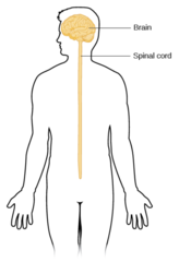
Central nervous system
Integrates, processes, and coordinates sensory data and motor commands.
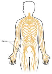
Peripheral nervous system
All neural tissue outside of the CNS; relays messages into the CNS and back out to the rest of the body for a response.
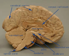
Cerebrum
large part of the brain containing the cerebral cortex (of the two cerebral hemispheres)

Cerebral hemispheres
Right and left hemispheres

Corpus callosum
Connects the hemispheres

Cerebral cortex
layer surrounding the entire cerebrum (can't see it)
Gyri (singular; gyrus)
Prominent fold/ridge on surface of cerebrum (FLESH)

Sulci (singular; sulcus)
Shallow groove

Fissures
deeper than sulci; elongated

Longitudinal fissure
separates the two hemispheres

Lateral sulcus
on the side; separates temporal lobe & parietal

Parieto-occipital sulcus
On back; separates occipital and parietal lobe

Central sulcus
On top; separates the frontal and parietal lobe

Precentral gyrus
Before central sulcus (reddish w/ stripes); closer to frontal lobe

Postcentral gyrus
After central sulcus (purple); closer to occipital lobe

Frontal lobe
Forehead

Parietal lobes (2)
.Middle (left and right)

Temporal lobes (2)
By the ears

Occipital lobe
Back of the head

Insula
underneath the lobes; (see on small model right on top)4

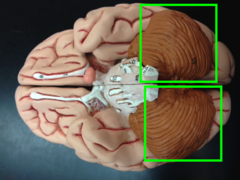
Cerebellum
Dark brown portion under the cerebrum.

Cerebellar hemispheres
Right and left
Cerebellar cortex
layer surrounding the cerebellum; cannot be seen

Folia
all of the ridges and lines on the cerebellum
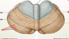
Vermis
where the two hemispheres come together in the cerebellum

Arbor vitae
all of the white nerves in the cerebellum; split in two
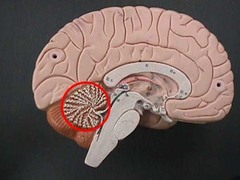
Diencephalon
"head of sea horse"

Thalamus
contains the yellow dot; whole region; "brain"

Hypothalamus
right before the optic nerve (outside of it); "mouth"

Pineal gland
Part above the thalamus; pink on model; "ponytail"

Anterior commissure
"eye of seahorse"

Interthalamic adhesion
yellow dot in the thalamus

Optic chiasm
where the two optic nerves come together

Optic nerve, II
also known as cranial nerve II, is a paired nerve that transmits visual information from the retina to the brain.

Optic tract
the track leading back to the brain; a continuation of the optic nerve that relays information

Pituitary gland
A pea-sized body attached to the base of the brain; pink blob on model

Infundibulum
the hollow stalk that connects the hypothalamus and the posterior pituitary gland; "straw"

Brainstem
Connects the cerebrum to the spinal cord

Tegmentum
a small central part of the brain-stem

Tectum
in the dorsal part of the midbrain; "backpack"

Corpora quadrigemina
is the four colliculi—two inferior, two superior—located on the tectum of the dorsal aspect of the midbrain

Superior colliculi
the tops ones (2); a structure in the midbrain that is part of the brain circuit for the transformation of sensory input into movement output

Inferior colliculi
the bottom ones (2); an important auditory center and may participate in the integration of hearing reflexes

Aqueduct of midbrain
canal separating the tectum and tegmentum

Midbrain
a general area within the brainstem; "internal neck"

Cerebral peduncles
outside portion of the "neck"; structures at the front of the midbrain which arise from the front of the pons

Pons
if the seahorse was "pregnant"; the part of the brainstem that links the medulla oblongata and the thalamus.

Transverse fibers
lines on pons; Nerve fibers crossing the midline and connecting two corresponding parts or regions of the nervous system

Medulla oblongata
"tail region"; the continuation of the spinal cord within the skull, forming the lowest part of the brainstem

Olive
exterior; "pink blob" right next to the hypoglossal nerve

Central canal
next to the medulla oblongata; aqueduct leads to it.

Lateral ventricle
"wings"; large top portion; the two largest cavities of the ventricular system of the human brain and contain cerebrospinal fluid

Septum pellucidum
just below the corpus callosum; a thin, triangular, vertical double membrane separating the anterior horns of the left and right lateral ventricles of the brain.

Interventricular foramina
"arms"; small portion leading up to the lateral ventricle

Third ventricle
"dragons face"; It is in the midline, between the left and right lateral ventricles.

Choroid plexus
all of the pink on the black model; veins and arteries. Also purple on the other model.

Aqueduct of midbrain
"spine"; connects the third ventricle in the diencephalon to the fourth ventricle

Fourth ventricle
"tail"; tent-shaped, with the peak of the tent protruding into the cerebellum and the floor of the tent being formed by the pons and medulla

Dural folds
falx cerebri
tentorium cerebelli
falx cerebelli

Falx cerebri
whole top portion; separates two cerebral hemispheres

Tentorium cerebelli
"tent"; wings; separates cerebrum from cerebellum

Falx cerebelli
small projection; separates two hemispheres of cerebellum; a small sickle shaped fold of dura mater, projecting forwards into the posterior cerebellar notch

Dural sinuses
are venous channels found between the endosteal and meningeal layers of dura mater in the brain.

Superior sagittal sinus
Top blue line; on top of falx cerebri

inferior sagittal sinus
across the bottom; below falx cerebri

Transverse sinus
along the edges of the tentorium cerebelli

Amygdala
round end points (large and white on model)

Cingulate gyrus
region surrounding the limbic system; orange on model

Fornix
most medial tubes; below septum pellucidum

Hippocampus
large portion leading away from amygdala; the elongated ridges on the floor of each lateral ventricle of the brain

Mammillary bodies
most medial circular folds; right in the middle
"horse's adam's apple"

Olfactory nerve , I
white line underneath cranium

Olfactory tracts

Olfactory bulbs

Optic nerve, II
"tip of the nose"

Oculomotor nerve, III
third of the cranial nerves and arises from the midbrain.

Trochlear nerve, IV
lateral to the oculomotor nerve; fourth cranial nerve and is the motor nerve of the superior oblique muscle of the eye

Trigeminal nerve, V
side of pons; the fifth paired cranial nerve & also the largest cranial nerve.

Abducens nerve, VI
where the "abs" would be

Facial nerve, VII
lateral; emerges from the brainstem between the pons and the medulla

Vestibulocochlear nerve, VIII
lateral to the facial nerves (VII); Arising from the brainstem between the pons and medulla

Glossopharyngeal nerve, IX
just below the vestibulocochlear nerve (VIII); exits the brainstem out from the sides of the upper medulla, just above the vagus nerve

Vagus nerve, X
below the glossopharyngeal nerve (IX); the longest cranial nerve

Accessory nerve, XI
skinny line below vagus nerve

Hypoglossal nerve, XII
medial to IX & X; controls all tongue movements
