GENETICS MIDTERM 2

0.0(0)
Studied by 0 people
call kaiCall Kai
learnLearn
examPractice Test
spaced repetitionSpaced Repetition
heart puzzleMatch
flashcardsFlashcards
GameKnowt Play
Card Sorting

1/34

encourage image

There's no tags or description

Looks like no tags are added yet.

35 Terms
1
New cards
<p>X linked genes are easy to map in Drosophila males</p>

X linked genes are easy to map in Drosophila males

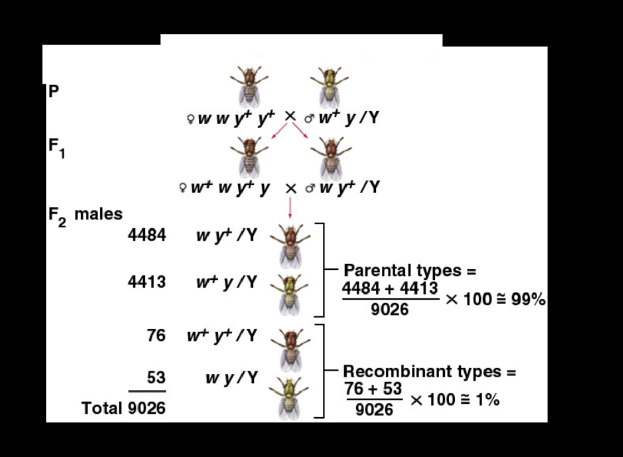
recombination frequency tells you how far apart two linked genes are, and in Drosophila males it’s very easy to measure.

Since a male has only one X, his phenotype directly shows which X he got from his mother, so you can simply count how many sons are recombinant versus parental. Recombinant sons divided by total sons (129 / 9026 ≈ 1%) is the recombination frequency, and 1% recombination = about 1 map unit (1 cM) between w and y


In Drosophila, crossing over happens in females but not in males, so the recombination you’re measuring comes entirely from the F₁ mother’s meiosis.


  1. Crossing over between w and y happens in the F₁ mother’s meiosis, where her two different X’s can swap pieces.

  2. Each son gets his father’s Y (no alleles for these genes) and one of his mother’s X’s, which may be parental or recombinant.

  3. With no second X to mask it, his phenotype shows exactly which type he got.

  4. So recombination frequency = recombinant sons ÷ total sons = 129 / 9026 ≈ 1.4% (rounded to 1% on the slide), or about 1 map unit.

2
New cards
<p>a) What are the parental chromosomes?</p><p>b) Which gene is in the middle? Write the genotype of the parental female reflecting this gene</p><p>order.</p><p></p><p></p><p><strong>a) What are the parental chromosomes?</strong></p><p>They are the mother’s two original X chromosomes, which show up as the two most common classes of sons (2278 and 2157):</p><ul><li><p><code>W Y m</code> (2278 sons)</p></li><li><p><code>w y M</code> (2157 sons)</p></li></ul><p><strong>b) Which gene is in the middle? Write the genotype of the parental female reflecting this gene order.</strong></p><p>The middle gene is <code>w</code>. The rarest classes (the double crossovers, 2 and 1 sons) are the parental X’s with only <code>w</code> switched, so <code>w</code> sits between <code>y</code> and <code>m</code>.</p><p>The gene order is <code>y – w – m</code>, so the parental female’s genotype is:</p><p><code>Y W m / y w M</code></p><p>(The slash separates her two X chromosomes. Each X lists its alleles in gene order.)</p>

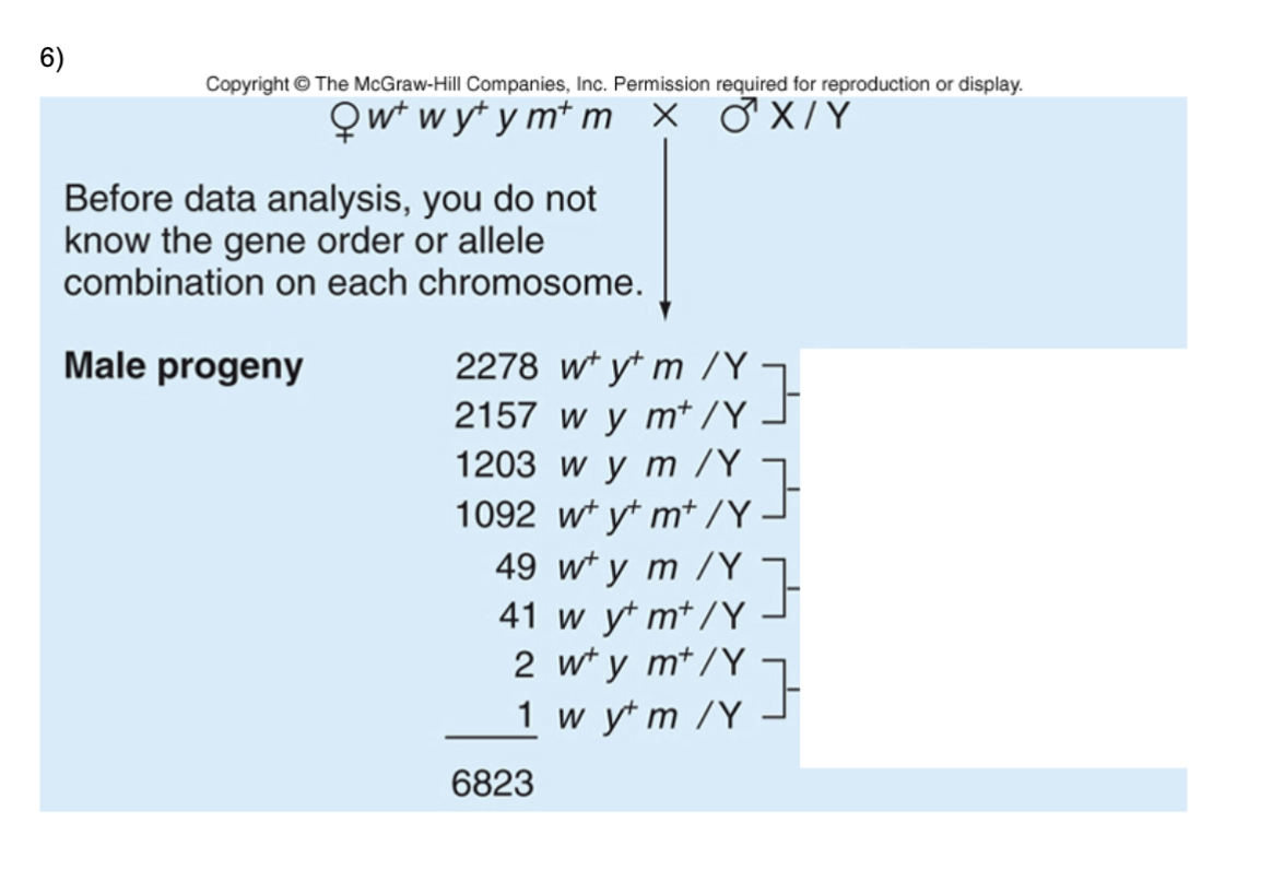
a) What are the parental chromosomes?

b) Which gene is in the middle? Write the genotype of the parental female reflecting this gene

order.


a) What are the parental chromosomes?

They are the mother’s two original X chromosomes, which show up as the two most common classes of sons (2278 and 2157):

  • W Y m (2278 sons)

  • w y M (2157 sons)

b) Which gene is in the middle? Write the genotype of the parental female reflecting this gene order.

The middle gene is w. The rarest classes (the double crossovers, 2 and 1 sons) are the parental X’s with only w switched, so w sits between y and m.

The gene order is y – w – m, so the parental female’s genotype is:

Y W m / y w M

(The slash separates her two X chromosomes. Each X lists its alleles in gene order.)

finding the total map distance of the three x linked genes

<p>finding the total map distance of the three x linked genes</p>
3
New cards

7) Genes A and B are X linked and are located 7 map units apart on the X chromosome in

Drosophila. A female of genotype A b/ a B is mated with a wildtype male.

a) What is the probability that one of her sons will be phenotypically wildtype for A and B? Her

daughter?

knowt flashcard image
4
New cards
<p>2 genes can independently influence the same trait:</p>

2 genes can independently influence the same trait:

the two genes sort independently, so the cross gives the same 9:3:3:1 genotype classes as any dihybrid cross. The only difference is how you read the result. In a normal dihybrid cross, each gene controls its own trait (like seed color and seed shape). Here, both genes feed into one trait, snake color, so you read the phenotype by asking which pigments the snake can make.


polygenic!

<p>the two genes sort independently, so the cross gives the same 9:3:3:1 genotype classes as any dihybrid cross. The only difference is how you read the result. In a normal dihybrid cross, each gene controls its own trait (like seed color and seed shape). Here, both genes feed into one trait, snake color, so you read the phenotype by asking which pigments the snake can make.</p><p></p><p>polygenic! </p>
5
New cards
<p>2 genes in two different pathways can influence the same trait</p>

2 genes in two different pathways can influence the same trait

The two arrows both point to snake skin color. The snake’s color is the combined result of whichever pigments its cells are making:

  • Both pathways working gives the camouflage pattern.

  • Only the B pathway working gives black.

  • Only the O pathway working gives orange.

  • Neither working gives albino


Each gene has its own pathway (gene → mRNA → pigment), and the two pathways don’t interact. Both feed into one trait, snake skin color, and the snake’s color is whatever combination of pigments it ends up making


6
New cards
<p>What about genes that function in a common pathway?</p>

What about genes that function in a common pathway?

Here, both genes work in the same pathway, one step after the other.

The pathway

  1. Colorless precursor 1 is converted by protein A (made from gene A) into colorless precursor 2.

  2. Colorless precursor 2 is converted by protein B (made from gene B) into the purple pigment.

The flower only turns purple if both steps happen. Protein B can only work on precursor 2, and precursor 2 only exists if protein A made it. I’m assuming that capital letters (A, B) are the working dominant alleles and lowercase (a, b) are non-working, which is the same convention as before.

<p>Here, <strong>both genes work in the same pathway, one step after the other.</strong></p><p><strong>The pathway</strong></p><ol><li><p>Colorless precursor 1 is converted by <strong>protein A</strong> (made from gene A) into colorless precursor 2.</p></li><li><p>Colorless precursor 2 is converted by <strong>protein B</strong> (made from gene B) into the <strong>purple pigment</strong>.</p></li></ol><p>The flower only turns purple if both steps happen. Protein B can only work on precursor 2, and precursor 2 only exists if protein A made it. I’m assuming that capital letters (<code>A</code>, <code>B</code>) are the working dominant alleles and lowercase (<code>a</code>, <code>b</code>) are non-working, which is the same convention as before.</p>
7
New cards
<p>PATHWAY EXAMPLE </p>

PATHWAY EXAMPLE

Answer: 9 black : 3 brown : 4 tan

Reading the pathway

tan → (G) → brown → (H) → black

  • G is the working allele for the enzyme that turns tan into brown. H is the working allele for the enzyme that turns brown into black. Tan is the starting color. A fly or animal with no working conversion just stays tan.

  • Step 2 only works if step 1 happened, since the H enzyme needs brown to act on, and brown only exists if G worked.


9 G/-; H/- black

3 G/-; h/h brown

4 g/g; -/- tan

8
New cards
<p>Genetic interactions affect the phenotypes displayed</p>

Genetic interactions affect the phenotypes displayed

Situation

Genotype ratio

Phenotype ratio

Why

Independent genes

9:3:3:1

9:3:3:1

Each genotype class looks different

Pathway, similar intermediates

9:3:3:1

9:7

Three classes (A/-; b/b, a/a; B/-, a/a; b/b) all look white

Pathway, different intermediates

9:3:3:1

9:3:4

Two classes (g/g; H/-, g/g; h/h) both look tan

dihybrid cross will always have the same genotypic ratio when genes assort independently

<table style="min-width: 100px;"><colgroup><col style="min-width: 25px;"><col style="min-width: 25px;"><col style="min-width: 25px;"><col style="min-width: 25px;"></colgroup><tbody><tr><th colspan="1" rowspan="1"><p>Situation</p></th><th colspan="1" rowspan="1"><p>Genotype ratio</p></th><th colspan="1" rowspan="1"><p>Phenotype ratio</p></th><th colspan="1" rowspan="1"><p>Why</p></th></tr><tr><td colspan="1" rowspan="1"><p>Independent genes</p></td><td colspan="1" rowspan="1"><p>9:3:3:1</p></td><td colspan="1" rowspan="1"><p>9:3:3:1</p></td><td colspan="1" rowspan="1"><p>Each genotype class looks different</p></td></tr><tr><td colspan="1" rowspan="1"><p>Pathway, similar intermediates</p></td><td colspan="1" rowspan="1"><p>9:3:3:1</p></td><td colspan="1" rowspan="1"><p><strong>9:7</strong></p></td><td colspan="1" rowspan="1"><p>Three classes (<code>A/-; b/b</code>, <code>a/a; B/-</code>, <code>a/a; b/b</code>) all look white</p></td></tr><tr><td colspan="1" rowspan="1"><p>Pathway, different intermediates</p></td><td colspan="1" rowspan="1"><p>9:3:3:1</p></td><td colspan="1" rowspan="1"><p><strong>9:3:4</strong></p></td><td colspan="1" rowspan="1"><p>Two classes (<code>g/g; H/-</code>, <code>g/g; h/h</code>) both look tan</p></td></tr></tbody></table><p>dihybrid cross will always have the same genotypic ratio when genes assort independently </p>
9
New cards
<p>another example of genetic interaction: epistasis</p><p></p><p>The presence of an allele at one gene masks the phenotype of another</p><p>e.g. coat color in labrador retrievers controlled by two genes:</p><p>B controls production of pigment</p><p>E controls deposition of pigment</p>

another example of genetic interaction: epistasis


The presence of an allele at one gene masks the phenotype of another

e.g. coat color in labrador retrievers controlled by two genes:

B controls production of pigment

E controls deposition of pigment

1. Understanding the two genes

Labrador retriever coat color is controlled by two genes:

Gene B: Controls which pigment is produced.

  • B/B or B/b → Black pigment

  • b/b → Brown pigment

Gene E: Controls whether that pigment gets deposited in the fur.

  • E/E or E/e → Pigment can be deposited

  • e/e → Pigment cannot be deposited in the fur


10
New cards
<p>Example of epistasis in humans</p><p>Bombay allele (h) in determining blood type</p><p></p><ul><li><p>The H gene is epistatic to the ABO gene.</p></li><li><p>The ABO gene is hypostatic to the H gene (the gene being masked).</p></li></ul><p>More specifically, the <code>hh</code> genotype masks the expression of the ABO gene.</p><table style="min-width: 75px;"><colgroup><col style="min-width: 25px;"><col style="min-width: 25px;"><col style="min-width: 25px;"></colgroup><tbody><tr><th colspan="1" rowspan="1"><p>ABO genotype</p></th><th colspan="1" rowspan="1"><p>H genotype</p></th><th colspan="1" rowspan="1"><p>Observed ABO phenotype</p></th></tr><tr><td colspan="1" rowspan="1"><p>\(I^AI^B\)</p></td><td colspan="1" rowspan="1"><p><code>H/-</code></p></td><td colspan="1" rowspan="1"><p>AB</p></td></tr><tr><td colspan="1" rowspan="1"><p>\(I^AI^B\)</p></td><td colspan="1" rowspan="1"><p><code>hh</code></p></td><td colspan="1" rowspan="1"><p>Bombay (appears O)</p></td></tr><tr><td colspan="1" rowspan="1"><p>\(I^Ai\)</p></td><td colspan="1" rowspan="1"><p><code>H/-</code></p></td><td colspan="1" rowspan="1"><p>A</p></td></tr><tr><td colspan="1" rowspan="1"><p>\(I^Ai\)</p></td><td colspan="1" rowspan="1"><p><code>hh</code></p></td><td colspan="1" rowspan="1"><p>Bombay (appears O)</p></td></tr><tr><td colspan="1" rowspan="1"><p>\(I^Bi\)</p></td><td colspan="1" rowspan="1"><p><code>H/-</code></p></td><td colspan="1" rowspan="1"><p>B</p></td></tr><tr><td colspan="1" rowspan="1"><p>\(I^Bi\)</p></td><td colspan="1" rowspan="1"><p><code>hh</code></p></td><td colspan="1" rowspan="1"><p>Bombay (appears O)</p></td></tr><tr><td colspan="1" rowspan="1"><p>\(ii\)</p></td><td colspan="1" rowspan="1"><p><code>H/-</code></p></td><td colspan="1" rowspan="1"><p>O</p></td></tr><tr><td colspan="1" rowspan="1"><p>\(ii\)</p></td><td colspan="1" rowspan="1"><p><code>hh</code></p></td><td colspan="1" rowspan="1"><p>Bombay (appears O)</p></td></tr></tbody></table><p></p>

Example of epistasis in humans

Bombay allele (h) in determining blood type


  • The H gene is epistatic to the ABO gene.

  • The ABO gene is hypostatic to the H gene (the gene being masked).

More specifically, the hh genotype masks the expression of the ABO gene.

ABO genotype

H genotype

Observed ABO phenotype

\(I^AI^B\)

H/-

AB

\(I^AI^B\)

hh

Bombay (appears O)

\(I^Ai\)

H/-

A

\(I^Ai\)

hh

Bombay (appears O)

\(I^Bi\)

H/-

B

\(I^Bi\)

hh

Bombay (appears O)

\(ii\)

H/-

O

\(ii\)

hh

Bombay (appears O)


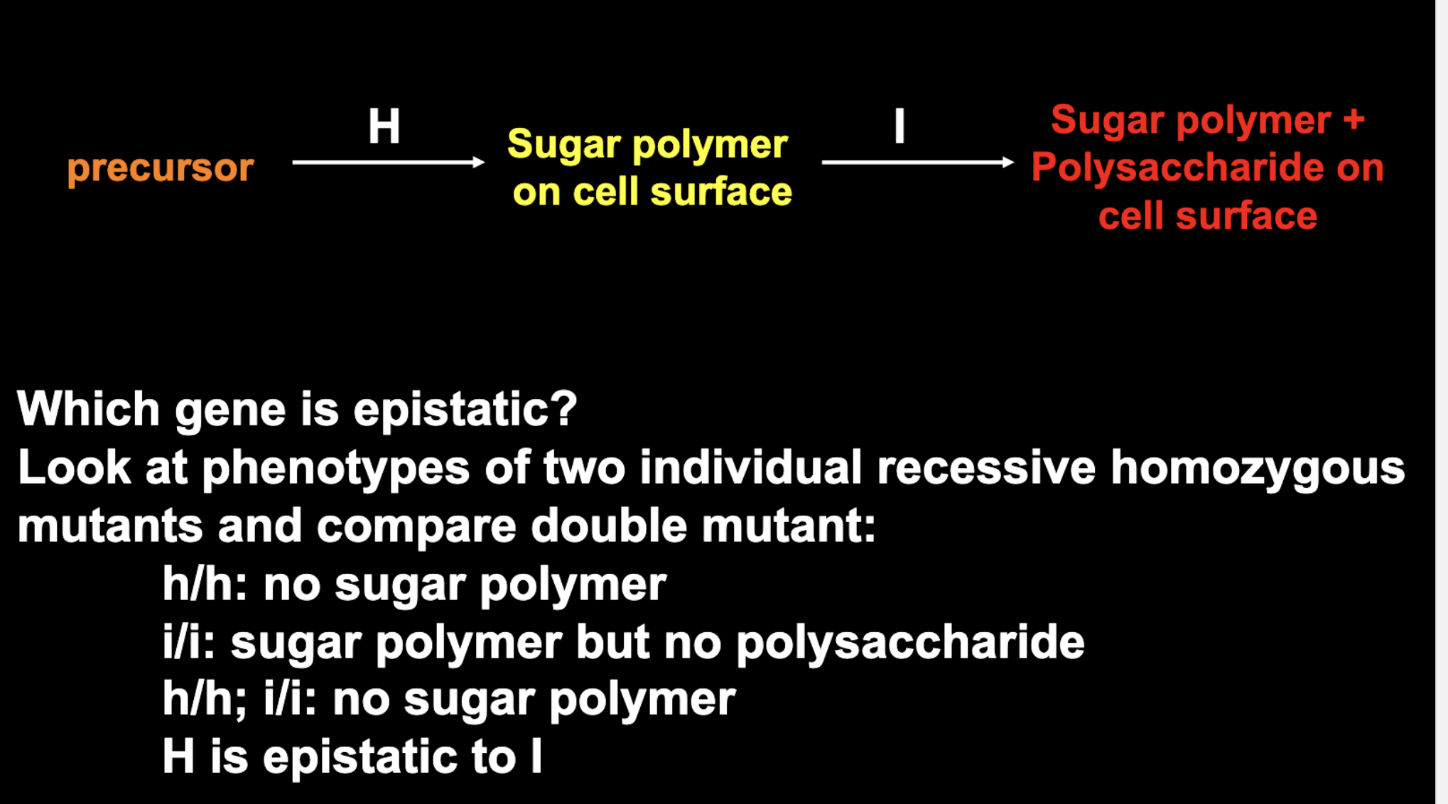
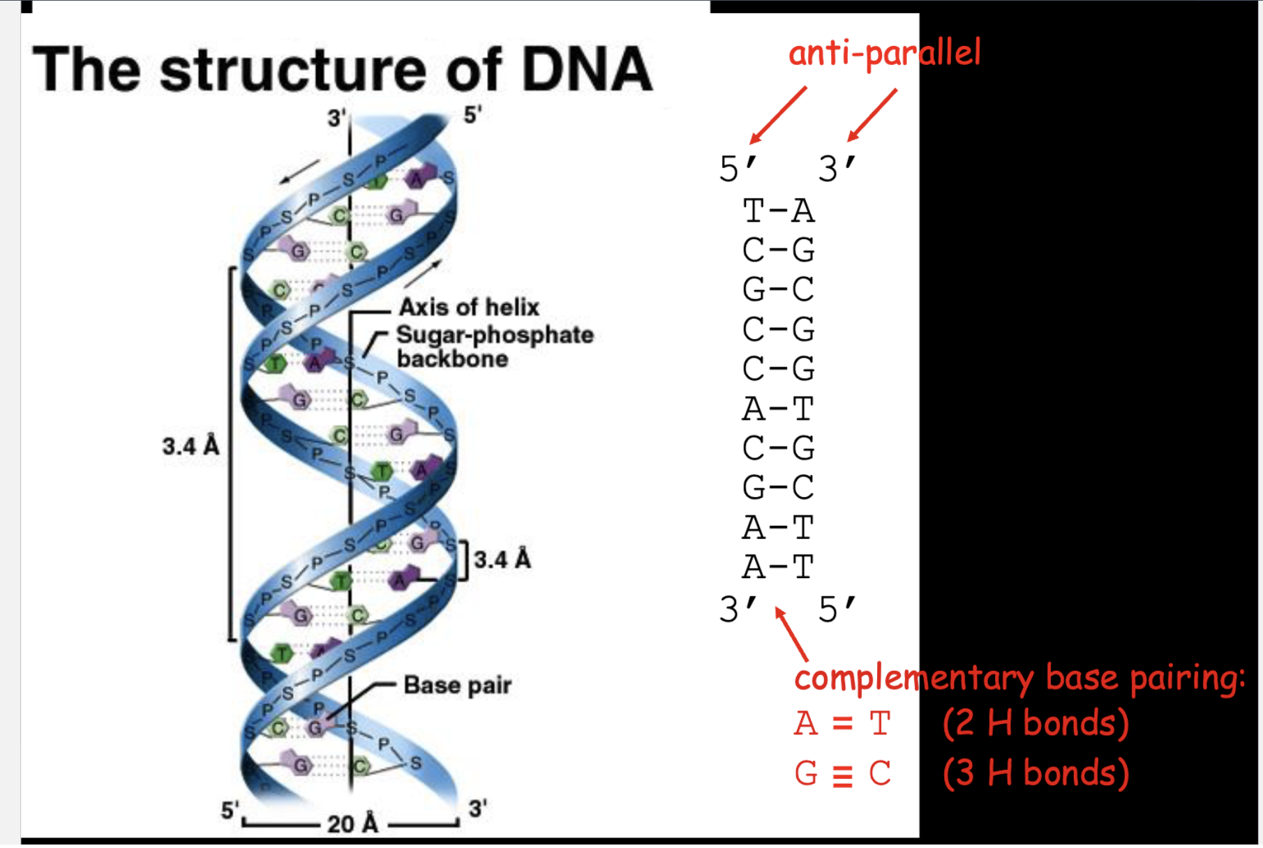
We can depict the relationship between the H and I genes

as a biochemical pathway

  1. Step 1 (H gene): Determines whether the H substance is produced on the red blood cell surface. (if hh then the H sugar polymer is not present and nothing can attach)

  2. Step 2 (ABO gene): Determines whether A and/or B sugars are added to the H substance. (if they are not but H is present, then it is regular type O)


  • The H gene is functional (HH or Hh). This means the individual can produce the H substance (sugar polymer) on the surface of their red blood cells.

  • The ABO genotype is ii. Both alleles are nonfunctional, so neither A nor B sugars are added to the H substance.

  • The result is normal type O blood, because the red blood cells have H substance but neither A nor B antigens.

<p>We can depict the relationship between the H and I genes</p><p>as a biochemical pathway</p><ol><li><p>Step 1 (H gene): Determines whether the H substance is produced on the red blood cell surface. (if hh then the H sugar polymer is not present and nothing can attach) </p></li><li><p>Step 2 (ABO gene): Determines whether A and/or B sugars are added to the H substance. (if they are not but H is present, then it is regular type O) </p></li></ol><p></p><p></p><ul><li><p>The H gene is functional (<code>HH</code> or <code>Hh</code>). This means the individual can produce the H substance (sugar polymer) on the surface of their red blood cells.</p></li><li><p>The ABO genotype is <code>ii</code>. Both alleles are nonfunctional, so neither A nor B sugars are added to the H substance.</p></li><li><p>The result is normal type O blood, because the red blood cells have H substance but neither A nor B antigens.</p></li></ul><p></p>
11
New cards
<p>EPISTASIS TEST </p><p></p><p>How do we figure out the order of gene action in a</p><p>biochemical pathway?</p>

EPISTASIS TEST


How do we figure out the order of gene action in a

biochemical pathway?

9 G/-; H/- black

3 G/-; h/h brown

4 g/g; -/- tan

<p>9 G/-; H/- black</p><p>3 G/-; h/h brown</p><p>4 g/g; -/- tan</p>
12
New cards
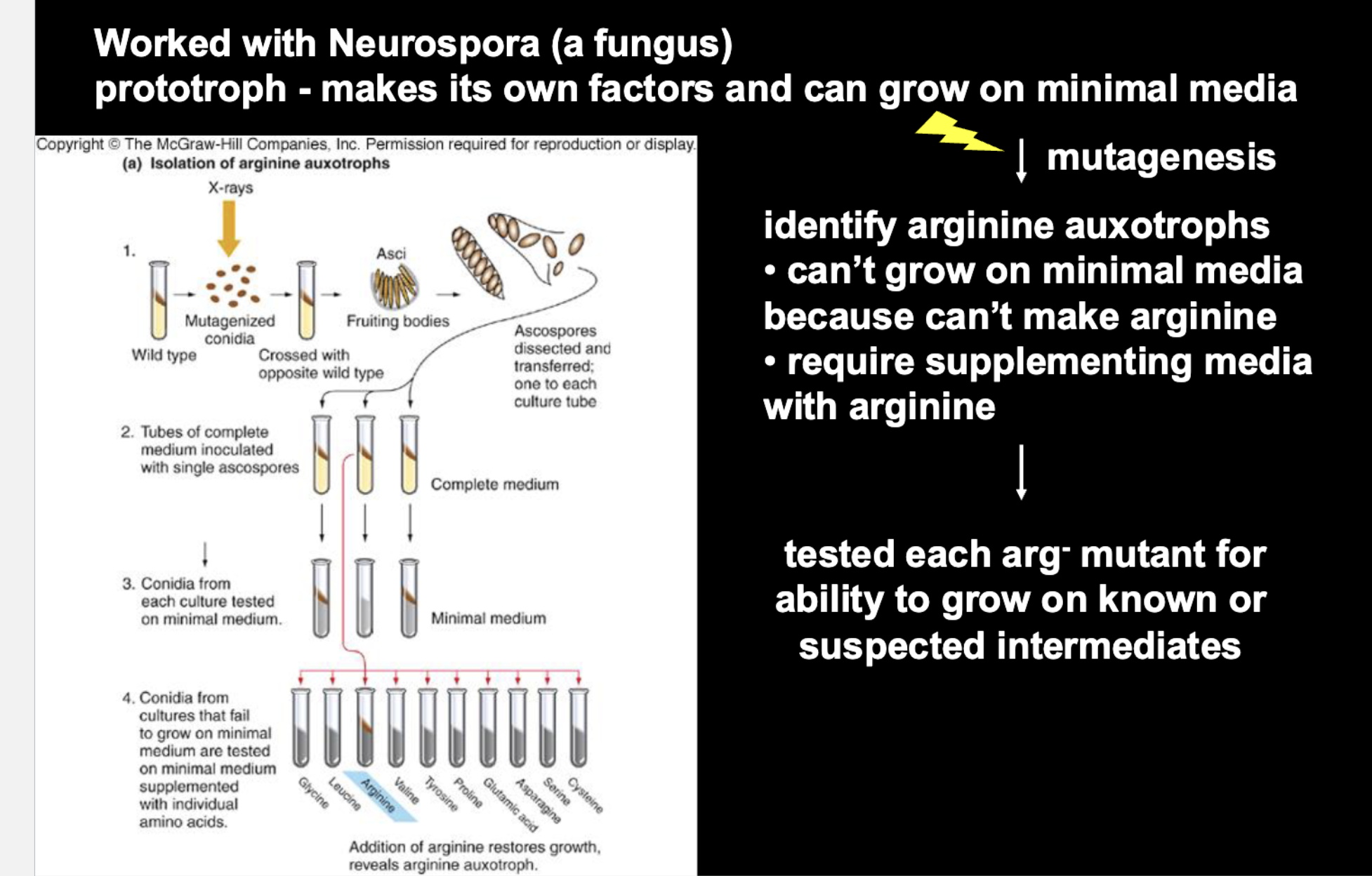
<p>Work of Beadle and Tatum in the 1940’s</p><p></p><p><strong>Steps that identified the arginine auxotroph</strong></p><ol><li><p><strong>Mutagenize:</strong> Wild-type <em>Neurospora</em> conidia were exposed to X-rays to induce random mutations.</p></li><li><p><strong>Cross:</strong> The mutagenized strain was crossed with the opposite mating type wild type, producing fruiting bodies with asci containing ascospores.</p></li><li><p><strong>Isolate single spores:</strong> Individual ascospores were dissected out and each placed in its own tube of <strong>complete medium</strong>, so every mutant, even a nutrient-dependent one, stayed alive.</p></li><li><p><strong>Test on minimal medium:</strong> Conidia from each culture were transferred to <strong>minimal medium</strong>. Cultures that failed to grow were auxotrophs, since they had lost the ability to make something essential.</p></li><li><p><strong>Test on supplemented media:</strong> Conidia from the non-growers were tested on minimal medium plus <strong>one amino acid per tube</strong> (glycine, leucine, arginine, valine, etc.).</p></li><li><p><strong>Conclude:</strong> Growth was restored only in the tube supplemented with <strong>arginine</strong>. This showed the mutant couldn’t synthesize arginine, so it was an <strong>arginine auxotroph (arg⁻)</strong>.</p></li></ol><p></p>

Work of Beadle and Tatum in the 1940’s


Steps that identified the arginine auxotroph

  1. Mutagenize: Wild-type Neurospora conidia were exposed to X-rays to induce random mutations.

  2. Cross: The mutagenized strain was crossed with the opposite mating type wild type, producing fruiting bodies with asci containing ascospores.

  3. Isolate single spores: Individual ascospores were dissected out and each placed in its own tube of complete medium, so every mutant, even a nutrient-dependent one, stayed alive.

  4. Test on minimal medium: Conidia from each culture were transferred to minimal medium. Cultures that failed to grow were auxotrophs, since they had lost the ability to make something essential.

  5. Test on supplemented media: Conidia from the non-growers were tested on minimal medium plus one amino acid per tube (glycine, leucine, arginine, valine, etc.).

  6. Conclude: Growth was restored only in the tube supplemented with arginine. This showed the mutant couldn’t synthesize arginine, so it was an arginine auxotroph (arg⁻).


TESTING ARG- MUTANT GROWTH from specific intermediates


  • Start with a collection of arg⁻ mutants (arginine auxotrophs), each a separate strain with one mutation.

  • Make tubes of minimal medium, each with one added compound: ornithine, citrulline, or arginine.

What growth means

Since these mutants need arginine to grow, growth means the cell ended up with arginine. Either it was added directly, or the cell made it from the compound you gave it.

How to read the results

  • A compound rescues a mutant only if it comes after the block. The working enzymes downstream then convert it to arginine.

  • Growth: the block is somewhere before that compound.

  • No growth: the block is at or after that compound (the cell gets stuck before making arginine).

  • Arginine rescues every arg⁻ mutant because it’s the end product, so it skips any block.

  • The later the block, the fewer compounds rescue the mutant.

  • One tube only gives before-or-after. The exact position comes from comparing the whole row of results.


<p>TESTING ARG- MUTANT GROWTH from specific intermediates </p><p></p><ul><li><p>Start with a collection of arg⁻ mutants (arginine auxotrophs), each a separate strain with one mutation.</p></li><li><p>Make tubes of <strong>minimal medium</strong>, each with one added compound: ornithine, citrulline, or arginine.</p></li></ul><p><strong>What growth means</strong></p><p>Since these mutants need arginine to grow, growth means the cell ended up with arginine. Either it was added directly, or the cell made it from the compound you gave it.</p><p><strong>How to read the results</strong></p><ul><li><p>A compound rescues a mutant only if it comes <strong>after the block</strong>. The working enzymes downstream then convert it to arginine.</p></li><li><p><strong>Growth:</strong> the block is somewhere <em>before</em> that compound.</p></li><li><p><strong>No growth:</strong> the block is <em>at or after</em> that compound (the cell gets stuck before making arginine).</p></li><li><p><strong>Arginine</strong> rescues every arg⁻ mutant because it’s the end product, so it skips any block.</p></li><li><p>The later the block, the fewer compounds rescue the mutant.</p></li><li><p>One tube only gives before-or-after. The exact position comes from comparing the whole row of results.</p></li></ul><p></p>
13
New cards
<p>ARGININE EXPERIMENT </p>

ARGININE EXPERIMENT

Starting point

  • All arg⁻ mutants are arginine auxotrophs: they can’t make arginine, so they can’t grow on minimal medium.

  • Each mutant has a mutation in a different gene (ArgE, F, G, or H). Each gene encodes one enzyme for one step of the pathway.

The test

  • Grow each mutant on minimal medium plus one compound: ornithine, citrulline, arg-succ, or arginine.

  • Growth means the mutant ended up with arginine, either added directly or made from the compound.

How to read the results

  • Growth on a compound means every step after it works, so the block is before it.

  • No growth means a broken step is at or after it, so the cell can’t reach arginine.

  • Arginine rescues every mutant because it’s the end product.

  • The more compounds a mutant grows on, the earlier its block. The fewer, the later.

Mutant

Orn

Cit

Arg-succ

Arg

Block is

ArgE⁻

yes

yes

yes

yes

before ornithine

ArgF⁻

no

yes

yes

yes

ornithine → citrulline

ArgG⁻

no

no

yes

yes

citrulline → arg-succ

ArgH⁻

no

no

no

yes

arg-succ → arginine

Pathway

ArgE⁺ → ornithine → ArgF⁺ → citrulline → ArgG⁺ → arg-succ → ArgH⁺ → arginine

Conclusion

  • The mutants are blocked at different single steps, which gives the order of the pathway.

  • Each mutant lost exactly one step, so each gene controls one enzyme (one gene, one enzyme).


<p><strong>Starting point</strong></p><ul><li><p>All arg⁻ mutants are arginine auxotrophs: they can’t make arginine, so they can’t grow on minimal medium.</p></li><li><p>Each mutant has a mutation in a <strong>different gene</strong> (ArgE, F, G, or H). Each gene encodes one enzyme for one step of the pathway.</p></li></ul><p><strong>The test</strong></p><ul><li><p>Grow each mutant on minimal medium plus <strong>one</strong> compound: ornithine, citrulline, arg-succ, or arginine.</p></li><li><p><strong>Growth</strong> means the mutant ended up with arginine, either added directly or made from the compound.</p></li></ul><p><strong>How to read the results</strong></p><ul><li><p>Growth on a compound means every step <strong>after</strong> it works, so the block is <strong>before</strong> it.</p></li><li><p>No growth means a broken step is <strong>at or after</strong> it, so the cell can’t reach arginine.</p></li><li><p>Arginine rescues every mutant because it’s the end product.</p></li><li><p>The more compounds a mutant grows on, the <strong>earlier</strong> its block. The fewer, the <strong>later</strong>.</p></li></ul><table style="min-width: 150px;"><colgroup><col style="min-width: 25px;"><col style="min-width: 25px;"><col style="min-width: 25px;"><col style="min-width: 25px;"><col style="min-width: 25px;"><col style="min-width: 25px;"></colgroup><tbody><tr><th colspan="1" rowspan="1" style=""><p>Mutant</p></th><th colspan="1" rowspan="1" style=""><p>Orn</p></th><th colspan="1" rowspan="1" style=""><p>Cit</p></th><th colspan="1" rowspan="1" style=""><p>Arg-succ</p></th><th colspan="1" rowspan="1" style=""><p>Arg</p></th><th colspan="1" rowspan="1" style=""><p>Block is</p></th></tr><tr><td colspan="1" rowspan="1"><p>ArgE⁻</p></td><td colspan="1" rowspan="1"><p>yes</p></td><td colspan="1" rowspan="1"><p>yes</p></td><td colspan="1" rowspan="1"><p>yes</p></td><td colspan="1" rowspan="1"><p>yes</p></td><td colspan="1" rowspan="1"><p>before ornithine</p></td></tr><tr><td colspan="1" rowspan="1"><p>ArgF⁻</p></td><td colspan="1" rowspan="1"><p>no</p></td><td colspan="1" rowspan="1"><p>yes</p></td><td colspan="1" rowspan="1"><p>yes</p></td><td colspan="1" rowspan="1"><p>yes</p></td><td colspan="1" rowspan="1"><p>ornithine → citrulline</p></td></tr><tr><td colspan="1" rowspan="1"><p>ArgG⁻</p></td><td colspan="1" rowspan="1"><p>no</p></td><td colspan="1" rowspan="1"><p>no</p></td><td colspan="1" rowspan="1"><p>yes</p></td><td colspan="1" rowspan="1"><p>yes</p></td><td colspan="1" rowspan="1"><p>citrulline → arg-succ</p></td></tr><tr><td colspan="1" rowspan="1"><p>ArgH⁻</p></td><td colspan="1" rowspan="1"><p>no</p></td><td colspan="1" rowspan="1"><p>no</p></td><td colspan="1" rowspan="1"><p>no</p></td><td colspan="1" rowspan="1"><p>yes</p></td><td colspan="1" rowspan="1"><p>arg-succ → arginine</p></td></tr></tbody></table><p><strong>Pathway</strong></p><p>ArgE⁺ → ornithine → ArgF⁺ → citrulline → ArgG⁺ → arg-succ → ArgH⁺ → arginine</p><p><strong>Conclusion</strong></p><ul><li><p>The mutants are blocked at different single steps, which gives the <strong>order</strong> of the pathway.</p></li><li><p>Each mutant lost exactly one step, so each gene controls one enzyme (<strong>one gene, one enzyme</strong>).</p></li></ul><p></p>
14
New cards
<p>Complementation</p><p></p><p>complementation is a test that asks whether two mutations are in the <strong>same gene</strong> or in <strong>different genes</strong>. It works because two mutants can “cover” for each other, but only if each one has a working copy of what the other lacks.</p><p><strong>The test</strong></p><ol><li><p>Take two <strong>homozygous recessive mutants</strong> (both parents show the mutant phenotype).</p></li><li><p>Cross them.</p></li><li><p>Look at the F1 phenotype.</p></li></ol><p><strong>The two outcomes</strong></p><table style="min-width: 75px;"><colgroup><col style="min-width: 25px;"><col style="min-width: 25px;"><col style="min-width: 25px;"></colgroup><tbody><tr><th colspan="1" rowspan="1" style=""><p>F1 phenotype</p></th><th colspan="1" rowspan="1" style=""><p>Meaning</p></th><th colspan="1" rowspan="1" style=""><p>Why</p></th></tr><tr><td colspan="1" rowspan="1"><p><strong>Wild type (+)</strong></p></td><td colspan="1" rowspan="1"><p>Different genes, the mutations <strong>complement</strong></p></td><td colspan="1" rowspan="1"><p>Each parent supplies the working copy of the gene the other has broken</p></td></tr><tr><td colspan="1" rowspan="1"><p><strong>Mutant (−)</strong></p></td><td colspan="1" rowspan="1"><p>Same gene, the mutations <strong>fail to complement</strong></p></td><td colspan="1" rowspan="1"><p>Both copies of that gene are broken, so no working copy exists</p></td></tr></tbody></table><p></p>

Complementation


complementation is a test that asks whether two mutations are in the same gene or in different genes. It works because two mutants can “cover” for each other, but only if each one has a working copy of what the other lacks.

The test

  1. Take two homozygous recessive mutants (both parents show the mutant phenotype).

  2. Cross them.

  3. Look at the F1 phenotype.

The two outcomes

F1 phenotype

Meaning

Why

Wild type (+)

Different genes, the mutations complement

Each parent supplies the working copy of the gene the other has broken

Mutant (−)

Same gene, the mutations fail to complement

Both copies of that gene are broken, so no working copy exists


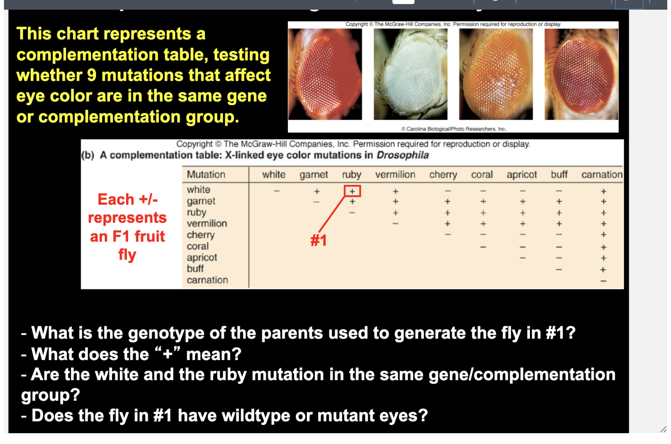
1. Genotype of the parents

  • White parent: w/w, R/R (broken white gene, normal ruby gene)

  • Ruby parent: W/W, r/r (normal white gene, broken ruby gene)

  • Both parents have mutant eyes.

  • F1 fly in #1: w/W, R/r

2. What does the “+” mean?

  • The F1 fly has wild-type eyes, so the two mutations complement each other.

  • Each parent supplies the working copy of the gene the other parent has broken. The F1 has one W and one R, and both mutations are recessive, so the pathway works.

3. Are white and ruby in the same gene/complementation group?

  • No. A “+” means the mutations are in different genes.

  • If they were in the same gene, neither parent could supply a working copy, and the F1 would be mutant (−).

4. Does the fly in #1 have wild-type or mutant eyes?

  • Wild type. It has at least one working copy of both the white gene (W) and the ruby gene (R).

Quick rule

  • + = wild-type F1 = different genes (complement)

  • − = mutant F1 = same gene (fail to complement)

<p><strong>1. Genotype of the parents</strong></p><ul><li><p><strong>White parent:</strong> w/w, R/R (broken white gene, normal ruby gene)</p></li><li><p><strong>Ruby parent:</strong> W/W, r/r (normal white gene, broken ruby gene)</p></li><li><p>Both parents have mutant eyes.</p></li><li><p><strong>F1 fly in #1:</strong> w/W, R/r</p></li></ul><p><strong>2. What does the “+” mean?</strong></p><ul><li><p>The F1 fly has <strong>wild-type eyes</strong>, so the two mutations <strong>complement</strong> each other.</p></li><li><p>Each parent supplies the working copy of the gene the other parent has broken. The F1 has one W and one R, and both mutations are recessive, so the pathway works.</p></li></ul><p><strong>3. Are white and ruby in the same gene/complementation group?</strong></p><ul><li><p><strong>No.</strong> A “+” means the mutations are in <strong>different genes</strong>.</p></li><li><p>If they were in the same gene, neither parent could supply a working copy, and the F1 would be mutant (−).</p></li></ul><p><strong>4. Does the fly in #1 have wild-type or mutant eyes?</strong></p><ul><li><p><strong>Wild type.</strong> It has at least one working copy of both the white gene (W) and the ruby gene (R).</p></li></ul><p><strong>Quick rule</strong></p><ul><li><p><strong>+</strong> = wild-type F1 = different genes (complement)</p></li><li><p><strong>−</strong> = mutant F1 = same gene (fail to complement)</p></li></ul><p></p>
15
New cards
<p><strong>1. Genotype of the parents</strong></p><ul><li><p><strong>White parent:</strong> w/w (broken white gene)</p></li><li><p><strong>Coral parent:</strong> w^c/w^c (a different mutation in the same gene)</p></li><li><p>Both parents have mutant eyes, and neither carries a W.</p></li><li><p><strong>F1 fly in #2:</strong> w/w^c</p></li></ul><p><strong>2. What does the “−” mean?</strong></p><ul><li><p>The F1 fly has <strong>mutant eyes</strong>, so the two mutations <strong>fail to complement</strong>.</p></li><li><p>Neither parent can supply a working copy of the gene, so the F1 has no W.</p></li></ul><p><strong>3. Are white and coral in the same gene/complementation group?</strong></p><ul><li><p><strong>Yes.</strong> A “−” means the mutations are in the <strong>same gene</strong>, so they belong to the same complementation group.</p></li><li><p>They are different alleles of that gene.</p></li></ul><p><strong>4. Does the fly in #2 have wild-type or mutant eyes?</strong></p><ul><li><p><strong>Mutant.</strong> Both copies of the gene are broken (w and w^c), so no working enzyme is made.</p></li></ul><p></p>

1. Genotype of the parents

  • White parent: w/w (broken white gene)

  • Coral parent: w^c/w^c (a different mutation in the same gene)

  • Both parents have mutant eyes, and neither carries a W.

  • F1 fly in #2: w/w^c

2. What does the “−” mean?

  • The F1 fly has mutant eyes, so the two mutations fail to complement.

  • Neither parent can supply a working copy of the gene, so the F1 has no W.

3. Are white and coral in the same gene/complementation group?

  • Yes. A “−” means the mutations are in the same gene, so they belong to the same complementation group.

  • They are different alleles of that gene.

4. Does the fly in #2 have wild-type or mutant eyes?

  • Mutant. Both copies of the gene are broken (w and w^c), so no working enzyme is made.


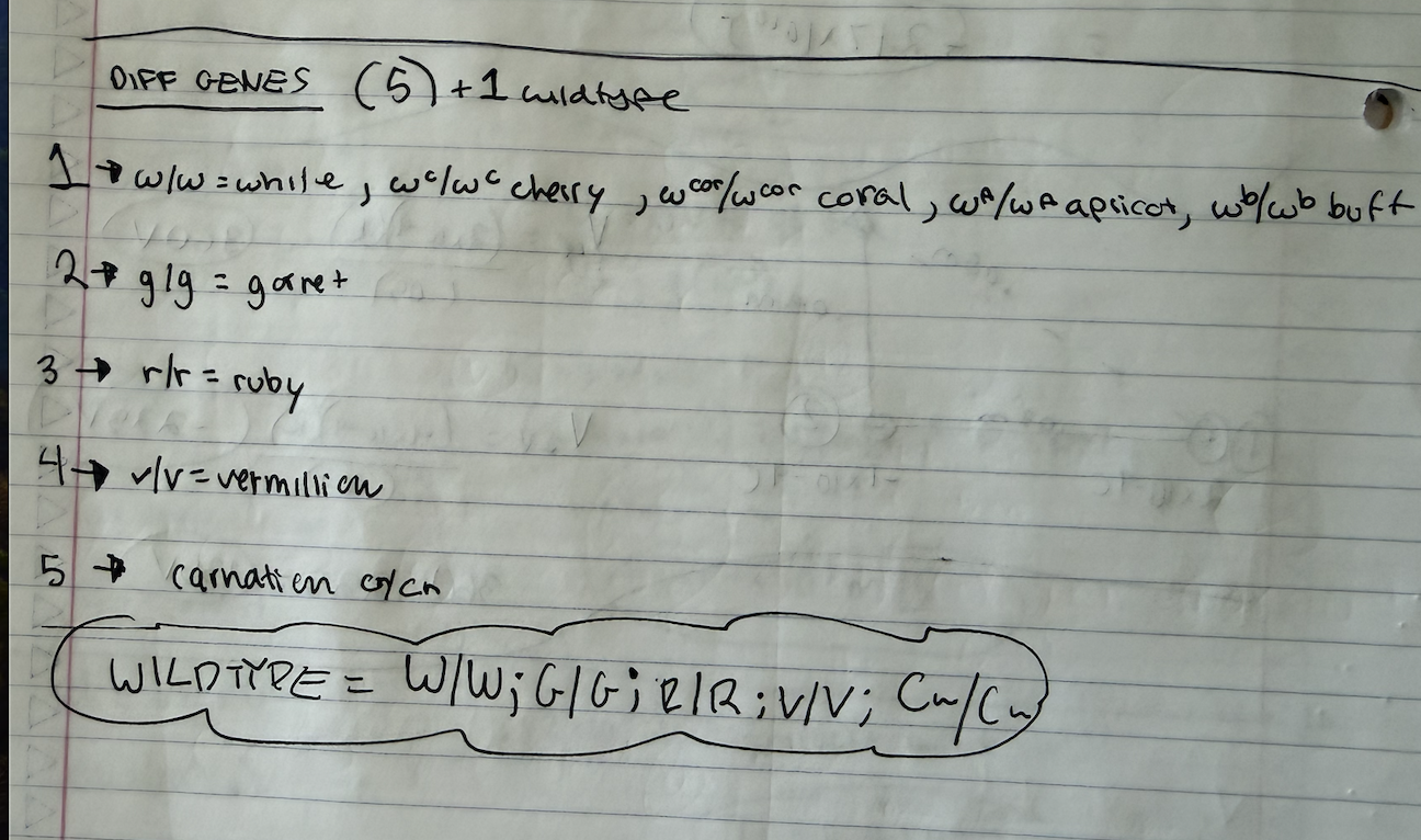
finding the total number of genes.

the wild type genotype = all the genes with at least one dominant allele
<p>finding the total number of genes. </p><p>the wild type genotype = all the genes with at least one dominant allele </p>
16
New cards
<p>Understanding Genetic Interactions</p>

Understanding Genetic Interactions

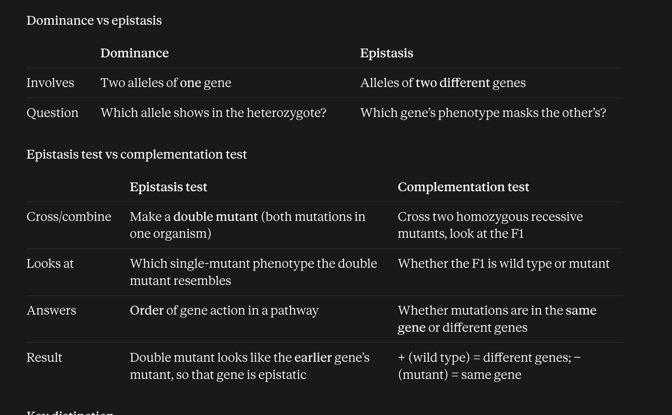
  • Epistasis (recessive): one gene’s phenotype masks another’s. In a pathway, the earlier gene is epistatic (G is epistatic to H in the tan → brown → black pathway).

  • Complementation: tests whether two mutations are in the same gene or different genes


Key distinction

  • Epistasis assumes you already have different genes and asks how they relate in a pathway.

  • Complementation asks whether two mutations are in the same gene in the first place.

<ul><li><p><strong>Epistasis (recessive):</strong> one gene’s phenotype masks another’s. In a pathway, the <strong>earlier</strong> gene is epistatic (G is epistatic to H in the tan → brown → black pathway).</p></li><li><p><strong>Complementation:</strong> tests whether two mutations are in the same gene or different genes</p></li></ul><p></p><p></p><p><strong>Key distinction</strong></p><ul><li><p>Epistasis assumes you already have <strong>different genes</strong> and asks how they relate in a pathway.</p></li><li><p>Complementation asks whether two mutations are in the <strong>same gene</strong> in the first place.</p></li></ul><p></p>
17
New cards
<p>Some traits are discrete or</p><p>discontinuous</p>

Some traits are discrete or

discontinuous
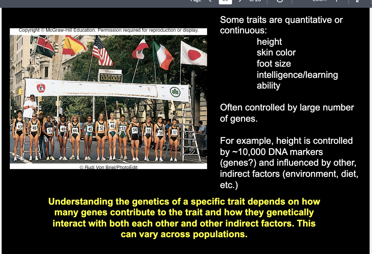
knowt flashcard image
18
New cards
<p>How do mutations affect protein function and produce phenotypes?</p>

How do mutations affect protein function and produce phenotypes?

Dominant and recessive describe the heterozygote’s phenotype.

  • Recessive: the heterozygote looks wildtype. The mutant phenotype appears only in the homozygote.

  • Dominant: the heterozygote shows the mutant phenotype. One mutant copy is enough.


Recessive mutations (loss of function)

Most genes are haplosufficient: one wildtype copy makes ~50% of the normal protein, which is enough for a normal phenotype. A loss-of-function allele is therefore covered by the wildtype copy in a heterozygote, and the mutant phenotype only appears when both copies are mutant.

Type

Mutant protein function

Homozygote phenotype

Null / amorphic

None

Full mutant phenotype with no function

Hypomorphic

Reduced

Milder, “leaky” mutant phenotype


Dominant mutations

The wildtype copy can’t rescue the phenotype. This happens in one of two ways: there is too little normal protein (haploinsufficient), or the mutant allele adds something the wildtype copy can’t cancel.

Type

Mutant allele

Why the heterozygote is mutant

Haploinsufficient

Loss of function

50% of normal protein isn’t enough

Hypermorphic

Gain of function (more activity)

Excess activity the wildtype copy can’t cancel

Neomorphic (gain of function)

Gain of function (new activity, or expressed in the wrong place or time)

New activity the wildtype copy can’t cancel


<p>Dominant and recessive describe the <strong>heterozygote’s phenotype</strong>.</p><ul><li><p><strong>Recessive:</strong> the heterozygote looks wildtype. The mutant phenotype appears only in the homozygote.</p></li><li><p><strong>Dominant:</strong> the heterozygote shows the mutant phenotype. One mutant copy is enough.</p></li></ul><p></p><p> Recessive mutations (loss of function) </p><p>Most genes are <strong>haplosufficient</strong>: one wildtype copy makes ~50% of the normal protein, which is enough for a normal phenotype. A loss-of-function allele is therefore covered by the wildtype copy in a heterozygote, and the mutant phenotype only appears when both copies are mutant.</p><table style="min-width: 75px;"><colgroup><col style="min-width: 25px;"><col style="min-width: 25px;"><col style="min-width: 25px;"></colgroup><tbody><tr><th colspan="1" rowspan="1"><p>Type</p></th><th colspan="1" rowspan="1"><p>Mutant protein function</p></th><th colspan="1" rowspan="1"><p>Homozygote phenotype</p></th></tr><tr><td colspan="1" rowspan="1"><p><strong>Null / amorphic</strong></p></td><td colspan="1" rowspan="1"><p>None</p></td><td colspan="1" rowspan="1"><p>Full mutant phenotype with no function </p></td></tr><tr><td colspan="1" rowspan="1"><p><strong>Hypomorphic</strong></p></td><td colspan="1" rowspan="1"><p>Reduced</p></td><td colspan="1" rowspan="1"><p>Milder, “leaky” mutant phenotype</p></td></tr></tbody></table><p> </p><p></p><p>Dominant mutations </p><p>The wildtype copy can’t rescue the phenotype. This happens in one of two ways: there is <strong>too little</strong> normal protein (haploinsufficient), or the mutant allele <strong>adds</strong> something the wildtype copy can’t cancel.</p><table style="min-width: 75px;"><colgroup><col style="min-width: 25px;"><col style="min-width: 25px;"><col style="min-width: 25px;"></colgroup><tbody><tr><th colspan="1" rowspan="1"><p>Type</p></th><th colspan="1" rowspan="1"><p>Mutant allele</p></th><th colspan="1" rowspan="1"><p>Why the heterozygote is mutant</p></th></tr><tr><td colspan="1" rowspan="1"><p><strong>Haploinsufficient</strong></p></td><td colspan="1" rowspan="1"><p>Loss of function</p></td><td colspan="1" rowspan="1"><p>50% of normal protein isn’t enough</p></td></tr><tr><td colspan="1" rowspan="1"><p><strong>Hypermorphic</strong></p></td><td colspan="1" rowspan="1"><p>Gain of function (more activity)</p></td><td colspan="1" rowspan="1"><p>Excess activity the wildtype copy can’t cancel</p></td></tr><tr><td colspan="1" rowspan="1"><p><strong>Neomorphic</strong> (gain of function)</p></td><td colspan="1" rowspan="1"><p>Gain of function (new activity, or expressed in the wrong place or time)</p></td><td colspan="1" rowspan="1"><p>New activity the wildtype copy can’t cancel</p></td></tr></tbody></table><p></p>
19
New cards
<p>Genes encode proteins and proteins are responsible for phenotype</p>

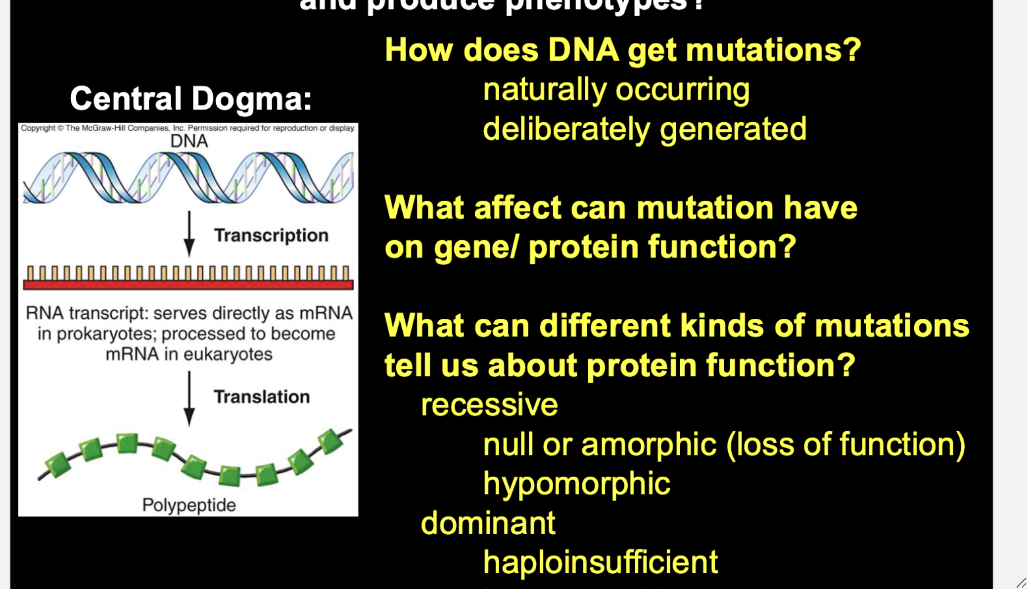
Genes encode proteins and proteins are responsible for phenotype

The phenotype you see is the end result of a chain: gene, then mRNA, then protein, then phenotype. A mutation in the gene can change the protein, and a changed protein can change the phenotype.


  • Loss of function: the protein is missing or doesn’t work (null) or works less well (hypomorphic).

  • Gain of function: the protein does more (hypermorphic) or something new (neomorphic).


Applying it to the red gene

  • A null mutation in the red gene means no working red protein, so no red pigment.

  • A heterozygote with one working copy usually still makes enough protein for red pigment, which is why that loss of function is typically recessive.

  • If the gene were haploinsufficient, 50% of the protein wouldn’t be enough, and the heterozygote would show reduced or missing red.


20
New cards
<p>Types of mutations</p>

Types of mutations

a) Substitution: one base pair is replaced by a different base pair. Total length doesn’t change.

  • Transition: purine → purine or pyrimidine → pyrimidine (A↔G, C↔T).

  • Transversion: purine ↔ pyrimidine (e.g., A→C, G→T).


(b) Deletion: one or more base pairs are removed from the sequence. The DNA gets shorter. In the slide, a GCA/CGT segment is cut out.


(c) Insertion: one or more base pairs are added into the sequence. The DNA gets longer. In the slide, an AA/TT pair is added.


(d) Inversion: a segment of DNA is cut out, flipped 180°, and reinserted in the same place. The bases are all still there, but their order and orientation in that region are reversed (the strands swap, so the 5'→3' direction of the segment is reversed).


(e) Reciprocal translocation: two different (nonhomologous) chromosomes break and swap segments with each other. No DNA is gained or lost overall, but pieces end up on the other chromosome.
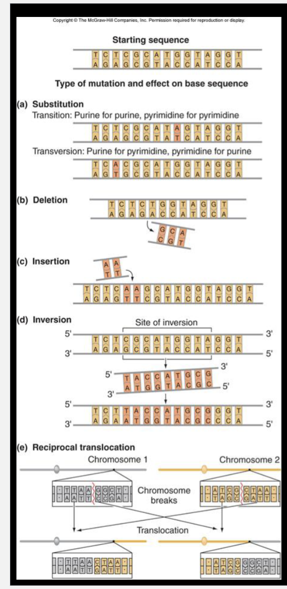
<p><strong>a) Substitution:</strong> one base pair is replaced by a different base pair. Total length doesn’t change.</p><ul><li><p><strong>Transition:</strong> purine → purine or pyrimidine → pyrimidine (A<span data-name="left_right_arrow" data-type="emoji">↔</span>G, C<span data-name="left_right_arrow" data-type="emoji">↔</span>T).</p></li><li><p><strong>Transversion:</strong> purine <span data-name="left_right_arrow" data-type="emoji">↔</span> pyrimidine (e.g., A→C, G→T).</p></li><li><p></p></li></ul><p><strong>(b) Deletion:</strong> one or more base pairs are removed from the sequence. The DNA gets shorter. In the slide, a GCA/CGT segment is cut out.</p><p></p><p><strong>(c) Insertion:</strong> one or more base pairs are added into the sequence. The DNA gets longer. In the slide, an AA/TT pair is added.</p><p></p><p><strong>(d) Inversion:</strong> a segment of DNA is cut out, flipped 180°, and reinserted in the same place. The bases are all still there, but their order and orientation in that region are reversed (the strands swap, so the 5'→3' direction of the segment is reversed).</p><p></p><p><strong>(e) Reciprocal translocation:</strong> two <strong>different (nonhomologous) chromosomes</strong> break and swap segments with each other. No DNA is gained or lost overall, but pieces end up on the other chromosome.</p>
21
New cards
<p>Mutation Types </p><p></p>

Mutation Types


Substitution (one base pair replaced by another)

  • Transition: purine → purine, or pyrimidine → pyrimidine

  • Transversion: purine ↔ pyrimidine

  • Silent: same amino acid produced

  • Missense: different amino acid

  • Nonsense: codon becomes a stop codon and ends early


Insertion (base pairs added)

  • Frameshift: not a multiple of 3, so the reading frame shifts

  • In-frame: a multiple of 3, so the frame is kept still can change protein sequence


Deletion (base pairs removed)

  • Frameshift: not a multiple of 3

  • In-frame: a multiple of 3


Inversion (a segment is flipped 180° within the same chromosome)


Reciprocal translocation (two nonhomologous chromosomes swap segments)

22
New cards
<p>Mutagens - substances that alter or mutate DNA</p>

Mutagens - substances that alter or mutate DNA

1. Starting DNA (far left)
A normal double helix. Look at the third base pair from the top: it’s a G-C pair.

2. DNA damage (second helix)
The C in that pair becomes a U (uracil, in red). This can happen when cytosine loses an amino group (deamination), which turns it into uracil. The pair is now G-U, which is a mismatch, because U is normally found in RNA and not in DNA. This is DNA damage, not yet a mutation, because the cell could still detect and repair it.

3. DNA replication (arrow)
If the damage isn’t repaired before replication, the two strands separate and each one serves as a template (yellow highlighted strands) for a new complementary strand.

4. Two sister chromatids (right)

  • Left chromatid: the template is the strand with the original G. The new strand gets a C across from it, so you get G-C again, which is normal. This chromatid is fine.

  • Right chromatid: the template is the strand carrying the U. DNA polymerase reads U like a T and pairs it with A. That’s the A-U pair in the third row. The partner strand, which still had the G, is now paired with the new C... but the key point is the U-containing strand gets an A opposite it.

Why this becomes a mutation

The A-U pair is on the right chromatid. At the next round of replication, the U (read as T) templates another A, and that A in turn gets a T partner, so this chromatid’s lineage ends up with a normal A-T base pair where there used to be G-C.


<p><strong>1. Starting DNA (far left)</strong><br>A normal double helix. Look at the third base pair from the top: it’s a <strong>G-C</strong> pair.</p><p><strong>2. DNA damage (second helix)</strong><br>The <strong>C</strong> in that pair becomes a <strong>U</strong> (uracil, in red). This can happen when cytosine loses an amino group (deamination), which turns it into uracil. The pair is now <strong>G-U</strong>, which is a mismatch, because U is normally found in RNA and not in DNA. This is DNA <em>damage</em>, not yet a mutation, because the cell could still detect and repair it.</p><p><strong>3. DNA replication (arrow)</strong><br>If the damage isn’t repaired before replication, the two strands separate and each one serves as a <strong>template</strong> (yellow highlighted strands) for a new complementary strand.</p><p><strong>4. Two sister chromatids (right)</strong></p><ul><li><p><strong>Left chromatid:</strong> the template is the strand with the original <strong>G</strong>. The new strand gets a <strong>C</strong> across from it, so you get <strong>G-C</strong> again, which is normal. This chromatid is fine.</p></li><li><p><strong>Right chromatid:</strong> the template is the strand carrying the <strong>U</strong>. DNA polymerase reads U like a T and pairs it with <strong>A</strong>. That’s the <strong>A-U</strong> pair in the third row. The partner strand, which still had the G, is now paired with the new C... but the key point is the U-containing strand gets an A opposite it.</p></li></ul><p> Why this becomes a mutation </p><p>The <strong>A-U</strong> pair is on the right chromatid. At the next round of replication, the U (read as T) templates another <strong>A</strong>, and that A in turn gets a <strong>T</strong> partner, so this chromatid’s lineage ends up with a normal <strong>A-T</strong> base pair where there used to be <strong>G-C</strong>.</p><p></p>
23
New cards
<p>Causes of different mutations </p><p></p><p>Causes of substitutions </p><table style="min-width: 75px;"><colgroup><col style="min-width: 25px;"><col style="min-width: 25px;"><col style="min-width: 25px;"></colgroup><tbody><tr><th colspan="1" rowspan="1" style=""><p>Cause</p></th><th colspan="1" rowspan="1" style=""><p>What happens</p></th><th colspan="1" rowspan="1" style=""><p>Result</p></th></tr><tr><td colspan="1" rowspan="1"><p><strong>Depurination</strong></p></td><td colspan="1" rowspan="1"><p>A purine base (A or G) is lost from the DNA backbone, leaving a gap. During replication, polymerase puts the wrong base across from the gap.</p></td><td colspan="1" rowspan="1"><p>Substitution</p></td></tr><tr><td colspan="1" rowspan="1"><p><strong>Deamination</strong></p></td><td colspan="1" rowspan="1"><p>A base loses an amino group and becomes a different base. The main example is C → U, as on your last slide. The U is read as T, so G-C becomes A-T.</p></td><td colspan="1" rowspan="1"><p>Substitution (transition)</p></td></tr><tr><td colspan="1" rowspan="1"><p><strong>Ethylating and methylating agents</strong></p></td><td colspan="1" rowspan="1"><p>Chemicals add ethyl or methyl groups to bases, which changes how they pair, so the wrong base is inserted during replication.</p></td><td colspan="1" rowspan="1"><p>Substitution</p></td></tr></tbody></table><p></p>

Causes of different mutations


Causes of substitutions

Cause

What happens

Result

Depurination

A purine base (A or G) is lost from the DNA backbone, leaving a gap. During replication, polymerase puts the wrong base across from the gap.

Substitution

Deamination

A base loses an amino group and becomes a different base. The main example is C → U, as on your last slide. The U is read as T, so G-C becomes A-T.

Substitution (transition)

Ethylating and methylating agents

Chemicals add ethyl or methyl groups to bases, which changes how they pair, so the wrong base is inserted during replication.

Substitution


Causes of deletions, insertions and inversions

X-rays followed by non-homologous end joining (same chromosome)

  • X-rays break the DNA, including double-strand breaks.

  • The cell repairs the break by gluing the ends back together (non-homologous end joining), which is error-prone.

  • If a piece is lost, you get a deletion. If a piece rejoins flipped, you get an inversion.

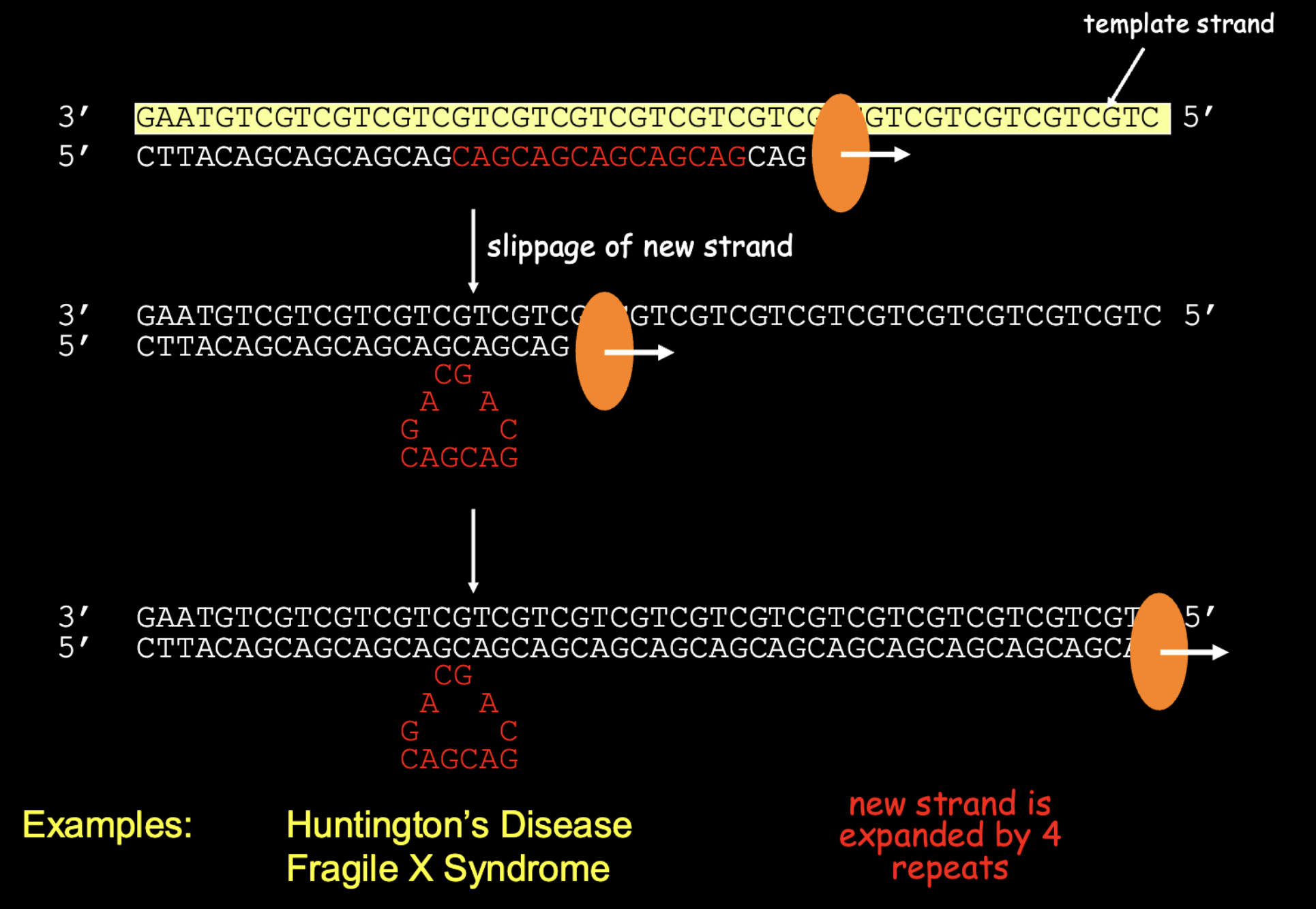
Replication slippage in a repetitive region

  • In a repetitive stretch of DNA (like CAGCAGCAG), the new strand and template can slip out of alignment during replication, forming a small loop.

  • If the new strand loops, you get an insertion (extra repeats). If the template loops, you get a deletion (fewer repeats).


Cause of reciprocal translocation:

X-rays followed by non-homologous end joining (between different chromosomes)

  • X-rays break two different chromosomes at the same time.

  • The ends are joined to the wrong partner, so the two chromosomes swap pieces. This is a reciprocal translocation.

24
New cards
<p>Expansion of repeats may occur by slippage during DNA replication</p>

Expansion of repeats may occur by slippage during DNA replication

1. DNA replication begins. DNA polymerase uses a DNA template to synthesize a complementary DNA strand. The DNA contains repetitive sequences, such as CAG CAG CAG CAG.

2. The new strand slips out of alignment. Because the sequences repeat, the newly synthesized strand can unpair and re-pair at the wrong location. The repeated sequences look the same, so they can pair in an incorrect alignment.

3. A loop forms. The newly synthesized strand loops out, leaving some repeats unpaired with the template.

4. DNA polymerase continues replication. The enzyme continues synthesizing DNA using the template strand.

5. The repeat expansion is retained. If the loop in the new strand is retained and becomes part of the completed DNA, the new strand contains extra repeat units that were in the loop


this repeat expansion is a type of insertion mutation

25
New cards
<p>Geneticists deliberately introduce mutations to affect protein function and study the effects</p><p></p><ol><li><p>X-rays damaged the DNA of wild-type Neurospora prototrophs.</p></li><li><p>Some mutations disrupted genes responsible for producing functional enzymes needed for arginine synthesis.</p></li><li><p>These fungi could no longer synthesize arginine and therefore could not grow on minimal media.</p></li><li><p>Scientists added individual amino acids to the growth media to determine which nutrient was missing.</p></li><li><p>When arginine supplementation restored growth, they identified the fungi as arginine auxotrophs.</p></li></ol><p></p>

Geneticists deliberately introduce mutations to affect protein function and study the effects


  1. X-rays damaged the DNA of wild-type Neurospora prototrophs.

  2. Some mutations disrupted genes responsible for producing functional enzymes needed for arginine synthesis.

  3. These fungi could no longer synthesize arginine and therefore could not grow on minimal media.

  4. Scientists added individual amino acids to the growth media to determine which nutrient was missing.

  5. When arginine supplementation restored growth, they identified the fungi as arginine auxotrophs.


Mutagen

How it causes mutations

X-rays (used in experiment)

Damage DNA, causing breaks and rearrangements

Ethylating/methylating agents

Chemically modify DNA bases, potentially causing incorrect base pairing

Transposable elements

Insert into new DNA locations and potentially disrupt genes


DNA mutation from X RAY → defective enzyme → disrupted arginine synthesis → arginine auxotrophy.


26
New cards
<p>How do mutations affect protein function?</p><p></p><p>If a mutation affects protein expression, it can interfere with different stages of gene expression:</p><ul><li><p>Transcription: The gene may not be transcribed into mRNA.</p></li><li><p>Splicing: The mRNA may be incorrectly processed, affecting the protein that gets made.</p></li><li><p>Translation: The mRNA may not be translated properly into a protein.</p></li></ul><p>So there are two important distinctions in your slide:</p><ol><li><p>Protein function: The protein is produced, but it may not work properly.</p></li><li><p>Protein expression: The mutation affects whether, when, or how much protein is produced.</p></li></ol><p></p>

How do mutations affect protein function?


If a mutation affects protein expression, it can interfere with different stages of gene expression:

  • Transcription: The gene may not be transcribed into mRNA.

  • Splicing: The mRNA may be incorrectly processed, affecting the protein that gets made.

  • Translation: The mRNA may not be translated properly into a protein.

So there are two important distinctions in your slide:

  1. Protein function: The protein is produced, but it may not work properly.

  2. Protein expression: The mutation affects whether, when, or how much protein is produced.


. Transcription (DNA → mRNA)

Location: Nucleus (in eukaryotic cells)

  1. RNA polymerase binds to the DNA at the promoter.

  2. DNA unwinds, exposing the template strand.

  3. RNA polymerase reads the template DNA strand and builds a complementary mRNA strand.

  4. The mRNA is processed (5' cap, poly-A tail, and splicing to remove introns).

  5. Mature mRNA leaves the nucleus and enters the cytoplasm.


2. Translation (mRNA → Protein)

Location: Ribosome in the cytoplasm (or on the rough ER).

  1. The ribosome attaches to the mRNA.

  2. The ribosome reads the mRNA in groups of three nucleotides called codons.

  3. tRNA molecules bring the corresponding amino acids.

  4. Each tRNA has an anticodon that pairs with the complementary mRNA codon.

  5. The ribosome joins amino acids together through peptide bonds, forming a polypeptide.

  6. Translation ends when the ribosome reaches a stop codon.

  7. The polypeptide folds into a functional protein (sometimes with additional processing).

27
New cards
<p>Substitutions, insertions, or deletions can impact protein sequence and protein function</p>

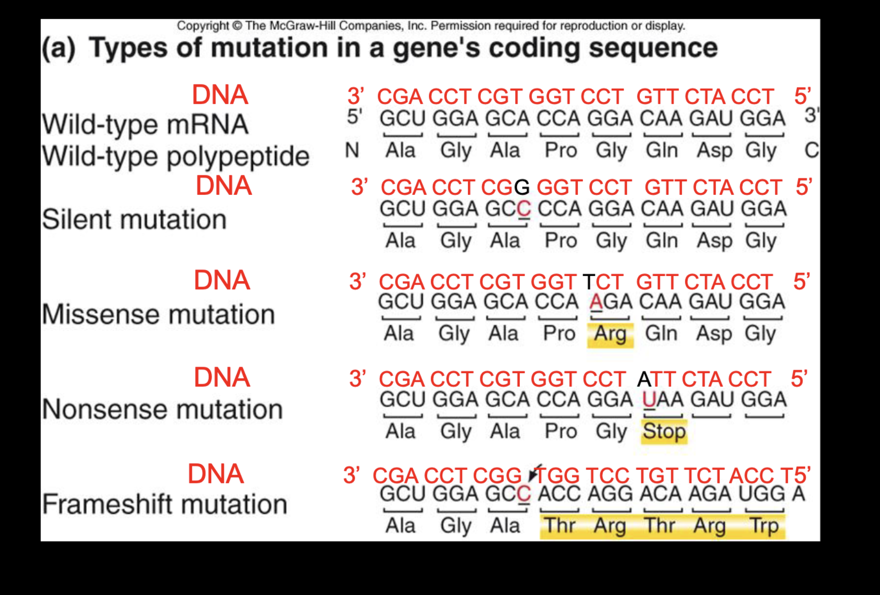
Substitutions, insertions, or deletions can impact protein sequence and protein function

. Silent mutation

  • A nucleotide changes, but the codon still codes for the same amino acid.

  • No change to the amino acid sequence of the protein.

2. Missense mutation

  • A nucleotide substitution changes a codon, resulting in a different amino acid.

  • Can alter protein structure and function —> therefore phenotype

3. Nonsense mutation

  • A nucleotide substitution changes a codon into a premature stop codon.

  • Causes translation to end early, potentially producing a shortened, nonfunctional protein.

4. Frameshift mutation

  • Caused by an insertion or deletion of nucleotides in numbers not divisible by 3.

  • Shifts the reading frame, changing the grouping of downstream codons.

  • Often changes many amino acids and can severely disrupt protein function or make it nonfunctional


28
New cards
<p>Mutations at several points of PROTEIN SYNTHESIS </p><p></p><p>Mutations do not have to occur within the protein-coding sequence to affect protein production or function. Mutations in regulatory and processing regions can also change the phenotype by affecting the proteins function </p><p>Important regions shown in the diagram</p><table style="min-width: 50px;"><colgroup><col style="min-width: 25px;"><col style="min-width: 25px;"></colgroup><tbody><tr><th colspan="1" rowspan="1"><p>Region</p></th><th colspan="1" rowspan="1"><p>Function</p></th></tr><tr><td colspan="1" rowspan="1"><p>Promoter</p></td><td colspan="1" rowspan="1"><p>DNA sequence where RNA polymerase and transcription factors bind to initiate transcription.</p></td></tr><tr><td colspan="1" rowspan="1"><p>Untranslated leader sequence</p></td><td colspan="1" rowspan="1"><p>Transcribed into mRNA but not translated into protein; can influence translation.</p></td></tr><tr><td colspan="1" rowspan="1"><p>Splicing sites</p></td><td colspan="1" rowspan="1"><p>Identify where introns are removed and exons are joined during mRNA processing.</p></td></tr><tr><td colspan="1" rowspan="1"><p>Stop codon</p></td><td colspan="1" rowspan="1"><p>Signals the end of translation.</p></td></tr><tr><td colspan="1" rowspan="1"><p>Transcription termination region</p></td><td colspan="1" rowspan="1"><p>Signals the end of transcription.</p></td></tr></tbody></table><p></p>

Mutations at several points of PROTEIN SYNTHESIS


Mutations do not have to occur within the protein-coding sequence to affect protein production or function. Mutations in regulatory and processing regions can also change the phenotype by affecting the proteins function

Important regions shown in the diagram

Region

Function

Promoter

DNA sequence where RNA polymerase and transcription factors bind to initiate transcription.

Untranslated leader sequence

Transcribed into mRNA but not translated into protein; can influence translation.

Splicing sites

Identify where introns are removed and exons are joined during mRNA processing.

Stop codon

Signals the end of translation.

Transcription termination region

Signals the end of transcription.


  • Promoter mutation: Can reduce or prevent transcription, resulting in less or no protein.

  • Untranslated leader mutation: Can affect how efficiently mRNA is translated into protein.

  • Splicing-site mutation: Can cause incorrect removal of introns or joining of exons, producing abnormal mRNA.

  • Stop codon mutation: Can interfere with normal termination of translation, potentially producing an abnormally long protein.

  • Transcription termination mutation: Can interfere with proper termination of transcription and RNA processing.


29
New cards
<p>What is the “protein explanation” for why most mutations are recessive?</p>

What is the “protein explanation” for why most mutations are recessive?

  • Most mutations impair or eliminate protein function.

  • If an individual is heterozygous (one wild-type allele and one loss-of-function mutant allele), the wild-type allele can produce enough functional protein.

  • Therefore, the individual still displays the wild-type phenotype, and the mutation is recessive.


30
New cards
<p>What are some “protein explanations” for why some mutations are dominant?</p><p></p><table style="min-width: 50px;"><colgroup><col style="min-width: 25px;"><col style="min-width: 25px;"></colgroup><tbody><tr><th colspan="1" rowspan="1"><p>Mutation DOMINANT</p></th><th colspan="1" rowspan="1"><p>What the mutant allele does</p></th></tr><tr><td colspan="1" rowspan="1"><p>Neomorphic</p></td><td colspan="1" rowspan="1"><p>Produces a protein with a new function.</p></td></tr><tr><td colspan="1" rowspan="1"><p>Hypermorphic</p></td><td colspan="1" rowspan="1"><p>Produces too much protein or an overly active protein.</p></td></tr><tr><td colspan="1" rowspan="1"><p>Dominant negative</p></td><td colspan="1" rowspan="1"><p>Produces an abnormal protein that interferes with the normal protein.</p></td></tr></tbody></table><p></p>

What are some “protein explanations” for why some mutations are dominant?


Mutation DOMINANT

What the mutant allele does

Neomorphic

Produces a protein with a new function.

Hypermorphic

Produces too much protein or an overly active protein.

Dominant negative

Produces an abnormal protein that interferes with the normal protein.


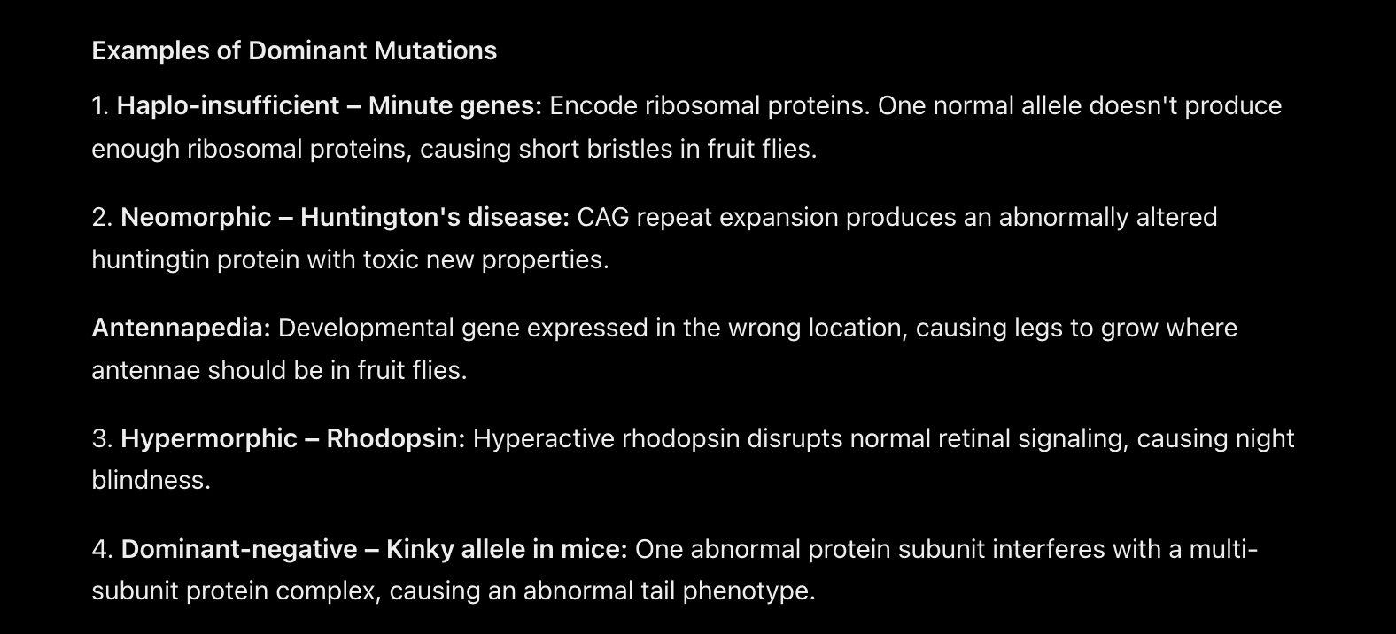
Haplo-insufficient gene

  • One functional wild-type allele does not produce enough protein to maintain the normal phenotype.

  • Even though the other allele is functional, the total amount of functional protein is insufficient.

  • Therefore, a heterozygote can display a mutant phenotype.

2. Neomorphic mutation

  • The mutation causes the protein to acquire a new function that it did not normally have.

  • Alternatively, the protein may be expressed or function at the wrong time or in the wrong location.

  • This can cause an abnormal phenotype because the protein is doing something it normally wouldn't.

3. Hypermorphic mutation

  • The mutation causes the protein to have increased activity or be produced in excessive amounts.

  • The protein is essentially doing its normal job too much.

  • This excessive activity disrupts normal cellular processes.

4. Dominant-negative mutation

  • The mutation produces an abnormal protein that interferes with the function of the normal, wild-type protein.

  • This commonly occurs when proteins work together in multi-subunit complexes.

  • Even though one allele produces functional protein, the mutant protein interferes with the normal complex.


dominant negative

  • The wild-type allele makes normal subunits.

  • The mutant allele makes defective subunits.

  • The defective subunits combine with normal subunits.

  • When even one defective subunit enters the complex, it can prevent the entire complex from functioning.
<p>Haplo-insufficient gene</p><ul><li><p>One functional wild-type allele does not produce enough protein to maintain the normal phenotype.</p></li><li><p>Even though the other allele is functional, the total amount of functional protein is insufficient.</p></li><li><p>Therefore, a heterozygote can display a mutant phenotype.</p></li></ul><p>2. Neomorphic mutation</p><ul><li><p>The mutation causes the protein to acquire a new function that it did not normally have.</p></li><li><p>Alternatively, the protein may be expressed or function at the wrong time or in the wrong location.</p></li><li><p>This can cause an abnormal phenotype because the protein is doing something it normally wouldn't.</p></li></ul><p>3. Hypermorphic mutation</p><ul><li><p>The mutation causes the protein to have increased activity or be produced in excessive amounts.</p></li><li><p>The protein is essentially doing its normal job too much.</p></li><li><p>This excessive activity disrupts normal cellular processes.</p></li></ul><p>4. Dominant-negative mutation</p><ul><li><p>The mutation produces an abnormal protein that interferes with the function of the normal, wild-type protein.</p></li><li><p>This commonly occurs when proteins work together in multi-subunit complexes.</p></li><li><p>Even though one allele produces functional protein, the mutant protein interferes with the normal complex.</p></li></ul><p></p><p></p><p>dominant negative </p><ul><li><p>The wild-type allele makes normal subunits.</p></li><li><p>The mutant allele makes defective subunits.</p></li><li><p>The defective subunits combine with normal subunits.</p></li><li><p>When even one defective subunit enters the complex, it can prevent the entire complex from functioning.</p></li></ul><p></p>
31
New cards
<p>Mitosis produces daughter cells with an exact copy of the genome</p>

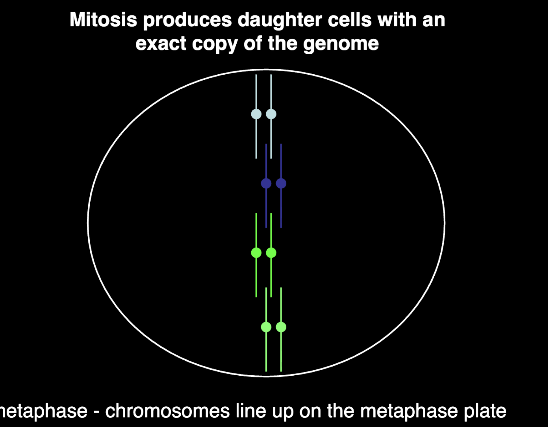
Mitosis produces daughter cells with an exact copy of the genome

Replication produces a sister chromatid

metaphase - chromosomes line up

anaphase - chromosomes separate and segregate

cytokinesis - production of two daughter cells

<p>Replication produces a sister chromatid</p><p>metaphase - chromosomes line up</p><p>anaphase - chromosomes separate and segregate</p><p>cytokinesis - production of two daughter cells</p>
32
New cards
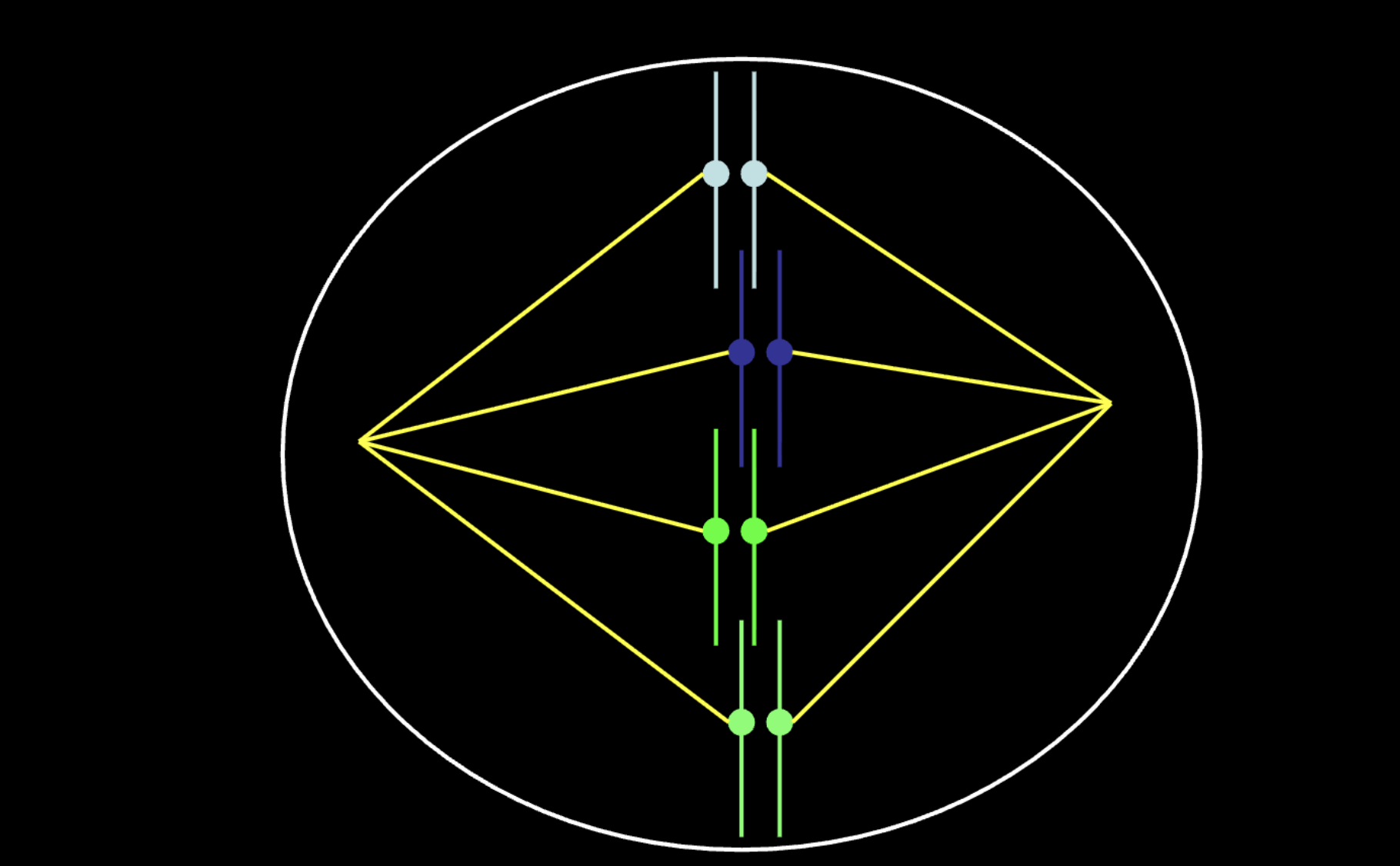
<p>What segregates the sister chromatids?</p><p></p><p>The spindle - a molecular machine that is highly dynamic and pulls</p><p>sister chromatids apart during anaphase</p>

What segregates the sister chromatids?


The spindle - a molecular machine that is highly dynamic and pulls

sister chromatids apart during anaphase

Microtubules attach to the chromosome via the kinetochore


The kinetochore assembles on the region of the chromosome

called a centromere


Sister chromatid cohesion opposes the pulling forces of the

spindle and makes sure that chromosomes are aligned correctly

with both kinetochores facing opposite poles


At anaphase sister cohesion is deliberately dissolved and sisters

chromatids can be pulled apart by the spindle

<p>Microtubules attach to the chromosome via the kinetochore</p><p></p><p>The kinetochore assembles on the region of the chromosome</p><p>called a centromere</p><p></p><p>Sister chromatid cohesion opposes the pulling forces of the</p><p>spindle and makes sure that chromosomes are aligned correctly</p><p>with both kinetochores facing opposite poles</p><p></p><p>At anaphase sister cohesion is deliberately dissolved and sisters</p><p>chromatids can be pulled apart by the spindle</p>
33
New cards
term image

B

34
New cards
<p>Mitotic cell division also referred to as the cell cycle</p><p></p><p>The cell cycle is regulated by signals that either promote or inhibit cell division. This regulation ensures cells divide at the appropriate time.</p><p></p>

Mitotic cell division also referred to as the cell cycle


The cell cycle is regulated by signals that either promote or inhibit cell division. This regulation ensures cells divide at the appropriate time.


The cell cycle is controlled by proteins called cyclins and CDKs, which regulate when a cell progresses through each stage.

  • G₁: Cell grows and prepares for DNA replication.

  • S phase: DNA is replicated.

  • G₂: Cell prepares for division.

  • M phase: Mitosis occurs; chromosomes are separated and the cell divides.

  • G₀: Cell exits the active cell cycle and is not actively dividing. Many cells in the body remain in G₀.


Important control points

  • Restriction point (late G₁): The cell-cycle commitment point. Once the cell passes it, it is generally committed to completing the cycle and dividing.

  • Cyclins + CDKs: Proteins that promote cell-cycle progression by activating processes needed to move through the stages.

  • Inhibitory proteins: Proteins that slow down or stop cell-cycle progression when conditions are not appropriate.


35
New cards
<p>Genetics of Cancer</p><p></p><p>Cancer is often the result of the accumulation of mutations that affect</p><p>a cell’s ability to control its division</p>

Genetics of Cancer


Cancer is often the result of the accumulation of mutations that affect

a cell’s ability to control its division

cancer usually comes from accumulated mutations that disrupt control of cell division. Since mutations build up over a lifetime, the longer someone lives, the more likely a single cell has collected enough of the right ones to become cancerous. Cancer typically needs multiple mutations (“hits”), which is why incidence is so low early on and accelerates later.


  • Effects stack up. As more layers fail in the same cell, the cell divides more readily, ignores stop signals, and survives damage it should die from. Once enough protections are gone, it divides essentially unchecked.