Hip Imaging
 Call Kai
 Learn
 Practice Test
 Spaced Repetition
 Match
 Flashcards
 Knowt Play
1/57

There's no tags or description
Looks like no tags are added yet.
58 Terms
In general, the male pelvis is __________, with a round inlet, and the female pelvis is ___________ with a more oval inlet
In general, the male pelvis is narrower, with a round inlet, and the female pelvis is broader with a more oval inlet
What are the two standard views to view the hip and pelvis with imaging
Anteropostior view
Frog-leg view
frog-leg view

anteroposterior view

The anteroposterior view provides several advantages, as it allows viewing of
the entire pelvis, sacrum and coccyx as well as lumboscaral and femoral joints
besides providing a great full view of the pelvis the anteroposterior view also provides insight on
proximal third of shaft, femorl head and neck and angle of inclincation
Anteroposterior Patient Position The lower extremities are internally rotated ___ to ___ degrees to place the femoral neck plane parallel to the image receptor
15 to 20 degrees
The advantages of the frog leg view
Demonstrates the femoral head, neck, and proximal third of the femoral shaft and the greater and lesser trochanters from the medial aspect as well as the femoral head is exposed
disadvantages of the frog leg view
angle of inlincation of femoral neck is not visible
the posterior and anterior columns of the pelvis are represented by...
ilioischial and iliopubic lines
ilioischial line

iliopubic line
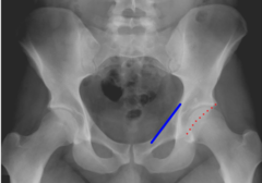
the angle of wiberg measures..
measures the extent to which the acetabulum cover the head of the femur
angle of wiberg

the angle formed must be greater than ___ degrees in an adult or dysplasia of the hip exists
25 degrees
normal angle of wiberg
25-35 degrees
DDH
shentons line

when observing shentons line, one side is..
always compaired to the other
interruption of Shenton's line can indicate
fracture neck of femur
DDH
a rapidly growing tumor may show more of a "____________" pattern like an osteosarcoma
sunburst
sunburst pattern

wards triangle

wards triangle is
Zone of weakness; area on neck of the femur where the trabeculae are weak and non-overlapping
decreased joint space bilaterally on weight-bearing surfaces may also be accompanied with...
osteophytes and increased density on the weight bearning portion of the acetabulum
The cartilage spacing at the symphysis pubis - the space should be less than __ mm wide
5 mm
Diastasis of the pubic symphysis is not uncommon following...
delivery (birth)
Calcifications in soft tissues are a result of dystrophic causes ___% to ___% of the time
95% to 98% of the time
the remaining __% to __% are the result of chondrocalcinosis, metastatic, or tumoral calcifications, and very unlikely, osteosarcoma metastases
2 to 5%
avulsion fractures of the ASIS, AIIS, ischial tuberosity, illiac wing, sacral, and ischiopubic fractures all fall into the broad category of
stable fractures
avulsion fracture of ASIS

avulsion fracture of AIIS

avulsion fracture of ischial tuberosity

iliac wing fracture

sacral fracture

ischiopubic rami fractures

vertical fractures through the body of the sacrum, L transverse sup ramus fracture w sup displacement, L comminuted inf pubic ramus fx and diastasis of the symphysis pubis are all considered
unstable fractures
Vertical fracture through the body of the sacrum

L transverse superior pubic ramus fx with superior displacement

Diastasis of the symphysis pubis

subcapital neck fracture
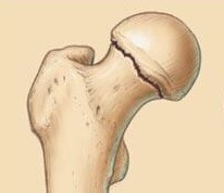
transcervical neck fracture

intertrochanteric fracture
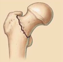
subtrochanteric fracture

fracture of the greater trochanter

fracture of the lesser trochanter

some infants are born with chronically dislocating
hips
The age group at risk for chronic dislocation of the hips
2-3 years of age
chronic dislocation of the hips is found in females more frequently than males at a __ to __ ratio
4 to 1
Legg-Calvé-Perthes Disease
Avascular necrosis of the femoral head in children.
Legg-Calvé-Perthes Disease occurs more in boys than girls at a __ to __ ratio
4 to 1
The most significant defining characteristic of Legg-Calvé-Perthes Disease
flattening of the femoral head
Slipped Capital Femoral Epiphysis
seen in children during growth-spurt years. the proximal epiphysis slips from its normal position on the femoral head
Slipped Capital Femoral Epiphysis imaging

traumatic hip dislocation
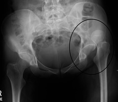
Osteonecrosis
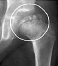
When viewing plain X-ray films for AVN, a careful review of the femoral head is required
femoral head
labral tear imaging
