Lecture 5 - Fungal and Yeast Infections
 Call Kai
 Learn
 Practice Test
 Spaced Repetition
 Match
 Flashcards
 Knowt Play
1/152

There's no tags or description
Looks like no tags are added yet.
153 Terms
Keratinized tissue like skin, hair, nails
Dermatophytes - tissue infected
Filamentous fungus
Dermatophytes - fungal type
Yeast
Candida - fungal type
Warm, moist, occluded areas like skin and mucosal surfaces
Candida - favored environment
Annular, scaly lesions
morphology - dermatophyte infection
Beefy red erythema
morphology - cutaneous candidiasis
Satellite lesions
lesion feature that strongly suggests Candida
Septate hyphae
Dermatophytes - KOH findings
Budding yeast and pseudohyphae
Candida - KOH findings
No
Is nystatin effective against dermatophytes?
Yes
Is nystatin effective against Candida?
Trichophyton, Microsporum, epidermophyton
three major types of dermatophytosis
Skin, hair, and nails
structures that Trichophyton typically infect
Skin and hair
structures that Microsporum typically infect
Skin and nails
Structures that Epidermophyton typically infect
Human-to-human transmission
anthropophilic transmission - definition
Animal-to-human transmission
zoophilic transmission - definition
Soil-to-human transmission
geophilic transmission - definition
Contaminated object or surface that transmits infection
fomite transmission - definition
Warm, humid climate
climate that increases dermatophyte infection risk
Contact sports, sweating, occlusive footwear, shared locker rooms and showers, immunosuppression
Personal risk factors that can increase risk of dermatophyte infection
Dermatophyte infection of the body
tinea corporis - definition
Dermatophyte infection of the groin
tinea cruris - definition
Dermatophyte infection of the feet
tinea pedis - definition
Dermatophyte infection of the hands
tinea manuum - definition
Dermatophyte infection of the scalp
tinea capitis - definition
Dermatophyte infection of the beard area
tinea barbae - definition
Dermatophyte infection of the face
tinea faciei - definition
Dermatophyte infection of the nails
tinea unguium - definition
By anatomical location NOT by the causative organism
How tinea infections are named
Annular, erythematous, and scaly with raised/active peripheral borders
shape and morphology of tinea corporis lesions
Pruritus
symptom that commonly accompanies tinea corporis
Central clearing
What is the term used to describe the middle of this dermatophyte lesion?
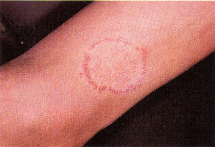
centrifugally
How dermatophyte lesions typically enlarge
Peripheral scale at the advancing edge
key clinical indicator that distinguishes tinea corporis from other rashes
Nummular dermatitis
granuloma annulare
psoriasis
pityriasis roseaa
contact derm
EM
d/d of tinea coporis that is NOT fungal
Upper medial thighs and inguinal folds
tinea cruris location
Pruritus
common symptom of tinea cruris
Erythematous, scaly plaques, advancing scaly border, MAY have central clearing
tinea cruris morphology
Interdigital - usually 4th and 5th toes
tinea pedis most common location
occlusion
sweating / hot / humid environment
communal showers
athletic facilities
Risk factors for tinea pedis
Interdigital
Moccasin
Vesiculobullous
3 clinical patterns of tinea pedis
Maceration, scaling, fissuring, pruritus
skin findings characteristic of interdigital tinea pedis
Secondary bacterial infection
complication that can occur with interdigital tinea pedis
Chronic diffuse scaling on the plantar surface and lateral foot - usually bilateral
characteristic pattern of moccasin-type tinea pedis
Vesicles or bullae frequently on the plantar or medial foot
characteristic pattern of vesiculobullous tinea pedis
Tinea pedis affecting both feet with unilateral tinea manuum
two feet-one hand syndrome
Typically unilateral, dry scaling w/ hyperkeratosis and palmar involvement
tinea manuum skin PE findings
Children
age group that most commonly develops tinea capitis
Scalp scaling, patchy alopecia, broken hairs w/ black dot appearance, pruritus
PE characteristics of tinea capitis
Cervical or occipital nodes
lymph nodes that may enlarge in tinea capitis
Intense inflammatory reaction to dermatophyte infection
kerion
Boggy inflammatory plaque w/ pustules, crusting, tenderness, and alopecia
PE characteristics of kerion
Abscess
Kerion is often mistaken as an _____.
Scarring alopecia
permanent complication of kerion
Systemic antifungal therapy; topicals do not adequately penetrate the affected hair shaft and follicle
Required Tx for tinea capitis - key fact
Griseofulvin (6-12 weeks), Terbinafine (4-6 weeks)
Systemic treatment options for tinea capitis
Liver function tests (LFTs)
laboratory tests requiredfor prolonged systemic antifungal therapy
Selenium sulfide or ketoconazole shampoo
Topical shampoo adjunctive tx options for tinea capitis
Beard or mustache area
anatomical region affected by tinea barbae
Follicular pustules, inflammatory plaques, broken hairs, and crusting
follicular findings that suggest tinea barbae
Bacterial folliculitis or acne
bacterial condition that can mimic tinea barbae
same as tinea capitis - d/t follicular involvement
Tx for tinea barbae
Onychomycosis
another name for tinea unguium
Yellow or white discoloration, thickening, subungual debris, onycholysis, dystrophy
nail changes that suggest onychomycosis
Toenails > fingernails
nails most commonly affected by onychomycosis
Tinea pedis
condition frequently concurrent w/ onychomycosis
Psoriasis, trauma, LP, dermatitis, age dystrophy
D/D's for onychomycosis
KOH preparation, culture, bx
Confirmatory tests for fungal infections
Terbinafine
first-line oral tx for onychomycosis
Efinaconazole (Jublia) - must be applied for 48 weeks QD - have very low cure rates
topical treatments for onychomycosis and limitations?
Vick's VapoRub, Tea Tree Oil
unconventional tx's for onychomycosis
Clinical assessment
How dermatophyte infection is typically Dx'd
KOH preparation
Preferred initial diagnostic for dermatophytes
1. identify active advancing border
2. Scrape scale with clade or curette
3. Place specimen on slide
4. Add KOH
5. Apply coverslip
6. Examine microscopically
Steps of KOH Prep
1. Scrape area/scales vigorously with handheld scalpel blade
2. Scales land on microscope slide
3. Push into heap into DTM culture
4. Place cap loosely to allow some air
5. Check in 2 weeks for color change of test medium
Steps of Fungal Culture
Fungal hyphae and spores
microscopic finding confirms dermatophyte infection
When diagnosis is uncertain, recurrent dz, tx failure
When to consider diagnositic testing for tinea
Topical antifungals - "azoles", terbinafine, butenafine
first-line therapy for localized tinea corporis, cruris, or pedis
Tx the lesions and surrounding skin BID for minimum of 2 weeks
Directions for topical antifungal medications
reduce moisture in area, avoid sharing towels and clothing
Non-pharmacologic tx education for localized dermatophyte
Extensive or refractory disease, capitis/barbae, ungium, selected immunocompromised pt's
type of dermatophyte infection often requires oral treatment
Steroid-modified dermatophyte infection - less scaly, less sharply demarcated, more extensive and more difficult to tx
tinea incognito
Moniliasis
another name for cutaneous candidiasis
Candida
Most common yeast that causes cutaneous candidiasis
DM, Obesity, Heat, Poor hygiene, Glucocorticoids (topical and PO), Abx use, debilitated state
Risk factors that predispose pt's to candidiasis
Beefy red erythema with satellite papules/pustules
PE appearance of candidal intertrigo
Burning/prurtitis, tenderness, maceration, fissuring, cracking
Symptom commonly associate w/ candidal intertrigo
Skin folds - inframammary, abdominal, groin, axillae, intergluteal cleft
where candidal intertrigo commonly occurs
present OUTSIDE the main affected area (unlike dermatophyte infections)
satellite lesions in Cutaneous Candidiasis
Summer (hot months)
During which season does candidal intertrigo commonly worsen
Candida infection of the diaper area - same presentation as candidal intertrigo
candidal diaper dermatitis
Inflammation at the corners of the mouth - commonly caused by candidiasis
Angular cheilitis (perleche)
Thrush
another name for oral candidiasis
White oral plaques on tongue and buccal mucosa/palate - removal of plaque may show an erythematous surface underneath
oral thrush PE presentation
inhaled corticosteroids, abx, dentures, diabetes, immunosuppression
Oral thrush risk factors
Nystatin, clotrimazole, fluconazole
oral antifungal tx's for oral thrush
microscopic finding confirms dermatophyte infection
Nystatin
topical polyene treats cutaneous candidiasis - key fact
Ketoconazole
topical azole treats cutaneous candidiasis - key fact