Week 9 ,, Protozoan Part 1
 Call Kai
 Learn
 Practice Test
 Spaced Repetition
 Match
 Flashcards
 Knowt Play
1/83

There's no tags or description
Looks like no tags are added yet.
84 Terms
Trophozoite
The motile feeding stage that is able to undergo cell division
Cyst
The dormant infective stage that is released to the environment
Acanthamoeba spp. Infective Stage
Ingestion of trophozoite
Acanthamoeba spp. Diagnostic Stage
Cysts and trophozoites in the tissues
Acanthamoeba spp. Clinical Features
Acanthamoeba spp. Laboratory Diagnosis
Keratitis of the eye ,, Granulomatous amebic encephalitis (GAE), and/or disseminated disease ,, Skin lesions
Naegleria fowleri is also known as what?
The brain-eating amoeba
Naegleria fowleri Diagnostic Stage
Trophozoites in CSF and brain tissue (flagellated forms occasionally in CSF)
Naegleria fowleri Infective Stage
Trophozoites enters the body through the nasal mucosa
Naegleria fowleri Clinical Features
Severe headaches, fever, vomiting, neurologic deficits that progresses rapidly and frequently to coma/death
Naegleria fowleri Laboratory Diagnosis
Microscopic examination of CSF ,, Immunofluorescence ,, PCR
Excystation
The process where a parasitic organism escapes from its protective cyst wall or envelope after being ingested by a host
Entamoeba histolytica Infective Stage
Ingestion of mature cysts and cysts/trophozoites passed in feces
Entamoeba histolytica Diagnostic Stage
Presence of mature cysts and cysts/trophozoites in feces
Entamoeba dispar
Trophozoites and cysts are indistinguishable from E. histolytica
Entamoeba histolytica/dispar Mature Cyst Features
1, 2, or 4 nuceli ,, Centrally located karyosomes ,, Fine, uniformly distributed peripheral chromatin ,, 1-2 chromatoid bodies with blunted ends
Entamoeba histolytica/dispar Trophozoite Features
Single nucelus ,, Centrally placed karysome ,, Uniformly distributed peripheral chromatin ,, “Ground-glass” cytoplasm appearance
What is indicative of Entamoeba histolytica?
Erythrophagocytosis
Commensal Protozoa
Nonpathogenic ,, Presence of parasite increases the likelihood of pathogens being present ,, Misidentification leads to unnecessary treatment
Entamoeba dispar
Trophozoite and cyst are indistinguishable from Entamoeba histolytica
Entamoeba hartmanni
Morphologically identical to E. hisolytica/dispar ,, Trophozoites and cysts are smaller in size
Entamoeba coli
Trophozoites : Coarse, unevenly distributed peripheral chromatin ,, Large, irregular eccentric karysomes ,, Coarse, granular cytoplasm with numerous vacuoles and inclusions // Cysts : 1-8 nuclei ,, Inclusions such as chromatoidal bars with splintered ends and glycogen vacuoles
Endolimax nana
Trophozoite : Single nucleus with no peripheral chromatin ,, Large, blot-like karyosome can be pushed to one side , Numerous vacuoles with bacteria, yeast, and organic debris // Cysts : Spherical or ovid ,, 1-4 nuclei ,, No peripheral chromatin and large blot-like karyosome
Iodamoeba bütschlii
Trophozoites : 1 nucleus with no peripheral chromatin ,, Large blot-like karyosome that may be surrounded by small granules ,, Vacuoles and debris in cytoplasm // Cysts : Single nucleus with the same appearance as the trophozoite ,, Oval or spherical ,, Characteristic large glycogen vacuole (brown on iodine stained wet prep)
Dientamoeba fragilis
Prodcues trophozoites only ,, 1 or 2 nuceli ,, Delicate nuclear membrane and no peripheral chromatin ,, Symmetrically arranged chromatin granule in the center of the nucleus
Blastocystis hominis
Blastocystis
Blastocystis hominis Infective Stage
Ingestion of thick-walled cysts
Blastocystis hominis Diagnostic Stage
Presence of thick-walled cysts
Blastocystis hominis Clinical Features
May be associated with watery diarrhea, abdominal pain, perianal pruritus, and excessive flatulence
Blastocystis hominis Laboratory Diagnosis
Microscopic examination of cyst-like stage in feces ,, Permanently stained smears are preferred over wet mount preparations because fecal debris may be mistaken for organisms in the latter ,, Do NOT wash the specimens in water because it will lyse the organism, resulting in false negatives
Balantidium coli
Balantidiasis
Balantidium coli Diagnostic Stage
Trophozoites and cysts
Balantidium coli Infective Stage
Trophozoites and cysts
Balantidium coli Geographical Distribution
Worldwide but more frequent in areas where pigs (main reservior) are raised
Balantidium coli Clinical Features
Most cases are asymptomatic ,, May cause persistent diarrhea, occasionally dysentery (blood and mucus), abdominal pain, and weight loss
Balantidium coli Microscopic Examination
Tropozoites in stool specimens or tissue collected during endoscopy ,, Cysts are less frequently encountered ,, Stool speicmens should be collected repeatedly
Balantidium coli Trophozoite Features
Bean shaped macronucleus
Cryptosporidium ssp
Cryptoporidiosis
Cryptosporidium ssp Diagnostic Stage
Thich-walled oocyst (sporulated) exits the host
Cryptosporidium ssp Infective Stage
Ingestion of thick-walled oocyst ingested by host
Cryptosporidium ssp Clinical Features
From asymptomatic to severe, life-threatening illness ,, Watery diarrhea, dehydration, weight-loss, abdominal pain, fever, nausea, and vomiting ,, Symptoms can be chronic and more severe in immunocompromised patients ,, Can also be found extraintestinally in the lungs, and possibly conjunctiva
Cryptosporidium ssp Microscopic Examination
Modified Acid Fast Staining will stain the oocysts bright red against a background of blue-green fecal debris and yeast
Cyclospora cayetanensis
Cyclosporiasis
Cyclospora cayetanensis Infective Stage
Ingestion of sporulated oocysts in contaminated food/water
Cyclospora cayetanensis Diagnostic Stage
Excretion of unsporulated oocysts in the stool
Cyclospora cayetanensis Clinical Features
Watery diarrhea can be severe ,, Anorexia, weight loss, abdominal pain, nausea, vomiting, myalgias, low-grade fever, and fatigue
Cyclospora cayetanensis Microscopic Examination
Similar in appearance to Cryptosporidium oocysts but is twice the size and has a much thicker wall
Cystoisospora belli
Least common intestinal coccidia that infect humans ,, Ellipsoidal large oocysts
Giardia intestinalis
Common intestinal infection
Giardia intestinalis Clinical Features
Varies from asymptomatic to severe diarrhea and malabsorption ,, Greasy stool that tends to float (steatorrhea) ,, Abdominal pain, bloating, nausea, and vomiting
Giardia intestinalis Infective Stage
Ingestion of cysts in contaminated food or water
Giardia intestinalis Diagnostic Stage
Cysts and trophozoites passed in the stool (trophozoites do not survive in the environment)
Giardia intestinalis Laboratory Diagnosis
Direct mounts and concentration procedures for identification of cysts or trophozoites in the feces ,, Excreted intermittently, so repeated sampling may be necessary ,, Samples of duodenal fluid or duodenal biopsy may demonstrate trophozoites
Chilomastix mesneli
Nonpathogenic flagellate ,, Generally considered a commensal residing in the cecum and/or colon
Chilomastix mesneli Trophozoite Features
Lemon-shaped trophozoite with anterior hyaline knob ,, One nucleus with a well developed membrane and karyosome (only seen in stained preparations)
Trichomonas vaginalis
GU tract infection
Trichomonas vaginalis Infective Stage
Trophozoite in the vagina or orifice of the urethra ,, Can spread through sexual intercourse
Trichomonas vaginalis Diagnostic Stage
Trophozoite in vaginal and prostatic secretions and urine
Trichomonas vaginalis Clinical Features in Women
Vaginitis with a purulent discharge ,, Vulvar and cervical lesions, abdominal pain, dysuria and dyspareunia
Trichomonas vaginalis Clinical Features in Men
Frequently asymptomatic ,, Urethritis, epididymitis, and prostatitis
Trichomonas vaginalis Laboratory Diagnosis
Wet mounts to detect actively motile organisms (rapid but insensitive) ,, DFA (more sensitive but complex) ,, Culture of parasite (more sensitive but takes 3-7 days) ,, Samples

Identify the Protozoa
Entamoeba histolytica/dispar cyst

Identify the Protozoa
Entamoeba hystolytica/dispar trophozoites

Identify the Protozoa
Entamoeba hystolytica/dispar trophozoites

Identify the Protozoa
Entamoeba hartmanni cyst

Identify the Protozoa
Entamoeba hartmanni trophozoite

Identify the Protozoa
Entamoeba coli trophozoite

Identify the Protozoa
Entamoeba coli cyst
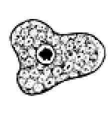
Identify the Protozoa
Endolimax nana trophozoite

Identify the Protozoa
Endolimax nana cyst

Identify the Protozoa
Iodamoeba bütschlii trophozoite

Identify the Protozoa
Iodamoeba bütschlii cyst

Identify the Protozoa
Dieniamoeba fragilis trophozoite

Identify the Protozoa
Blastocystic hominis

Identify the Protozoa
Balantidium coli

Identify the Protozoa
Balantidium coli

Identify the Protozoa
Cystoisospora belli

Identify the Protozoa
Cystoisospora belli

Identify the Protozoa
Giardia intestinalis

Identify the Protozoa
Giardia intestinalis trophozoite
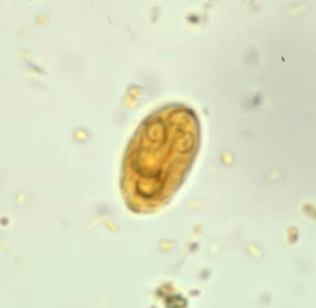
Identify the Protozoa
Giardia intestinalis cysts

Identify the Protozoa
Chilomastix mesneli trophozoite

Identify the Protozoa
Giardia intestinalis tropozoite
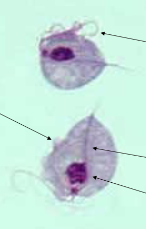
Identify the Protozoa
Trichomonas vaginalis trophozoite