Physiology 2.6: Vision
 Call Kai
 Learn
Learn Practice Test
 Spaced Repetition
 Match
Match Flashcards
 Knowt Play
1/58

There's no tags or description
Looks like no tags are added yet.
Name | Mastery | Learn | Test | Matching | Spaced | Call with Kai | Chat |
|---|
No analytics yet
Send a link to your students to track their progress
59 Terms
what is the visible spectrum
400 to 750 m
capable of stimulating the receptors of the eye
what is a wavelength
distance between successive peaks of electromagnetic radiation
eyes can sense both ____ and ____ of light
wavelength
intensity
optic nerve is considered to be the…..
blind spot
no photoreceptors
parasympathetic stimulation of iris leads to…
pupil constriction
less light entering
sympathetic stimulation of iris leads to…
dilatation of pupil
increased light entering
what is this
retina
contains photoreceptors
what is this
optic nerve
axons of ganglion neurons in retina that carry visual information to brain
how does the eye focus
lens and cornea
refract income light rays so they focus on the retina
where on the retina is there high focus
fovea centrallis
depression on macula lutea
highest photoreceptor density
what is the fovea centralis
greatest acuity image
highest photoreceptor density
depression in macula lutea
what is this
macula lutea
depression in retina with highest photoreceptor density
describe refraction
the bending of light rays as they pass from one transparent medium into another
the higher the density, the higher the refraction
visual information that reaches the retina is…
reverse right to left and is upside down

describe accommodation of the eye
eyes automatic ability to change the curvature and shape of the lens
helps focus on objects different distance
contraction or relaxation of ciliary muscles

what is responsible for the curvature of the lens
ciliary muscle
applies tension to zonular fibers
if you want to see far-away objects, you need [shape] lens
flat lens
less refraction
action
more tension on zonular fibers
relaxation of ciliary muscle

if you want to see close up objects, you need [shape] lens
thick lens
more refraction
action
less tension on zonular fibers
contraction of ciliary muscle

describe myopia
near-sightedness
can see nearsightedness
cannot see far
eyeball too long
focus is in front of retina

what lens do you use to fix myopia
concave/diverging lens

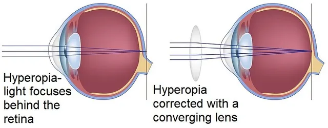
describe hyperopia
farsightedness
can see far objects
cannot see near objects
eyeball too short
focus is behind retina

how do you correct hyperopia
convex/converging lens
what is astigmatism
focusing programs in which there are irregularities in the curvature of the lens/cornea
multiple focal points

what is presbyopia
normal, age-related loss of close-up vision
lens get stiffer
what is cataracts
clouding of the lens
blurry vision

what is glaucoma
impaired drainage of aqueous humor
increased intra-ocular pressure
compression of vascular layer below retina
damage to retina and blindness

describe age-related macular degeneration
loss of visual information in center of visual field
damage to macula lutea

describe the histology of the retina
light passes all the way back to the retina and hits photoreceptor cells
rods and cons
passes through
horizontal cells
amacrine cells
describe rod photoreceptor cells
respond to low light
non-colored vision
have more photopigment
more sensitive
none in central fovea
low focus

describe cone receptor cells
respond to bright lights
colored vision
3 types of cones with different photopigment (blue, green, red)
less photopigments
high density in fovea
high focus

describe the 3 types of cones
red, green, and blue
detect different parts of the visible spectrum
colored vision requires at least 2

in cone photoreceptors, each cone types responds to…
each range of wavelength
for any given wavelength, the 3 cones are excited to different degrees

describe color blindness
1 or more cone mechanisms are lost
what is the purpose of horizontal and amacrine cells
lateral integration between neurons and retina
regulate and fine-tune retinal signaling
describe the components of photoreceptors
outer segment
membranous fold with photopigments
absorb light
inner segment
basic cell machinery
synaptic terminal
releases glutamate as neurotransmitter
what do photoreceptors release as neurotransmitter
glutamate
simple breakdown of photoreceptors in the dark
photoreceptors depolarize
release glutamate
bipolar cell activated
inhibits ganglion cell
no light is percieved
in visual transduction, glutamate activates…
bipolar cells
activated bipolar cell inhibits ganglion cell

simple breakdown of photoreceptors in the light
light causes photoreceptors to hyperpolarize
inhibited photoreceptor
inhibited bipolar cells
ganglion cell activated
transmits signals to perceive

describe photoreceptor synaptic activity in the dark
guanylyl cyclase converts GTP to cGMP
opens ligand-gated cation ion channels
influx of Na/Ca ions
depolarization
glutamate is released
describe photoreceptor synaptic activity in the lgiht
when light strikes photoreceptor, pigment activates G-protein transducin
activates enzyme that decreases intracellular cGMP
hyperpolarization
glutamate release decreases
describe the 5 neuronal types in the retina and which generate APs vs generate graded potentials
graded
photoreceptors
bipolar cells
horizontal cells
amacrine cells
action potentials
ganglion cells
axons form the optic nerve

give me a brief description of the type of neuron cells in the retina
photoreceptors
the rods/cones that detect light
bipolar cells
bridges that pass signals from photoreceptors
ganglion
send visual data to brain
horizontal and amacrine cells
lateral processing and modulation of visual signals
what is the direct pathway for transmission of visual information through the retina
photoreceptor → bipolar cells → ganglion cells → brain
light hits photoreceptor, bipolar cell activated
ganglion cell send image to brain
what is the indirect pathway for transmission of visual information through the retina
direct pathway but with editors
includes the direct pathway and horizontal and amacrine cells
extra cells alter the signal horizontally
sharpen the edges and enhance the contrast

what is the purpose of the on and off pathway of the retina
antagonistic arrangement acts as lateral inhibition
sharpens perception of edges and contrast

describe the on pathways
glutamate is inhibitory for bipolar cells
light strikes
photoreceptor cell hyperpolarized… less release of glutamate
bipolar cell depolarized and releases excitatory NT
ganglion neuron is depolarized… increased AP firing
visual signal sent to brain

describe the off pathway
glutamate is excitatory for bipolar cells
light strikes
photoreceptor cell is depolarized… less release of glutamate
bipolar cell hyperpolarizes and releases less excitatory NT
ganglion neuron is hyperpolarization… less AP firing
no visual signal sent to brain

What is the wavelength range of visible light. Name two aspects of light that are distinguished by the eye.
400 to 750nm
2 aspects
color
determined by intensity of wavelength
brightness
determined by amplitude of light wave process by photoreceptors

describe this
iris
regulates amount of light entering the eye
controls size of pupil


describe this
pupil
lets light enter the eye


describe this
retina
has photoreceptors that send visual signals to the brain


describe this
optic nerve
bundle of ganglion axons that connects retina to the visual cortex of the brain
carries sensory and visual information

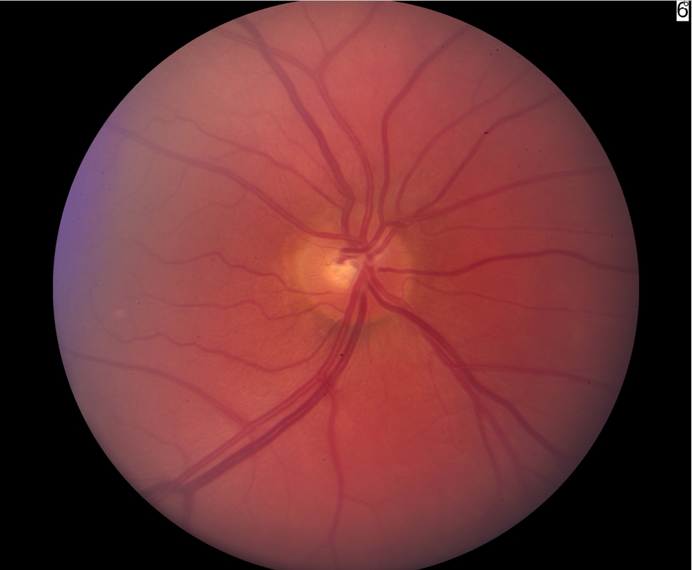
describe this
optic disc
point where all ganglion cell axons gather and exit to form the optic nerve
blind spot because it has no photoreceptors


describe this
lens
refracts light and focuses them directly onto retina
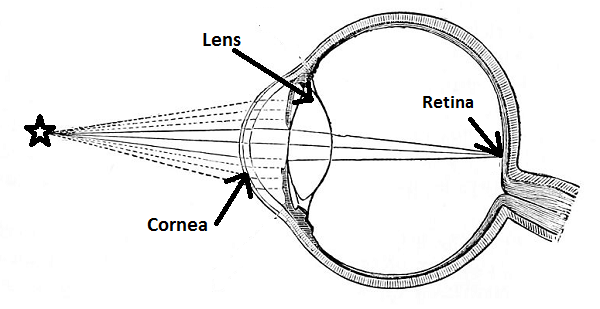

describe this
cornea
focuses light and protects the eye


describe this
macula lutea
pigmented area near center of retina
contains very high concentration of cone cells
responsible for color vision
exact center has fovea centralis

describe this
fovea centralis
depression in the retina (in macula lutea)
highest concentration of cone photoreceptors

Briefly describe the focusing system of the eye. What is the quality of image imposed at fovea centralis and the macula lutea?
the cornea and lens work to bend and refract incoming light rays directly onto the retina
focused light projects strait into macula and fovea
image is inverted and referred (brain processes it right-side up)