Lab practical final
 Call Kai
 Learn
 Practice Test
 Spaced Repetition
 Match
 Flashcards
 Knowt Play
1/164

There's no tags or description
Looks like no tags are added yet.
165 Terms

7
Frontal Lobe

1
Occipital lobe

8
Temporal Bone

10
Medulla Oblongata

9
Cerebellum


2
Pond

11
Corpus Callosum


17
Hypothalamus

3
Thalamus
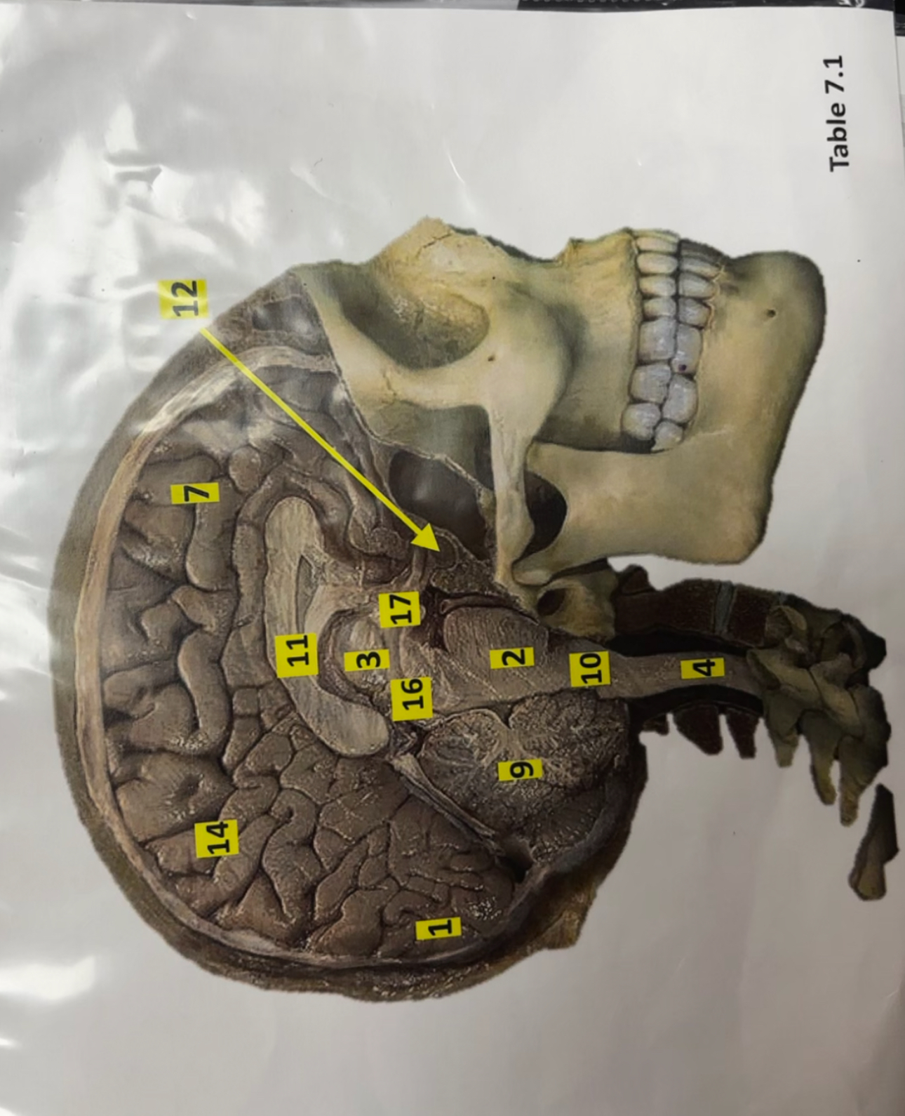
12
Pituitary gland

16
Midbrain

4
Spinal cord

14
Parietal Lobe


6
Dura meter; thick, tough, outer layer of the meninges

15
Arachnoid mater; delicate, web-like middle layer;acts as a shock absorber for your brain.

18
Pia mater; thin, delicate inner layer; rich with tiny blood vessels that nourishes brain

13
Gray matter

5
White matter

19
Dorsal root


21
Ventral roots

20
spinal nerve

22
vertebral body

23
Sclera

28
Iris

30
Ossicles

24
Tympanic membrane

31
Semicircular canals

25
Optic nerve

26
Cochlea

32
Retina

29
Pupil

27
Lens

33
Auditory canal

1
Right atrium

4
Right ventricle

3
Left atrium

5
Left ventricle

2
Coronary arteries

6
Pulmonary artery

8
Pulmonary Trunk

11
Aorta

9
Brachiocephalic (Trunk) |

10
Coronary sinus

7
Pulmonary Veins

13
Superior vena cava; second largest vein |

24
Inferior vena cava; largest vein |

29
Common carotid artery |

15
Tricuspid valve; |
the right side and has three leaflets
14
Chordae tendineae |

17 |
Bicuspid valve (mitral)

What slide is this?
Cardiac muscle; Found in walls of heart; Responsible for involuntary contraction of heart; Contains Intercalated discs(2 proteins gap junctions and desmosimes which hold cells tg)

What is in this slide?
Lung tissue; located inside thoracic cavity; responsible for gas exchange; alveoli is lined with simple squamous epithelium


what is on this slide? Location and function?
A. Artery B. Vein; found throughout body within circuitry system; 1.Arteries transport oxygenated blood away from heart. 2.Thick muscular walls; 1. Veins transport deoxygenated blood back to heart. 2. Thin walls/irregular shapes due to low pressure

what is on this slide?
Trachea; It’s extended from the larynx down to bronchi of lungs; Responsible for Air conduction; lined with pseudostrstified ciliated columnar epithelium


what is on this slide?
Stomach; located upper left portion of abdominal cavity; responsible for mechanical and chemical digestion


what is on this slide?
Small intestine; located distal to stomach; responsible for primary nutrient absorption


what is on this slide?
Large intestine/colon; framed around small intestine; responsible for recolonizing the colon with probiotic bacteria and white cells

what is on this slide?
KidneyA.Renal medulla B.Renal cortex; located in the back of abdominal cavity; responsible for filtering blood and remove metabolic waste


what is on this slide?
Testes; located within scrotum, outside of pelvic cavity; responsible for spermatogenesis


what is on this slide?
Ovary; located in pelvic cavity, lateral to uterus; responsible for oogenesis

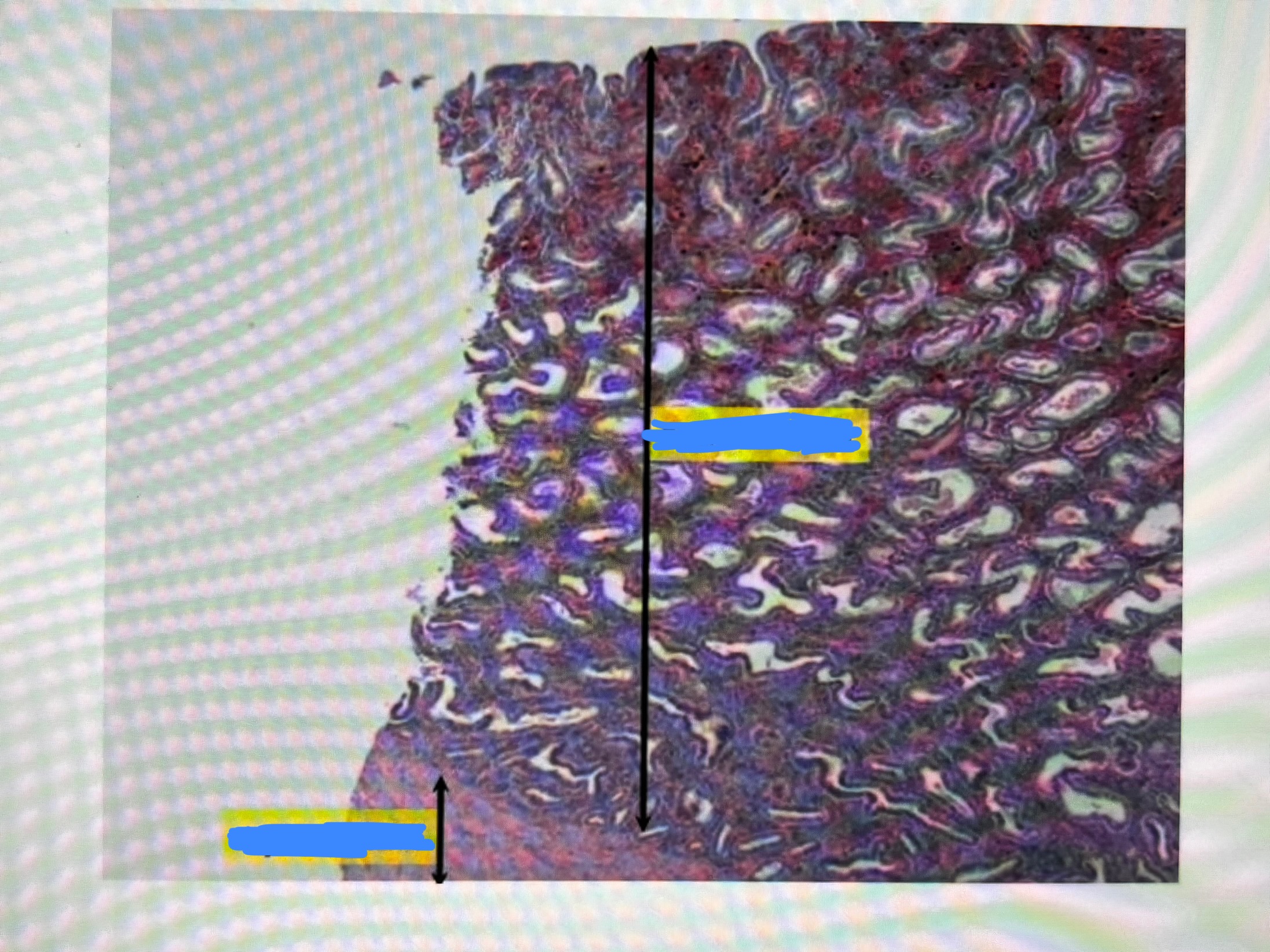
what is on this slide?
Uterus; located in pelvic cavity, superior to the vagina; responsible for site of implantation + development of fetus

What type of information pass through the dorsal and ventral horns and roots of the spinal cord?
Dorsal horns/roots: Sensory(afferent) info
Ventral horns/roots: Motor(efferent) info
What are intercalated discs and where are they found?
These are specialized junctions between cardiac muscle cells. They are found in cardiac muscle tissue within the heart wall
What are the 3 layers of blood vessels and what type of tissue are they of?
Túnica interna,inner layer made of simple squamous; Tunica Media, middle layer of smooth muscle and elastic fibers; Tunica Externa, outer layer made of connective tissue
What structure is found within the intervenntricular groove?
Coronary blood vessels
Where is the cartilage located throughout the respiratory tract?
Found throughout the trachea and primary bronchi (C-Shaped rings)
What are goblet cells? Where are they found and what do they secrete?
Found in the respiratory tract epithelium; they secrete mucus to deal dust and pathogens
How does hyperventilation effect CO2
Decreases CO2 levels in blood
How does excercise effect CO2?
Increases CO2
How does bag breathing effect CO2?
Increased CO2
How did we detect CO2 levels being expired in lab?
Calcium hydroxide
What is rugae and where is it found? Function?
Folds that allow the stomach to expand when full
What is villi and where is it found? Function?
Found in small intestine; finger like projections that increase surface area for nutrient absorption
Where in the digestive system do digestion and absorption occur?
Digestion: Occurs in the oral cavity, stomach, and small intestine
Absorption: small intestine
What are segmentation and peristalsis and where do they occur?
Peristalsis: wave-like contractions that propel food forward; occurs through the almimentary canal
Segmentation: Localized mixing contractions; occurs in the small intestine
What are the 4 layers of the alimentary canal?
Mucosa: innermost, absorbs nutrients
Submucosa: connective tissue layer containing blood/lymph vessels
Muscularis externa: smooth muscle responsible for peristalsis and segmentation
Serosa: outermost layer, protects and reduces friction

1
Lungs; primary organ for ventilation

17
Larynx; voice box


6
Uvula; soft appendage that hangs from roof of mouth

2
Epiglottis; prevents food from entering the larynx

11
Trachea;composed of hyaline cartilage

7
Pharnyx

4
Diaphragm; primary muscle of respiration

14
Intercostal muscles; respiratory muscles located in between ribs


13
Primary bronchus


3
Secondary bronchi


15
Alveoli sacs; location for external respiration

10
pulmonary veins


8
Pulmonary arteries


3
Parotid gland; largest salivary gland

4
Hard palate; composed of maxillary and palatine bones

1
Soft palate

15
Tongue; muscle for mechanical digestion

17
Pyloric sphincter; regulates the rate of food that enters small instestine

16
Liver; produced bile and cholesterol

25
Pancreas; produces insulin and glucagon


26 + 24
Duodenum; where pancreatic secretions enter


10
Transverse colon

22
Ascending colon

9
Descending colon