CAD and ACS - Unit 2 AH3
 Call Kai
 Learn
Learn Practice Test
 Spaced Repetition
 Match
Match Flashcards
 Knowt Play
1/25

There's no tags or description
Looks like no tags are added yet.
Name | Mastery | Learn | Test | Matching | Spaced | Call with Kai | Chat |
|---|
No analytics yet
Send a link to your students to track their progress
26 Terms
what are non-modifiable and modifiable cardiac risk factors
non-modifiable:
age
gender
family history
race
modifiable:
smoking
cholesterol/lipids
weight
diabetes mellitus
diet
physical activity
BP management
What is coronary artery disease (CAD)
atherosclerosis (plaque) forms and occludes coronary artery
causing
unstable angina
myocardial infarctions
sudden cardiac death
reduced blood flow through coronary microvasculature
coronary artery blood vessels don’t have plaque (occlusion), but damage to the inner walls of the blood vessels can lead to spasms and decrease blood flow to the heart muscle
ischemic symptoms - lack of blood flow and oxygen to the tissue leading to tissue death
CAD: clinical manifestations
stable angina
chest pain or discomfort that is associated w/ physical activity
linked to fixed plaque formations and is predictable
symptoms are often alleviated w/ rest and/or medications
unstable angina
refers to chest pain that can occur at rest
this type of angina can be brought on by physical activity as well
of the two types of angina, unstable angina is the most concerning
identified as the initial phase of acute coronary syndrome (ACS), defined as a disorder caused by an acute decrease in blood flow through the coronaries, to the myocardial tissue, and can be a precursor to MI
Should be treated as an emergency
prinzmetal’s angina
a variation of unstable angina is Prinzmetal’s, or variant, angina
the blockage of blood flow in this disorder is caused by coronary artery spasm rather than plaque formation, but atherosclerotic changes are commonly present
it typically occurs at rest and in clusters at night between midnight and 8 am
CAD: medical management - diagnosis
labs
total cholesterol, triglycerides, LDL, HDL, CK, CK-MB, troponin
electrocardiogram (ECG)
exercise or chemical stress test
coronary angiography (left sided heart cath)
gold standard for diagnosis
determines if percutaneous transluminal coronary angioplasty (PTCA) is an option (aka a cardiac stent placement)
CAD: medical management - medication
stop aggregation of blood components to endothelium
control factors leading to endothelial damage
relief or symptoms
so they don’t have blood clot next to the plaque buildup and cause cardiac event
CAD: surgical management
percutaneous transluminal coronary angioplasty (PTCA) aka percutaneous coronary intervention (PCI)
performed in the cardiac cath lab
monitored anesthesia care (formerly known as conscious sedation)
catheter with a small balloon on its tip is advanced under fluoroscopy through a suitable artery, commonly the femoral or radial, to the area with atherosclerotic plaque
the balloon inflated and deflated to open the lumen of the artery
once the lumen is open, a stent may be advanced to the location to hold the artery open and maintain adequate blood flow
will need 3 consent forms
PTCA consent form
PCI consent form
blood consent form
coronary artery bypass grafting (CABG)
performed in the operating room under general anesthesia
blockages in coronary arteries are bypassed using other arteries from the chest or arm or veins from the legs
a large incision through the sternum is made
cardiopulmonary bypass (CPB) is begun through large catheters in the vena cave or right atrium and aorta. a CPB provides continuous gas exchange and perfusion while the heart is stopped to provide a still, bloodless field for surgery
then arteries or veins being used as bypasses are surgically attached to the diseased coronary, creating an alternate path for blood to flow around the blockage
**Pathway to go around the blockage, leave the blockage alone
CAD: medical management - lifestyle management
maintain healthy body weight
diet
physical activity
smoking cessation
screening and treatment for depression
refraining from excessive alcohol use
cardiac rehabilitation
CAD: complications
myocardial infarction (acute coronary syndrome)
dysrhythmias
heart failure
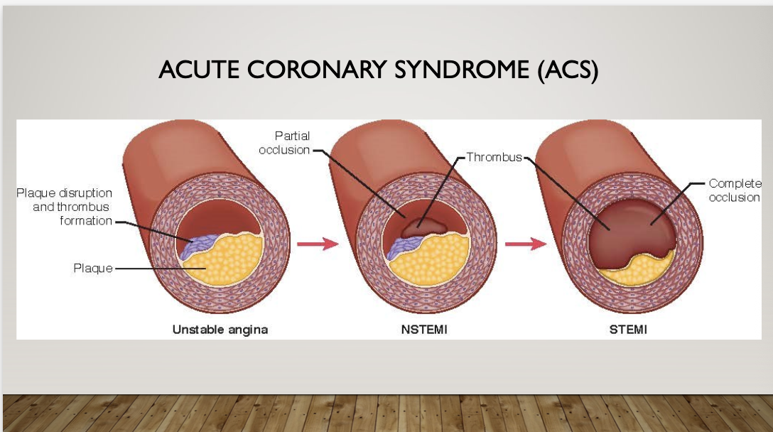
acute coronary syndrome
STEMI (ST elevation MI)
infarction with complete obstruction of blood flow
unstable angina
present w/ ischemic symptom → angina, dyspnea, vital sign changes
STEMI on 12 lead EKG, 1 millimeter or higher ST elevation in 2 or more leads is a MI w/ complete obstruction
Non-STEMI (non st elevation mi)
ischemia w/ partial obstruction of blood flow
unstable angina
both may present w/ unstable angina
myocardial infarction: clinical manifestations
chest pain (angina)
unstable
may start as stable and turn into unstable
be mindful of nonspecific symptoms:
epigastric discomfort
N/V
diaphoresis
syncope
SOB
pain between shoulders/jawline
females typically have nonspecific symptoms***
not always 10/10 crushing chest pain like in males
MI: medical management - diagnosis
lab tests
Troponin: cardiac damage (specific to the heart)
CK (Creatine kinase): enzyme found in heart and skeletal muscle, high levels indicate muscular injury (not specific to the heart)
CK-MB (Creatine kinase - myocardial band): specific to the heart
Myoglobin: protein found in skeletal muscle and heart (not specific to the heart)
diagnostic testing
ECG/EKG (electrocardiogram):
**gold standard for diagnosis of MI
echocardiogram: ultrasound of the heart looking at contractility and chambers
stress test: if active MI we will skip this step and go straight to cath lab
coronary angiography (aka heart cath lab)
difference between Troponin I and Troponin T
— always done in series, at least 3; then when positive, keep going until the peak
— first come in, then 1 hr, then 6 hr, then when they peak
— troponin does not rise for 3hr after MI
—NEED TROPONIN WHEN THEY FIRST COME IN, AN HOUR LATER AND THEN AGAIN BASED ON PROTOCOL AND IF POSITIVE KEEP TESTING UNTIL THEY PEAK
Troponin I
is specific to the heart muscle damage it only comes from myocardium
normal level: <0.04 ng/mL
MI: >0.04 ng/mL
Troponin T
comes from skeletal muscle and myocardium
normal level: <0.01 ng/mL
Emergent STEMI Treatment
aspirin
nitroglycerin
morphine
supplemental O2 if sats <90%
consider load w/ P2Y12 inhibitor
Aspirin
the 1st med prioritized in ACS
onset of action 1-7.5 min if chewed
initial dose 325 mg CHEWED, then 81 mg indefinitley
disables platelet aggregation (blood thinner)
POTENT!!!! (strong)
monitor for intolerance
s/s: flushing, itchy, rash, nassal congestion, asstham exacerbation
plavix can be used as an alternative for intolerance
shouldn’t take w/ other nsaids
can become thrombotic
will go on baby asprin everyday indefinitely
Nitroglycerin
potent vasodilator and venodilator
reduces preload and ventricular wall tension
decreases myocardial O2 consumption
sublingual, spray or intravenous
sublingual 0.4 mg Q 5 min x 3, then assess the need for IV NTG
monitor for hypotension, HA is common
do not give to pts w/ right ventricular infarction or use of other phosphodiesterase (PDE-5) inhibitors (VIAGRA OR CIALIS) {**can’t be given for erectile dysfunction drugs because they are vasodilators, so it will bottom out the BP}
caution w/ inferior wall MI
they are preload dependent, so if they don’t receive enough blood to return to the right side of the heart, they will go into cardiac arrest
Important to ask if pt had nitro prior to admission
BP goes down w/ nitro (keep checking BP), so if BP is low before giving nitro try to get it increased before giving nitro
Morphine
use as adjunct therapy (along with) to nitroglycerine
small incremental doses
1-2 mg IV q 5-15 min if CP unrelieved by NTG
potent analgesic (pain) and anxiolytic (anxiety)
causes venodilation and reduces preload, mild afterload reduction
decreases workload of heart
anxiolytic of choice
use cautiously in inferior wall MI
avoid in right ventricuclar MI
increased mortality in a large registry
more research needed
don’t use every time, but consider if nitro doesn’t work
oxygen
Administer supplemental oxygen only when:
the oxygen saturation falls below 90%
respiratory distress is present
other high risk features for hypoxemia are present
reserved for hypoxemia
hyperoxemia likely perpetuates oxidative injury after MI
Acute STEMI - PCI
gold standard for ST elevation MI: revascularization/PCI
door to balloon time goal: 90 min!!!
more trans-radial approach
monitor kidney (urine output, and BUN and Creatinine lvls) - dye load
there is so much dye put into pt during this procedure and can cause kidney failure
retroperitoneal bleed and hematoma (RBH)
femoral approach
retroperitoneal space can hold 4L of blood which is almost all pt blood in body so we need so look out for early s/s
gonna need blood or MTP
signs:
tachycardia
may not see d/t beta blockers
hypotension
back pain, groin pain, flank ecchymosis (late sign)
tx:
stat H/H, coags if applicable
may medically manage (hold pressure) or surgical repair (<10%)
percutaneous intervention w/ balloon tamponade
fluids, prepare to transfuse
fibrinolytic therapy
pt can get fibrinolytic therapy until they can get pt to chest pain center to get to cath lab, we are trying to hold them over
if unable to get to cath lab in a timely manner-
TNKase (tenecteplase)
same drug as for stroke, accept we are using it for emergency purposes in the coronary artery to bust the clot
rtPA (activase)
will still need to go to cath lab
indications:
pain < 6 hrs
ST elevation > 1 mm in 2 or more leads
nursing considerations:
bleeding is most common side effect
if bleeding occurs, discontinue all anticoagulants
monitor PT/INR/aPTT - prolongs both
monitor fibrinogen - decreased fibrinogen for up to 24*
reversal - cryoprecipitate and platelets
biggest area is cerebral hemorrhage
need frequent neuro assessments, monitor for bleeding, and avoid any additional needle sticks
frequent neurological assessments
avoid punctures
monitor urine output and BUN/creatinine
avoid invasive devices
avoid compressive devices
the “Big 5” discharge medications following ACS
ASA: (indefinitely)
P2Y12 receptor blocker: prevents clotting(at least 1 yr)
beta blocker: (indefinitely)
ACE-I (or ARB): EF <40% or anterior MI (indefinitely)
Statin (high intensity indefintely)
dual antiplatelet therapy is ASA and P2Y12 drugs
continuing dual antiplatelet therapy for 1 yr is critical - important for pt education
myocardial infarction: complications
heart failure
arrhythmias
MI: nursing interventions - assessments
vital signs and pulse oximetry:
tachycardia w/ a borderline low BP and decreased oxygenation saturation is a sign of inadequate CO and oxygen deliver
characteristics of pain
location, radiation, duration, intensity, precipitating or alleviating factors; use a 1-10 pain scale — chest pain is an indication of MI. continued or changing pain characteristics can be indicative of a worsening condition
ECG
ST-segment depression is indicative of ischemia
ST-segment elevation is indicative of injury
if present a Q wave is diagnostic for MI
restlessness
may be found in early stages, but progression to severe anxiety and sense of doom is late stage symptom
skin color and temp peripheral pulses, diaphoresis
decreased pulses and cold, clammy, pale skin are s/s of inadequate tissue perfusion and inadequate CO
activation of the sympathetic system w/ low co will stimulate diaphoresis
monitor urine output
decreased or absent urine output is a sign of decreased renal perfusion related to decreased CO
labs
assess troponin, CK, and CK-MB lvls
troponin is protein released from damaged cardiac muscle. it elevates w/in 4 hrs and can stay elevated for days
CK-MB, the CK isoenzyme marker specific to cardiac tissue, is released from the cells w/ cardiac muscle damage. Increased lvls can be seen at 3 hrs and remain elevated for as long as 36 hrs
MI: nursing interventions - actions
administer oxygen
insert two large-bore IVs
administer meds as ordered - meds are essential to be given in a timely manned
aspirin and heparin - are given to prevent new clot formation
nitroglycerin SL - dilates the coronary arteries, increasing blood flow and decreasing pain
morphine - narcotic given for pain relief if nitro is not effective
beta blockers - decrease the sympathetic response to an MI, decreasing cardiac workload and oxygen consumption
fibrinolytics work to dissolve clots
continuous ECG monitoring
electrocardiogram monitoring is essential to evaluate the evolution of the MI and the effectiveness of tx and to monitors for dangerous dysrhythmias that can occur
bed rest
the pt may require bed rest as well as emotional rest to decrease oxygen and cardiac demands
MI: nursing interventions - teaching
report s/s of MI
chest pain or discomfort or SOB
medication education
purpose, dose, and side effects of meds
prescribed meds are to treat the effects of MI and prevent future MI’s
american heart association “life’s simple 7”
no smoking
maintain a normal body weight
exercise for at least 150 min w/ moderate intensity activity, or 75 min of vigorous intensity activity, or a combination of each per week
eat healthy diet
maintain total cholesterol lvl less than 200 mg/dL
keep Bp less than 120/79 mm Hg
keep fasting blood glucose less than 100 mg/dL
EDUCATION!!!
medication compliance
don’t drink
don’t smoke
exercise
manage weight
heat healthy diet
lower cholesterol / lipids
reduce stress