Pathogenesis of Infectious Diarrhea – Bacterial causes
 Call Kai
 Learn
 Practice Test
 Spaced Repetition
 Match
 Flashcards
 Knowt Play
1/79

There's no tags or description
Looks like no tags are added yet.
80 Terms
Gram-negative bacterial causes of diarrhea
Salmonella spp.
Pathogenic Escherichia coli
Yersinia spp.
Campylobacter spp.
Lawsonia intracellularis
Gram-neg spirochetes bacterial causes of diarrhea
Brachyspira spp
Gram-positive bacterial causes of diarrhea
Clostridium spp. – Clostridium perfringens,
Clostridioides/Clostridium difficile
Acid-fast bacteria causes of diarrhea
Mycobacterium avium subsp. Paratuberculosis
Transmission and Predisposing Factors of Bacteria which cause Diarrhea
fecal-oral route → Can be present in the feed and water
Inadequate passive immunity (neonates)
Poor management practices: Overcrowding, Abrupt changes in feeding/diet, Poor hygiene
Stressing factors; co-infection with a virus, weaning, social hierarchy, intermingling, transportation, handling and routine husbandry practices.
Salmonella spp.

Salmonella spp: horses and poultry

Pathogenesis of salmonella spp.
ruffling of the cell membranes → uptake of the Salmonella into the Salmonella containing vesicles (SCVs)
Utilizes the Type III secretory system to inject virulence factors such as toxins into the host cells
Activation secretory pathways and alter ion balance → diarrhea
LPS responsible for endotoxic effect
The local inflammatory response damages epithelial cells - diarrhea
Diagnosis of salmonella spp.
Culture: Most Salmonella species – MacConkey – pale (non-lactose fermenter); Brilliant Green agar – red colonies; XLD – red colony black centers
Serology: Serological tests, Serotyping
Molecular testing: PCR, Gene sequencing
Treatment and control of salmonella spp.
Use of antimicrobials is controversial in clinically normal shedder and patients with the localized enteric form of the disease
Vaccination – commercial killed and live-attenuated vaccines
Competitive exclusion with pre- and probiotics
Good animal husbandry and biosecurity measures on farms
Virulence of E. coli
Endotoxin: LPS – Gram-negative released when the E. coli dies
Pyrogenic activity
Endothelial damage leads to intravascular coagulation and endotoxic shock
Capsules: Inhibits phagocytosis
Structures for adherence and colonization, Enterotoxins
Categories of pathogenic strains of E. coli in animals include
Enterotoxigenic E. coli (ETEC)
Attaching and Effacing E. coli (AEEC)
Enteropathogenic E. coli (EPEC)
Shiga toxin-producing E. coli (STEC)
Enterohaemorrhagic E. coli (EHEC)
Strains producing oedema disease
Enteroaggregative E. coli (EAEC)
Enteroinvasive E. coli (EIEC)
Diffusely adherent E.coli (DAEC)
Enterotoxigenic E. coli
Causes the majority of neonatal colibacillosis of piglets and other domestic animals
Colibacillosis refers to an E. coli infection characterised by one of the following
Diarrhea
Enteritis
Septicaemia
Bacteraemia
Enterotoxigenic E. coli Produce 2 important toxins: Heat-labile (LT)
antigenically related to the cholera toxin of Vibrio cholerae → affects adenylate cyclase system leading to hypersecretion of fluid into the intestines → diarrhea, hypovolemia and metabolic acidosis
Enterotoxigenic E. coli Produce 2 important toxins: Heat-stable (ST)
Two types
STa – a plasmid-encoded toxin affecting the guanylate cyclase system causing fluid accumulation in suckling mice and piglets.
STb – causes diarrhea and fluid accumulation in intestines of weaned pigs
Enterotoxigenic E. coli: Possess fimbrial antigens F4 (K88) and F5 (K99) (adhesins)

Diseases caused by E. coli in domestic animals: Pigs

E. coli in oedema disease (STEC)
Occurs in weaned pigs
E. coli colonize like ETEC – F18 pili
Shiga toxin (Stx2e) absorbed from the intestine → Stx2e damages blood vessels in target tissues - edema
Diseases caused by E. coli in domestic animals: Bovine

Diseases caused by E. coli in domestic animals: Ovine

Diseases caused by E. coli in domestic animals: Canine

Diseases caused by E. coli in domestic animals: Poultry

Lab diagnosis of E. Coli
Culture and Isolation: Pathogenic strains readily grow on ordinary media
Blood agar – often hemolytic
MacConkey agar – pink colonies
Eosin Methylene blue agar – characteristic metallic sheen
Identification: Biochemical tests – manual or automated
Laboratory diagnosis of E. coli: Serological demonstration of
Fimbrial antigens using commercial latex agglutination tests (Fimbrex K88, K99, 987P and F41)
Enterotoxins using a monoclonal antibody-based ELISA assays, DNA probes, tissue culture and suckling mice assays
Shigatoxins – Verocytoxin assays or PCR assays (stx1, stx2, eae and hlyA genes)
Specific serotypes, e.g. O139 and O141 associated with oedema disease
Control and treatment of E. Coli
Antimicrobial sensitivity tests due topresence of transmissible R plasmids which harbour antibiotic resistance genes.
Control: ETEC vaccination of dams to improve maternal levels of antibodies in colostrum
Poultry and cattle
Yersinia spp. virulence factors
Enter mucosa through M cells → bacteria are engulfed by macrophages → Produce toxins which can form pores and stimulate guanylate cyclase activity → increased cGMP levels and fluid accumulation in lumen
• Type 3 secretory system
Produce effector proteins called Yersinia outer proteins (Yops) – delivered to phagocytes-interfere with phagocytosis and produce reactive oxygen species.
Yersinia spp. Diagnosis
Histological examination of intestinal lesions – clusters of organisms in micro-abscesses in the mucosa
Culture and identification: Blood and MacConkey agar incubated aerobically at 37°C for 72 hours → Plate feces directly on selective plates
Serotyping: PCR-based methods
Treatment and control of yersinia spp.
Fluid replacement
Formalin killed vaccine
Good animal husbandry practices
Campylobacter species in dogs: Pathogenesis and Clinical Signs
Intermittent diarrhea may persist for up to 2 weeks sometimes up to months
Diarrhea is most severe in young dogs
Pregnant dogs can miscarry her inborn puppies
Virulence factors in campylobacteriosis in dogs
Cytolethal Distending Toxin
Lipopolysaccharides
Microcapsule or S layer (C. fetus subs fetus, and subsp. venerealis)
Outer membrane proteins
Flagella genes – for motility
Type IV and VI secretion system
Mucus-laden, watery, bile-streaked diarrhea (with or without blood)
Diagnosis of diseases caused by Campylobacter species: Sample
feces or blood - airtight container containing a transport medium like modified Cary- Blair medium
Diagnosis of diseases caused by Campylobacter species: culture and ID
require microaerophilic atmosphere for growth
Culture on selective medium (Campy agar: Skirrow, Butzler, Preston agar)
Strictly microaerophilic (5-10% O2, 1-10 CO2, 85% N2)
Stain smears from cultures and clinical specimens with Dilute Carbol Fuchsin (DCF) for 4 minutes
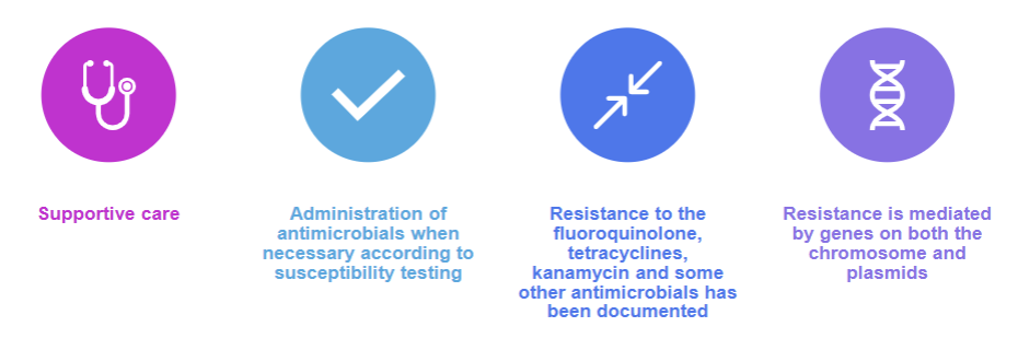
Treatment of campylobacteriosis
Lawsonia intercellularis pathogenesis

Lawsonia intracellularis clinical signs in weaned pigs(6-12 wks)
Porcine Intestinal Adenomatosis
• Chronic intermittent diarrhea with a reduction in weight gain
• Acute hemorrhagic enteropathy
Lawsonia intracellularis clinical signs in foals
Equine proliferative enteropathy (EPP)
Rapid weight loss
Diarrhea
Colic
Depression
Fever
Subcutaneous ventral edema
Lawsonia intracellularis: Treatment and Control
Antimicrobial agents such as tylosin or tiamulin may be used prophylactically or therapeutically in feed and water
Cleaning and disinfection of contaminated premises
Vaccination
Brachyspira spp.: General characteristics
Anaerobic intestinal bacteria, 6-14 spirals
Pathogenic Brachyspira spp. can be found in the intestines of both clinically affected and normal pigs
Can survive in moist feces for weeks (about 2 months) → Susceptible to desiccation
Brachyspira spp. and conditions

Brachyspira hyodysenteriae: Transmission
Fecal-oral route
Disease spreads slowly through pens
Dogs, rats, mice and flies may act as vectors
Brachyspira hyodysenteriae: Clinical signs
Weaned pigs 6 to 12 weeks old
Decreased appetite/Increased thirst
Emaciation/Loss of condition
Profuse muco-hemorrhagic diarrhea
Brachyspira hyodysenteriae: Virulence factors
Motility
Chemotactic capacities
Lipopolysaccharide
Haemolysin(s)
Enzymes such as NADH oxidase
Brachyspira hyodysenteriae: Pathogenesis

Brachyspira hyodysenteriae: Diagnosis
Darkfield microscopy on abnormal mucosa or fecal swabs
Culture: Blood agar with antibiotics incubated at 42°C for at least 3-5 days, anaerobically
Complete hemolysis around colonies
PCR: Immunofluorescence
Serological tests- ELISA
Brachyspira hyodysenteriae: Treatment and control
Improved sanitation and hygiene
Medication of drinking water → Tiamulin, lincomycin and nitroimidazoles
Depopulation, Cleaning and disinfection
Vaccination using the whole cell bacterin: Questionable efficacy
Clostridium perfringens virulence factors: α toxin
produced by all strains
acts on cell membrane & cause hemolysis/cell necrosis
Clostridium perfringens virulence factors: β toxin
produced by strains B&C
necrotizing enteritis & contraction of intestinal smooth muscle
Clostridium perfringens virulence factors: ε & ι toxin
ε (strains B&D), ι (strain E)
produced as a pro-toxin which is activated by proteolytic enzymes
Canine Hemorrhagic Gastroenteritis
Caused by overgrowth of Clostridium perfringens type A
Necrotic enteritis in chickens (rot gut) agent and transmission
C. perfringens type A and less frequently type C
fecal-oral route
Necrotic enteritis in chickens(rot gut)
Predisposing factors: Dietary, Mucosal damage, Coccidia usually occurs concurrently
Clinical signs: Sudden death
Depression, ruffled feathers, diarrhea
Hemorrhagic Enteritis in Pigs
C. perfringens type C
Distribution: Worldwide in newborn piglets
Pathogenesis: β-toxin → binds to vascular endothelial cells → vascular necrosis + necrosis of villi
Clinical signs: Sudden death
Dullness, anorexia, blood-stained feces, perianal hyperemia
Hemorrhagic Enteritis in Pigs: postmortem findings
Hallmark lesion-segmental mucosal necrosis with marked hemorrhage in the small intestines
Necrosis of the intestinal mucosa with blood-stained contents in the terminal small intestines, cecum and colon
Serosanguineous fluid in the pleural and peritoneal cavity
Pulpy kidney (Overeating disease) Thriving lambs 3 to 10 weeks of age
C. perfringens type D
Distribution: worldwide
Predisposition: Excessive consumption of high grain and low roughage diet
immunocompromised animals
GI parasites
Decreased intestinal motility
Pulpy kidney(overeating disease) pathogenesis
Gorging on high grain diet or succulent pasture
High starch content of the food is suitable for the rapid proliferation of clostridial organisms
Ingestion of excessive quantities of food leads to the entry of partially digested food from the rumen to the intestine
Production of toxins – mainly ε toxin
Pulpy kidney (Overeating disease) Clinical signs
Lambs often found dead
CNS – Dullness, opisthotonos, convulsions and terminal coma, blindness and head pressing
GI – bloating (later stages)
Hyperglycemia and glycosuria are constant features
Pulpy kidney postmortem finding
Acute disease: endocardial hemorrhage, excess pericardial fluid
Rapid kidney autolysis – pulpy (friable/soft) cortex
Focal symmetrical encephalomalacia – subacute effects of ε toxin → symmetrical hemorrhagic lesions in the basal ganglia and midbrain
Diagnosis of pulpy kidney
Clinical signs & post-mortem findings
ELISA assay for toxin
Toxin neutralization in mice
PCR for toxin producing genes
Diseases caused by various types of Clostridium perfringens

Diagnosis of C. perfringens enterotoxemias
Direct smears from the mucosa or intestinal from postmortem samples
Mouse inoculation tests
ELISA tests can be used as an alternative to in vivo assays
Culture and Isolation of C. perfringens
PCR can be used to detect toxin coding genes
Treatment and control of C. perfringens enterotoxaemias – Large animals
Hyperimmune serum
Acute nature of disease so antibiotic therapy is generally ineffective
Vaccination is the principal control method
Good animal husbandry practices
Avoid sudden dietary changes
Clostridioides difficile Habitat:
usually found as normal flora of the gut
Disease conditions associated with C. Diff
Pseudomembranous colitis
Chronic diarrhea in dogs
Hemorrhagic enterocolitis in foals without prior antimicrobial therapy
Neonatal diarrhea in pigs in the USA
Nosocomial infections in humans
Antibiotic associated: use of clindamycin, lincomycin, ampicillin, amoxicillin, cephalosporins and fluoroquinolones
Clostridioides difficile virulence factors: Toxin A (enterotoxin)
Stimulates fluid secretion
Diarrhea
C. diff virulence factors Toxin B (potent cytotoxin)
Damages enterocytes in the gut causes hemorrhage
Pathogenesis of C. diff
Antimicrobials causes a disruption in the normal flora of the intestine
Overgrowth of C. difficile from persistent spores
Toxin production and diarrhea
Clostridioides difficile overview

Pathogenesis of Johne’s disease

Paratuberculosis: Granulomatous proliferation of ileo-cecal mucosa (also in local
lymph nodes)

Diagnosis of paratuberculosis
Live animals
Observation of history and clinical signs confirmed by the demonstration of MAP in the feces by
Microscopy-scrapings or pinch biopsies from infected animals
Culture – special media required can take weeks, also rapid media with radiometric detection
If your not going to explain the rest of them idgaf
Diagnosis of paratuberculosis at necropsy
Finding of the pathognomonic lesions of the disease in the intestines, either grossly with the demonstration of typical acid-fast organisms in impression smears of the lesions or histologically
Isolation of MAP in culture
Control of Johnes
Isolate animals with clinical signs suggestive of Johne’s
Affected animals should be slaughtered promptly – shed large numbers of the organism
Bacteria can contaminate buildings and pasture
Detect and eliminate subclinical animals
Treatment of Johnes
Vaccines: Adjuvanted and live attenuated whole cell vaccines or subunit vaccines
Vaccinated animals become sensitive to tuberculin
Based on the immunodominant protein antigens – stimulate a strong Th1 response
Fungal causes of diarrhea in dogs and cats: Moldy food – mycotoxins
Consumption of contaminated food items including dairy products, walnuts, peanuts, stored grains, pasta
Rummage through garbage cans → Encountering decomposing substances
Clinical presentation of mycotoxins in dogs and cats
Early signs: Diarrhea, vomiting, agitation
Intermediate signs: Seizures, tremors, stiffness, weakness
Severe signs: Pneumonia, hypothermia, abnormal clotting
Types of Mycotoxins: Penitrem-A (tremortin)
Produced by Aspergillus, Claviceps and Penicillium
Most common type of mycotoxin frequently found in mold growing on bread, nuts, cheese and other spoiled products
Types of mycotoxins: alfatoxins
Produced by Aspergillus spp.,
Can also be a component of many types of molds
Can affect liver function
Types of mycotoxins: vomitoxin
A trichothecene mycotoxin produced by Fusarium spp. Which grows on grains such as those used to make bread or dog food
Infrequent cause of poisoning in dogs mainly caused by contaminated dog food
Types of mycotoxins: Roquefortine
A tremorgenic mycotoxin frequently found with penitrem-A