Ch 16 + 17 Anatomy- Yui Hisanaga
 Call Kai
 Learn
Learn Practice Test
 Spaced Repetition
 Match
Match Flashcards
 Knowt Play
1/9

There's no tags or description
Looks like no tags are added yet.
Name | Mastery | Learn | Test | Matching | Spaced | Call with Kai | Chat |
|---|
No analytics yet
Send a link to your students to track their progress
10 Terms
What are the 3 ways that the autonomic nervous system (ANS) differ from the somatic nervous system (SNS)?
Different effectors: the SNS effects skeletal muscle, and the ANS effects smooth, cardiac, and glands.
Different neurotransmitter effects: The SNS is always excitatory but the ANS can either be excitatory or inhibitory
Different efferent pathways: The SNS only has one motor neuron (the lower motor neuron) that is heavily myelinated, while the ANS has 2 neurons (preganglionic and postganglionic.) The post ganglionic neuron is lightly myelinated, while the postganglionic neuron is umyelinated.

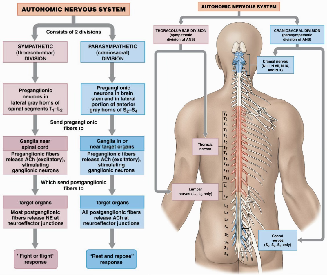
What are the two main divisions of the ANS? What roles are played by each division? What are the basic anatomical differences between the two divisions? What is the difference in what they innervate?
Sympathetic: “flight or fight” response
Has a short preganglionic neuron, meaning that it’s close to the spinal cord but has a veery long post ganglionic neuron.
Also called thoracolumbar, because it originates from thoracic and ends in the lumbar area.
“E division”: excitement, embarrassment, exercise, emergency
Innervates smooth muscle in blood vessels!!
Parasympathetic: “rest and digest” response
Has a long preganglionic neuron but has a short post ganglionic neuron.
Because it’s long, it can be very close to the target organ (terminal ganglia) or it can be inside of the actual organ (intramural ganglia)
Also called craniosacral, some of it is the top near the craniam and some of it is in the sacral area.
Vagus nerve (CNX)
“D division”: defecation, diurection, digestion
How are neurons organized within the ANS? You should know the basic organization of the various ganglia with respect to the spinal cord and the various interconnecting nerve rami.
Sympathetic:
Preganglionic neurons in the lateral gray horns
Release ACh (excitatory) close to the spinal cord
Post ganglionic fibers release NE, but epinephrine in the adrenal medulla.
Response: “Flight or fight”
Parasympathetic:
Preganglionic neurons in lateral gray horns
Release ACh (excitatory) but far from the spinal cord
Post ganglionic fibers release ACh.
Response: “Rest and digest”
What is the basic route of a preganglionic fiber in the sympathetic nervous system? What are the 3 different destinations possible for a preganglionic fiber entering the sympathetic chain ganglion? What are the routes a preganglionic fiber may take when exiting the sympathetic chain ganglion?
Basic Route: Starting at the lateral horn, it will travel out through the ventral root and then pass through the mixed nerve and finally pass through the white rami (bc the preganglionic neuron is lightly myelinated.) It’ll enter the sympathetic chain ganglion,
Different Destinations:
It can synapse with the post ganglionic neuron in the same chain ganglion. (so stays the same.)
It can descend/ascend and synapse in a different sympathetic ganglion chain
It can pass through the chain to go to the collateral ganglion (which is actually ventral)
Exit Routes:
Some exit the chain directly via sympathetic nerves
What is a collateral ganglia? What kind of neurons go there? How does it get there?
Collateral ganglia is the 3rd destination that is possible. Most fibers inferior to T4 will go to the collateral ganglia .
This creates a plexus!
Celiac
Superior Mesenteric
Inferior Mesenteric Ganglia
Go through sympathetic chain and travel through splanchnic nerves to get to the collateral ganglia.

What is unique about the sympathetic pathway to the adrenal medulla? What do chromafin cells act as?
The adrenal medulla is the exception to the the typical sympathetic pathway.
The preganglionic fibers will pass through the sympathetic chain and the collateral ganglion to synpase directly with chromafin cells in the adrenal medulla, which is responsible for creating epinephrine.
Chromaffin cells act as modified post ganglionic neurons that release epinerphrine and noepineprhine.

What cranial nerves carry preganglionic parasympathetic fibers?
CNs 3, 7, 11, 10.
But the majority of preganglionic fibers arise from vagus nerve CN X. Vagus means wander. This will send the branches to several autonomic plexuses.

How does the pattern of divergence differ between the sympathetic and parasympathetic division? What effect does this have on the types of receptors?
Sympathetic: Lots of divergence so it leads to a more amplified response, but it’s less specific so it’s more diffused.
Parasympathetic: Less divergence which leads to a more specific response
How does dual innervation work in the ANS? What are the different types of responses associated with each division?
Dual innervation means that both sympathetic and parasympathetic divisions are always at work in most organs, but sometimes one is more at play than the other.
What is vasovagal synscope? How are sympathetic and parasympathetic divisions involved in incidents of vasovagal synscope?
Vasovagal synscope is when someone faints at the sight of blood, needles, etc.
This is caused because the sympathetic division dropped and the parasympathetic went into overdrive. This caused the parasympathetic symptoms like slowed heart rate, blood pressure going down, which caused less blood flow into his brain which then caused him to faint.
As soon as the sympathetic division takes over again, someone will start to feel better.
