Complex Medical Lab/Practical
 Call Kai
 Learn
Learn Practice Test
 Spaced Repetition
 Match
Match Flashcards
 Knowt Play
1/54

Earn XP
Description and Tags
Heart/lung sounds, EKG reading, etc.
Name | Mastery | Learn | Test | Matching | Spaced | Call with Kai | Chat |
|---|
No analytics yet
Send a link to your students to track their progress
55 Terms
You are evaluating a patient with COPD, what do you expect to find when observing them sitting in a chair breathing?
barrel chest
less use of diaphragm, more use of scalenes
hypoxic drive to breathe
Normal lung sounds that are heard peripherally sound _____ while sounds closer to the trachea sound more _______
peripheral: softer
trachea: tubular
Crackling/rales lung sounds are associated with…
pneumonia
edema
fibrosis (fine crackles)
Rhonchi is another term for…
wheezing
_______ is a harsh, high-pitched breathing sound caused by a blocked or narrowed upper airway
stridor
Normal lung sounds when a person speaks sound muffled at the edges of the lungs. But with a person with __________ the sound will be loud and clear which signals _________
bronchophony
dense lung tissue
How do you confirm bronchophony in a patient? What are you expecting with a positive result?
have the patient say 99 (or another easy word) repeatedly as you listen to different points on the lung
positive result: increased sound in area of tissue consolidation
How does whispered pectoriloquy sound different in a patient with healthy lungs vs. abnormal lungs?
healthy: whisper sounds faint and unclear
abnormal: whisper sounds loud and clear
When a patient’s lung tissue is filled with fluid or solid matter instead of air, what will it sound like when they say the letter “E”? What is this called?
it will sound like an “A” in the area of consolidation
this is called egophony
You ask a patient to say the words “99” while palpating their upper back. You notice increased vibrations (more than usual) what does this mean and what is this called? What could be a cause of this?
there is solid or dense tissue in the lungs (sound travels better through solids/fluids than air)
this is called tactile fremitus
cause: pneumonia or lung tumor
_______ breath sounds are normal respiratory sounds with a medium pitch and intermediate intensity where inspiration and expiration last for equal amounts of time (1:1 ratio). Where are these breath sounds heard?
bronchovesicular
mid-chest area
posteriorly between the shoulder blades
Patient presents with the following symptoms:
chest pain when taking a deep breath or cough
abnormal sounds heard during both inspiration and expiration that are low-pitched and harsh grating
What sound is being heard and what is the likely condition?
pleurisy
pleural rub
A patient presents with pneumonia. What abnormal lung sounds do you expect to hear and why?
diminished breath sounds
this is due to lots of fluid in the lungs
the fluid is not allowing for proper gas exchange, so you won’t hear the air moving in and out
When performing a mediate percussion assessment on a patient with COPD, what would you expect and why?
dullness or higher resonance
due to barrel chest
_________ is a partial or total collapse of a lung or lobe of a lung
atelectasis
You are listening to the bases of the lungs on a patient with heart failure and here this: (listen to audio). What sound did you hear? What other conditions might this sound be associated with?
fine crackles (firewood on a campfire)
high pitched sound toward the end of inspiration
brief, short, discontinuous
won’t clear with coughing
location: bases of the lungs
conditions
CHF
atelectasis
pnemonia
pulmonary fibrosis
What is the reason for hearing fine crackles when a patient breathes (toward the end of inspiration)?
fine crackles occur as the deflated alveoli begin to pop open
when air rushes into the alveoli, it causes them to explode open or “crack” open which is why you hear that popping sound (at the end of inspiration)
What can you do for a patient who has congestive heart failure who is having trouble breathing (you hear the fine crackles upon auscultation)
Better breathing position with fowler’s position: head of the bed at 45-60 degrees
also helps to reduce venous return which helps to reduce fluid that is accumulating in their lungs
encourage coughing and deep breathing techniques
educate patient on monitoring daily weight for potential fluid accumulation
A patient presents with the following signs/symptoms:
fever: 102.2F
pain when breathing in deeply or coughing
SOB and dry cough
this lung sound (listen to audio)
What sound did you hear? What other conditions might this sound be associated with?
pleural friction rub (harsh grating noise)
can occur on inspiration and expiration
low pitch
continuous or discontinus depending on severity and cause
location: pleura layers
conditions
pleurisy
layers of pleura (parietal and visceral) are no longer gliding, but rubbing because they are inflamed which is why there is pain with breathing
pneumonia
pulmonary embolism
tuberculosis
lung cancer
With ________ the layers of the pleura are no longer gliding during breathing but are rubbing against one another due to being inflammed. This will likely cause what two things in a patient during breathing?
pleural friction rub
chest pain/pain with breathing on inspiration and expiration

You are seeing a patient in a hospital who was admitted with bronchitis. You ask the nurse to listen in to their lung sounds to get better with identifying different ones. This is what it sounds like (listen to audio). What did you hear? What other conditions might you hear this sound?
rhonchi (snoring, snorting sound)
heard on expiration
continuous
low pitch but loud
improves with coughing
this sound occurs because when air is leaving the trachea and bronchi, it hits secretions (mucus and fluid)
conditions
bronchitis
pneumonia
COPD
A _______ sound occurs in patients with bronchitis during ______ because when air is leaving the trachea and bronchi, it hits secretions such as mucus and fluid
rhonchi (wheezing)
expiration

A patient presents with the following signs/symptoms:
low-grade fever
increased respiratory rate
voice hoarseness
this lung sound (listen to audio)
What did you hear? What conditions might you hear this sound? What should you do if you hear this sound coming from a patient’s room?
stridor (screeching, squawking from throat area)
don’t even need stethoscope to hear
occurs with inspiration or expiration
high pitched sound
location: upper respiratory system
this sound occurs due to narrowing of the larynx and trachea from swelling or blockage (usually from an infection)
conditions
infection (epiglottitis)
anaphylaxis (allergic reaction)
object/obstruction
MEDICAL EMERGENCY
get nurse
A _______ sound might occur when a patient inhales and exhales due to narrowing of the larynx and trachea from swelling or a blockage
stridor
During a head to toe assessment you listen to the patient’s peripheral lung fields and hear the following sound (listen to audio)
What did you hear?
normal lung sound (vesicular lung sound)
heard at peripheral lung fields
soft, low-pitched
tends to be longer and louder on inspiration than expiration
A normal lung sound is also called a _________ sound
vesicular
A patient has SOB and a Hx of asthma. You hear the following sound upon auscultation (listen to audio)
What sound did you hear? What conditions might you hear this sound?
high-pitched wheezing (squeaky, whistling, musical sound)
mainly expiration (but can also occur in inspiration)
high pitch
continuous
location: throughout respiratory system but mainly lower
this occurs due to narrowing of the airways in the respiratory system
conditions
asthma
COPD
lung infections

A patient is receiving treatment for pneumonia. When auscultating the lungs, you hear this (listen to audio; it is hard to hear but focus on the inspiration part)
What sound did you hear? What conditions might you hear this sound?
coarse crackles (gurgling, bubbling)
start of inspiration (can extend into expiration)
low pitch
discontinuous (gurgling)
location: large airways (bronchi)
coughing doesn’t clear it
this occurs when the patient breathes through thick mucus/fluid in the larger airways
conditions
pneumonia
bronchiectasis
infections
heart failure due to pulmonary edema
You may hear __________ in a patient with CHF, pneumonia, or bronchiectasis during auscultation due to them breathing through thick mucus/fluid in the bronchi
course crackles
On an EKG, every big box is equal to _____ seconds
0.2
When reading an EKG, what are the 6 things you want to look at?
regularity
is the rhythm regular or irregular
look at R-R interval; top of one QRS to the next
equal # of boxes before every R-R segment = regular rhythm
rate
P wave before every QRS?
QRS after every P wave?
if no this means there is a conduction issue btw atria and ventricles
PR interval
< 0.20 seconds (1 big box) = conduction delay or heart block
QRS complex
duration/shape
normal: 0.1 sec (2-3 little boxes)
How do you calculate heart rate with a regular rhythm?
find one QRS
count ahead to the next QRS
every line goes like this: 300, 150, 100, 75, 60, 50

How do you calculate heart rate with an irregular rhythm?
count the number of R waves in 6 sec strip and multiple by 10
this is about 30 boxes
_______ is a HR under 60bpm w/ normal rhythm; when is this rhythm type an issue?
sinus bradycardia
an issue if cardiac output is low
determine if CO is low through checking BP!
HR 60 + hypooo tensive = issue!
______ is a HR above 100bpm w/ normal rhythm
sinus tachycardia
True or false: ALL sinus rhythms have normal P waves, PR interval (0.2 sec) and QRS duration (0.1 sec)
true

What is the following arrhythmia? How do you know?
Supraventricular tachycardia: SVT
high rate: 150-250
no P wave
QRS < 0.12 sec

What is the following arrhythmia? How do you know?
Atrial flutter
saw tooth P wave
high rate: 250-450

What is the following arrhythmia? How do you know?
Atrial fibrillation
irregular rhythm
P waves chaotic

What is the following arrhythmia? How do you know?
Premature ventricular contraction
no P waves
wide, early, random QRS
could be 1 for every beat (bigeminy) or 1 every third beat (trigeminy)

What is the following arrhythmia? How do you know?
ventricular tachycardia
rate > 100 bpm
3 or more PVC

What is the following arrhythmia? How do you know?
Torsades de pointes
prolonged QT interval

What is the following arrhythmia? How do you know?
Ventricular fibrillation
no organized rhythm

What type of heart block is this? How do you know?
1st degree
PR > 0.2 seconds

What type of heart block is this? How do you know?
Mobitz Type I
PR interval progressively gets longer
then QRS drops

What type of heart block is this? How do you know?
Mobitz Type II
regular dropped QRS every 2nd, 3rd, or 4th P wave

What type of heart block is this? How do you know?
Third degree heart block
Ps can be hidden in QRS
PVC
QRS > 1 sec

What is the following condition? How do you know?
ST segment elevation
likely due to MI (myocardial infarction)
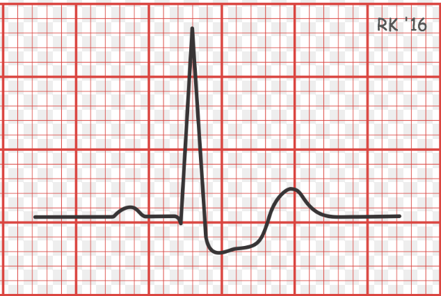
What is the following condition?
ST segment depression
myocardial ischemia

What is the following condition?
myocardial ischemia
inverted T waves
could also indicate old MI
systolic heart failure will typically be associated with which heart sound?
S3
diastolic heart failure will typically be associated with which heart sound?
S4
What are some breathing interventions for cardiopulm patients?
active cycle of breathing
normal 5-10 sec, deep inspr/relaxed expir 3-4 times, normal 5-10 sec, deep 3-4 times, forced expiratory to cough
pursed lip breathing
smell the flowers, blow out the birthday candles
What are some cough assistance interventions for cardiopulm patients?
butterfly technique
hands behind head
inhalation: elbows back, patient looks up
exhalation: elbows together, patient looks down
3x, then on 4th patient coughs during exhalation
Explain how you would instruct a patient to use an incentive spirometer
Purpose: the purpose of this intervention is to help increase lung volume and capacity
steps:
take a full deep breath through the mouth piece and keep the ball between the markers
repeat 10x every hour; repetitions don’t need to be back to back
if patient is unable to keep ball between marked numbers, see what the patient can do on the first attempt and then slowly progress