Anatomy and Physiology I - Practical #3
 Call Kai
 Learn
 Practice Test
 Spaced Repetition
 Match
 Flashcards
 Knowt Play
1/68

Earn XP
Description and Tags
Articulations & Body Movements, Microscopic Anatomy of Skeletal Muscle, Gross Anatomy of Skeletal Muscle. The terms in this flashcard set were those that were highlighted in the lab powerpoint. (WORK IN PROGRESS)
69 Terms
fibrous joints
generally don't move
cartilaginous joints
allow some movement

ligament - bone to bone connection

Synovial membrane - membrane lining the capsule of a joint

articular capsule - connective tissue structure that encloses the joint cavity of a synovial joint
pubic symphysis
cartilaginous joint at which two pubic bones fuse together
cranial sutures
fibrous joints that join the skull bones
synovial joints
freely movable joints

pivot joint - rotating bone turns around an axis; i.e. connection between radius/ulna and humerus

hinge joint

plane joint - short slipping or gliding movements; i.e. carpals

ball and socket joint
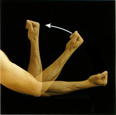
flexion - decreases the angle of a joint
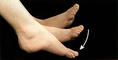
plantar flexion - bends the foot downward at the ankle

eversion - turning the sole of the foot outward

supination - Palm up, radius/ulna parallel

5
Dark A bands

z disc - Separates the sarcomeres from each other

transverse tubule

Terminal cisternae of sarcoplasmic reticulum - Structures within skeletal muscle cells that serve as reservoirs of calcium ions

7
Thick (myosin) filaments

sarcomere - Contractile unit of muscle
What are the boundaries of a sarcomere?
z disc to z disc
What are the two contractile proteins?
myosin (thick filament) and actin (thin filament)
What are the two regulatory proteins?
tropomyosin and troponin

epimysium - a sheath of fibrous elastic tissue surrounding a muscle.

tendon - connects muscle to bone

Perimysium - connective tissue surrounding a fascicle

What is a muscle fiber?
muscle cell, bundle of myofibrils

What is a myofibril?
Many of these units make up a single muscle fiber. These are made up of sarcomeres.

what is the structure labeled g.? (the structure resembling a sausage)
motor neuron

Endomysium - Connective tissue surrounding a muscle fiber

synaptic bulb - located at the end of the axon terminal, this releases neurotransmitters.

synaptic cleft - The narrow gap that separates the presynaptic neuron from the postsynaptic cell.

synaptic vesicles contain
neurotransmitter acetylcholine (ACh)

galea aponeurotica.
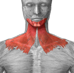
Platysma - tenses skin of neck

frontalis

orbicularis oculi

levator labil superioris

zygomaticus minor

epicranial aponeurosis (same as galea)

Sternocleidomastoid

buccinator

temporalis

Masseter (origin, insertion, action)
Origin: zygomatic arch, maxilla
Insertion: mandible
Action: mastication

Mylohyoid

pterygoid

Digastric

sternohyoid

sternothyroid

levator scapulae

internal intercostals

Aponerosis of External Oblique

rectus abdominis

internal oblique

transversus abdominis

Supraspinatus

Infraspinatus

Subscapularis

Teres minor

pectoralis major, action: Arm adduction, flexion, rotation
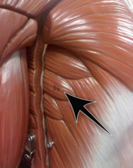
serratus anterior

rhomboideus major

levator scapulae

splenius capitis
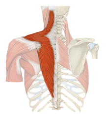
trapezius

latissimus dorsi

Deltoid (origin, insertion, action)
Origin: clavicle, scapula
Insertion: humerus - deltoid tuberosity
Action: abduction of arm