Digestive System
 Call Kai
 Learn
 Practice Test
 Spaced Repetition
 Match
 Flashcards
 Knowt Play
1/106

There's no tags or description
Looks like no tags are added yet.
107 Terms
Peritoneum
Lining of the abdomen
Parietal peritoneum
Lines abdominal walls
Visceral peritoneum
Covers organs
Intraperitoneal space
Only small amount of fluid; no organs in this space
Connecting peritoneum
Mesentery and omentum are examples of…
Mesentery
Peritoneum (sheets of connective tissue) that suspends the digestive tube in the abdomen from the dorsal body wall; contains blood, lymph, and nerves; delivers nutrients to intestines
Omentum
Peritoneum that attaches stomach to adjacent organs
Greater omentum
Peritoneum from greater curvature of the stomach to the dorsal body wall
Lesser omentum
Peritoneum from lesser curvature of stomach to liver and duodenum
Digestive system
Takes complex foods and breaks them down into simple nutrient molecules through the digestion process; absorbed into bloodstream; tube that runs from mouth to anus (with accessory organs for digestion and absorption)
Hindgut fermenters
Monogastric herbivores. Simple stomach with the ability to undergo microbial fermentation within the cecum.
Ruminants
Have mixing and fermentation compartments in addition to the stomach; have 3 forestomachs and one true stomach (abomasum)
Mouth, esophagus, stomach, small intestine, large intestine, anus
Digestive tract basic flow in order
Skeletal
Mouth, prehension, pharynx, cranial portion of the esophagus, external anal sphincter are examples of this muscle in the digestive system
Smooth
The wall of the digestive tube is an example of this muscle in the digestive system
Peristatic contractions
Smooth muscle contraction type that moves contents along the digestive tract

Segmental contractions
Smooth muscle contraction type that causes back-and-forth mixing movement; aids in digestion and absorption by mixing the digestive tract contents and slowing their movement through the tract

Oral (buccal) cavity
Bodily cavity where digestion starts; includes lips, tongue, teeth, salivary glands, hard palate, soft palate, and oropharynx
Lips (labia)
Highly sensitive organ; significant sense organ in horses, cattle, and other species; prehension
Parotid
Pair of salivary glands that is located ventral to the ear canals
Mandibular
Pair of salivary glands that is located central to the parotid
Sublingual
Pair of salivary glands that is located medial to the shafts of the mandible just under the base of the tongue
Salivary glands
Glands in animals that have ducts that carry saliva to the oral cavity
Teeth
Physically break down food into smaller pieces (mastication)
Maxilla and incisive bones
The upper arcade of the teeth contains the…
Mandible
The lower arcade of the teeth contains the…
Pointed, blunted
Carnivores have ______ teeth, while herbivores have _____ teeth
Occlusal surface
Surface where teeth come together
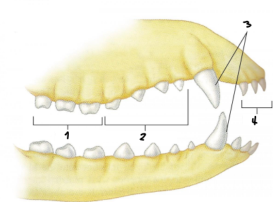
Incisors
Most rostal type of teeth; grasping teeth (4)

Canines
Type of teeth located in the corners of incisors; tearing teeth; tusks (3)

Premolars
Cheek teeth or cutting teeth (2)

Molars
Grinding teeth (1)
100
How are the teeth numbered in the right upper arcade?
200
How are teeth numbered in the left upper arcade?
400
How are teeth numbered in the right lower arcade?
300
How are teeth numbered in the left lower arcade?
Dental pad
Strengthened gum covering the incisive bone replacing the upper incisors in ruminants
Lingual Surface
“Inner surface” of the lower arcade that faces the tongue (3)

Palatal surface
“Inner surface” of the upper arcade that faces the hard palate (1)

Labial surface
“Outer surface” that faces the front of the mouth (4)
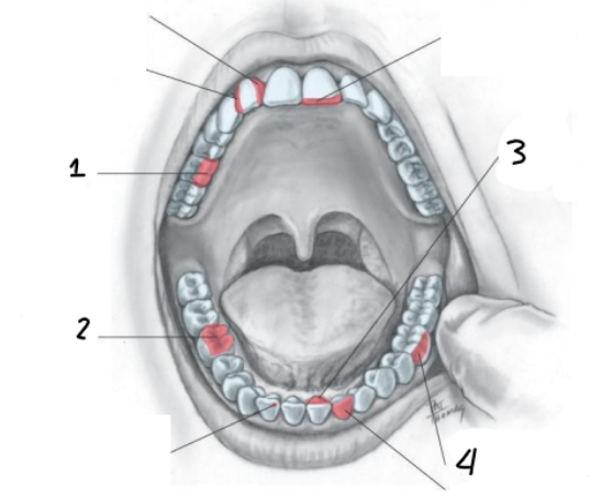
Buccal surface
“Outer surface” of both arcades that face the cheek (4)

R 3/3, L 3/3
Cat and dog incisor (upper/lower) dental formula
R 3/3, L 3/3
Horse incisor (upper/lower) dental formula
R 0/3, L 0/3
Cow incisor (upper/lower) dental formula
R 1/1, L 1/1
Cat and dog canine (upper/lower) dental formula
R 1/1, L 1/1
Horse canine (upper/lower) dental formula
R 0/1, L 0/1
Cow canine (upper/lower) dental formula
R 4/4, L 4/4
Dog premolar (upper/lower) dental formula
R 3/2, L 3/2
Cat premolar (upper/lower) dental formula
R 3/3, L 3/3
Cow premolar (upper/lower) dental formula
R 3-4/3, L 3-4/3
Horse premolar (upper/lower) dental formula
R 2/3, L 2/3
Dog molar (upper/lower) dental formula
R 1/1, L 1/1
Cat molar (upper/lower) dental formula
R 3/3, L 3/3
Cow and horse molar (upper/lower) dental formula
42
Dogs number of total teeth
30
Cats number of total teeth
32
Cow number of total teeth
40-42
Horses number of total teeth
Living
Teeth are (living/nonliving) structures
Crown
Portion of the tooth that projects above the gingiva (gums)
Root
Portion of the tooth embedded in the bone
Apex
Tip of the tooth root where blood vessels and nerves enter the tooth
Enamel
A thin layer of a white hard material that covers the crown of the tooth; hardest substance in the body
Dentin
Substance that makes majority of the tooth; surrounds the pulp cavity containing nerve and blood supply
Hypsodont
Pattern of dentition characterized by the continual growth of teeth; large amounts of crown under the gum line; examples of animals with this are horses, rabbits, and rats
Brachydont
Pattern of dentition characterized by the lack of continual growth of teeth; occludes animals with tusks; examples of organisms with this are humans, dogs, and cats
Esophagus
Muscular tube that extends from the pharynx to the stomach; enters the stomach at the cardia
Cardiac sphincter
Weak circle of smooth muscle that closes off an opening; stomach expands; fold of stomach against the esophagus acts as a natural valve
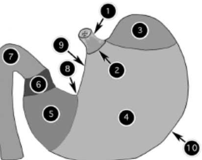
Cardia, fundus, body, pylorus
Portions of the stomach in order
Cardia (cardiac)
Portion of the monogastric stomach that blends with the esophagus, small in size; contains a sphincter (2)
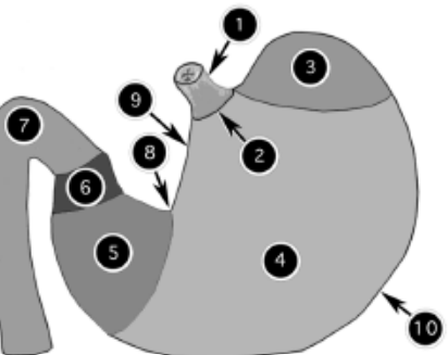
Fundus
Portion of the monogastric stomach that is located to the left and dorsal to the cardiac part (3)

Body
Portion of the monogastric stomach that is the large, central area between fundus and pyloric regions (4)
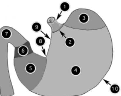
Pylorus
Portion of the monogastric stomach that joins body to the descending duodenum (5)

Pyloric sphincter
6
Ruggae
Multiple, long folds, curvatures in the stomach
Gastric Dilation and Volvulus (GDV)
Condition commonly associated with large meals and causes the stomach to dilate, due to food and gas; stomach can become dilated enough to rotate
Gastropexy
Treatment of GDV; surgical procedure that tacks a stomach to the right side of the inner abdominal wall
Forestomachs
Compartments that food pass through in rumanants before reaching the abomasum
Swallow, regurgitation, swallow, digest
Rumination in order simplified
Rumen
Large fermentative vat; processes plant material into useable energy; series of muscular sacs separated by pillars
Pilars
Long muscular folds or ruminal wall
Reticulum
Small and most cranial compartment; seperated from the rumen by the rumenoreticular fold; honeycomb appearance; surface area increases absorptive surface; portion of rumen
Omasum
When rumenoreticular contractions occur, they move ingesta here; muscular organ; breakdown food particles further; portion of rumen
Hindgut Fermenters
Stomach type of monogastric herbivores; have very large cecum
Small intestine
Site of majority of nutrient absorption into the bloodstream; divided cranial to caudal into duodenum, jejunum, ileum; boundaries cannot be seen grossly; always functioning
Cranial duodenal flexure, descending duodenum, caudal duodenal flexure, ascending duodenum
Portions of the duodenum (cranial to caudal)
Large intestine
Portion of the intestine containing the cecum, colon, and rectum; varies depending on species and diet Involved in water and electrolyte absorption Hind gut fermentation : rats, guinea pigs, horses Stores feces until defecation
Ileocecocolic junction, ascending colon, transverse colon, descending colon
Portions of the colon in order
Mouth, esophagus, stomach, duodenum, jejunum, ileum, ileocecocolic junction, cecum, colon, rectum, anus
Flow of ingesta in order
Mouth, esophagus, stomach, small intestine, cecum, large colon, small colon, rectum
Flow of equine ingesta
Rectum and anus
Portion of the digestive system that marks the end of ingesta; site of feces elimination
Rectum
Terminal end of the large intestine; has mucus secreting glands to lubricate and ease passage; can dilate (and store); contains sensory receptors that respond to expansion
Anus
Composed of internal and external muscular sphincters
Internal anal sphincter
Portion of the anus that is involuntary; responds to stretch receptors; opens to allow passage of feces into the anal canal to stimulate defecation
External anal sphincter
Portion of the anus that is voluntary; opens to complete the elimination process
Anal glands
Musk gland (powerful, foul smelling secretion); located at 5’ and 7’ o’clock positions; connected to the lateral margin of the anus by a small, single duct; expressed when animal defecates or becomes frightened; functions to mark territory and attract mates; can become blocked
Liver
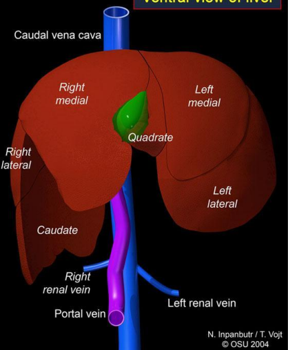
Organ that produces bile acids which aid in fat digestion; filters materials absorbed from GI tract before systematic circulation); largest gland in the body; divided into several hepative lobes (species dependant); hepatic lobes divided into microscopic hepatic lobules
Right lateral, right medial, caudate (next to right lateral lobe), left lateral, left medial, quadrate (between gallbladder and left medial)
Hepatic lobes
Monitors blood content
In the hepatic portal system, the liver…
Small intestine, hepatic portal vein, liver, hepatic vein
Hepatic portal system in order once products of digestion (nutrients) are absorbed