NU 236 Care of the Adult II: Genitourinary
 Call Kai
 Learn
 Practice Test
 Spaced Repetition
 Match
 Flashcards
 Knowt Play
1/102

There's no tags or description
Looks like no tags are added yet.
103 Terms
Micturation
urination
Dysuria
painful or difficult urination
Frequency
need to void often
Hesitancy
difficulty starting urine flow
Urgency
sudden intense feeling to void
Nocturia
awakened from sleep to void
Anuria
output < 100 cc/day
Oliguria
decreased urine output (100-400cc/day)
Polyuria
excessive urination (>2000 ml/day)
Azotemia
increased BUN, creatinine in the blood; indicative of renal impairment
Uremia
presence of urinary waste in the blood; sign of renal failure

Adrenal Glands
a pair of endocrine glands that sit just above the kidneys
Urinary Changes with Aging
Decreased blood flow to kidneys
Decreased filtration
Decreased bladder capacity
Weakened sphincter muscles
Shortened urethra (women)
If a patient is making
Kidneys not making urine
Bladder is not letting urine out
Nursing Interventions: Elderly
Encourage urination 2-4 hours
Observe for atypical UTI signs (confusion)
Nocturia
Provide privacy, help, and voiding stimulants
___________ is a safety issue in the elderly.
nocturia
New onset confusion in an elder, think?
UTI
Diagnostics: Labs
Creatinine
BUN
Glomerular filtration rate
Serum Creatinine
blood test measuring the end product of protein/muscle metabolism; increase indicates renal disease, as it only increases when 50% of renal function is impaired
Blood Urea Nitrogen (BUN)
blood test measuring the kidney's ability to excrete nitrogen, a byproduct of liver protein metabolism;
abnormal is not specific to kidney disease
Urinalysis
the examination of urine to determine the presence of abnormal elements
Obtaining a Urinalysis
Instruct pt. to wipe 3x, front to back
Start void, discard first few seconds, then obtain mid-void sample
Also can be obtained by catheterization
Urine Culture/Sensitivity
used to identify causative organisms and antibiotics in suspected infections; can take 24-48 hr
Obtaining a Urine C&S
Must be mid-stream, clean catch, or catheter and should be done BEFORE antibiotics are given
Clean Catch
a method of obtaining a urine specimen using a special cleansing technique; also called midstream urine
How are catheter specimens collected?
sterile aspiration from the catheter PORT, not collection bag
24 Hour Urine
all urine within the designated time frame is collected to measure a variety of things (creatinine clearance, nitrogen, sodium, etc.) to rule in or out various disease
Collecting 24 Hour Urine
At the start time, have patient void and discard then collect ALL urine and keep on ice
Put up signs in BR to remind staff
At 24 hr point, patient can void or empty catheter to add that urine to specimen and send it labeled to the lab

Ultrasonography
ultrasound uses sound waves to identify kidney structures; no contrast and is not painful
Bladder Scan
non-invasive bedside test the RN can perform with/without order at bedside; helpful for checking post-void residual
Kidney, Ureter, and Bladder X-Ray (KUB)
measures size of organs and can identify obvious stones, stictures, obstructions; check pregnancy status

Intravenous Pyelography (IVP)
series of x-rays that give information about the kidney size, shape, and rate of excretion with radiopaque dye injected IV
Nursing Interventions: IVP
Bowel prep and NPO midnight
Assess kidney function (Nephrotoxic) and allergy to contrast
Education
Contraindications: IVP
Pregnancy
Contrast allergy
Recent renal surgery or severe renal impairment
What must be considered with any procedure/test using contrast media?
History of renal insufficiency
Metformin in the last 48 hrs; must be d/c due to risk of lactic acidosis
Hydration status
NPO

Renal Arteriography
invasive procedure that assesses renal blood flow by injecting contrast into the renal arteries
Interventions: Renal Arteriography
SAME for all contrast procedures/bowel prep.
Assess for bleeding and check site often
Strict bedrest for 6 hrs to prevent bleeding

Renal Biopsy
invasive procedure that requires a permit and can be closed or open to check for kidney cancer
Interventions: Renal Biopsy
Patient is at major risk for bleeding!
Monitor bleeding, VS, urine output, H&H, shock symptoms
Strict bedrest for 6 hours after
Monitor urine character; hematuria is okay but NO clots
No strenous activity for 1-2 weeks
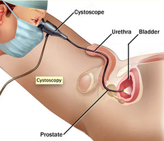
Cystoscopy
invasive or operative procedure that requirres consent; scope is inserted through the urethra and is used to diagnose urethral/bladder trauma, tumors, stones, or enlarged prostate OR treat these and can be done OR/office
Nursing Interventions: Cystoscopy
Monitor airway, VS, urine output, bleeding, and signs of infecton
May have catheter after, monitor urine characteristics (should be pink)
Encourage fluids to clear urine
Urinary Tract Infection
infection of one or more organs of the urinary tract
Upper urinary tract infection
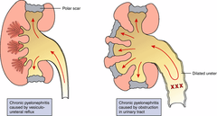
pyelonephritis
Lower urinary tract infections
Cystitis
Urethritis
Prostatitis
Bladder Bundle
Make catheter policies and protocols and assess and document the NEED for catheters
When should Foleys be used?
Operative ONLY as necessary
Urine output monitoring in the critically ill
Acute retention/obstruction
Assistance in pressure ulcer healing for incontinence
At patient request, to improve comfort at EOL
Interventions: Foley Catheters
Remove ASAP (preferably 24 hrs)
Foley care
Keep bag below bed
Empty bag and monitor/document output
Assess need for catheter daily
Suprapubic Catheter
indwelling catheter inserted directly in the bladder through an abdominal incision above the pubic bone; used in patients requiring long-term catheterization and should be washed with soap and water daily and changed 1x/month
Cystitis
inflammation of the bladder that can be infectious or non-infectious (chemicals/radiation)
Causes: Cystitis
E-coli
Foleys!
Assessment: Cystitis
Frequency
Urgency
Dysuria
Backache
Incontinence
Fullness
Urosepsis
spread of infection from the urinary tract to the bloodstream that results in a systemic infection; elderly are at HIGH risk with 15% fatality and can present with fever, tachycardia, and confusion
Labs: Cystitis
Urinalysis for bacteria/WBC/RBC
Urine culture/sensitivity for bacterial type
Nursing Interventions: Bladder Hygiene
Encourage 8-10 glasses of fluid a day
Wipe front to back
Good glucose control
Vitamin C/cranberry juice to acidify
Voiding after intercourse
Cotton underwear
Avoid douches, bubble baths, powder, or sprays
Nursing Interventions: Cystitis
Manage pain and treat infection
Regular bladder hygiene
> 3 infections/year = further workup for an immune or structural cause
Medications: Cystitis
Abx after culture
Anesthetic (pyridum) makes urine red
Anticholinergics (antispasmotics)
Pyelonephritis
bacterial infection of the upper urinary tract that often requires hospitalization
Assessment: Pyelonephritis
High risk patients
Fever
Chills
Tachycardia
Flank pain
CVA tenderness
N/V
Malaise
Urinalysis for WBC/bacteria
KUB or cystogram
Interventions: Pyelonephritis
Manage infection, pain, and fever
Analgesics and antibiotics
Encourage fluid
Surgery for structural causes
Monitor for renal failure
Hydronephrosis
the dilation (swelling) of one or both kidneys

Urolithiasis
kidney stones; cause not entirely understood
Assessment: Urolithiasis
Assess for renal colic
May have nausea/vomiting, sweating
Hematuria
Dysuria
Fever
KUB, IVP, or CT
Risks: Urolithiasis
Infection
Urinary stasis/retention
Dehydration
Increased uric acid
Family history
Renal Colic
excruciating spasmodic pain caused by kidney stones
Nursing Interventions: Urolithiasis
Manage pain and risk for infection
Strain all urine
Narcotics IVP for quick relief
IV fluids
If unable to pass a kidney stone, what can be done?
Extracorporeal Shock Wave Lithotripsy
Ureteroscopy/stent
Percutaneous nephrostoureterolithotomy
Open surgery if large
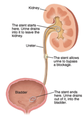
Ureteral Stent
insertion of a tube into the ureter to prevent/treat obstruction of urine flow; used for 3-7 days and fever should be reported, patient can remove if MD says string is showing

Nephrostomy Tube
a tube inserted through the abdominal wall into the renal pelvis to drain urine; change sterile dressing every 2-3 days or when wet, patient can shower if they wrap but NO baths
Renal Cell Carcinoma
cancer of the kidneys
Causes: Renal Cell Carcinoma
tobacco/chemical exposure
Assessment: Renal Cell Carcinoma
Usually asymptomatic
Dull, aching flank pain
Hematuria
Fever
Anemia
Hypercalcemia
Diagnosed with CT scan
Treatment: Renal Cell Carcinoma
nephrectomy

Nephrectomy
surgical removal of a kidney that can be radical or laparascopic
Risks: Nephrectomy
BLEEDING
Adrenal insufficiency
Respiratory complications
Nursing Interventions: Nephrectomy
Check for bleeding UNDER patient
Check for BP drop indicating bleeding
Check for abdominal distention
Watch for adrenal insufficiency and treat with IV fluid/blood if needed
Vitals often
Vigilant turn, cough, deep, breathe due to high risk for pneumonia
Adrenal Insufficiency
condition in which the adrenal glands underproduce necessary hormones; indicated by large urinary output followed by oliguria/hypotension

Testicular Cancer
rare cancer common among age 15-35 with often unknown cause, but can be undescended testes, family hx, or caucasian
Assessment: Testicular Cancer
Assess for unilateral lump or swelling
Patient should do TSE monthly
Treatment: Testicular Cancer
very treatable with orchiectomy; discuss sperm storage and or prosthesis
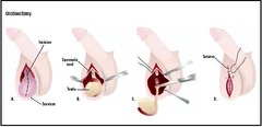
Orchiectomy
removal of one or both of the testes
Bladder Cancer
cancer of the bladder that is caused by smoking
Assessment: Bladder Cancer
Painless, intermittent hematuria with clots
Diagnosed with cystoscopy or CT for spread
Treatment: Bladder Cancer
Instill toxic substances into bladder (non-surgical)
TURBT (Partial cystectomy)
Complete cystectomy

TURBT
transurethral resection of bladder tumor
Complete cystectomy
removal of the entire bladder or surrounding muscle/tissue

Ileal conduit (Urostomy)
use of a small piece of small bowel to convey urine to the ureters and to a stoma in the abdomen; requires a ostomy bag for life to drain urine/mucus and is used for incontinent patients

Continent Internal Ileal Reservoir
small dimple in skin opens to. a bladder made of bowel; patient needs to self-cath intermittently for the rest of their life
Interventions: Ileal Conduit
Inspect stoma
Change flange/pouch every 3-5 days
Attach to large drainage bag at night
Benign Prostatic Hypertrophy
enlargement of the prostate gland that is very common in older men
Assessment: BPH
Weak stream
Distention
Frequency
Urgency
Nocturia
Retention
Treatment: BPH
Drug therapy
Frequent ejaculation
Avoid alcohol, diuretics, caffeine, fluid before bed
Avoid anticholinergics, antihistamines, and decongestants
Transurethral resection of the prostate
Trans-Urethral Resection of the Prostate (TURP)
an instrument is inserted up the urethra to remove the section of the prostate that is blocking urine flow
Interventions: TURP
May have CBI to keep catheter from clotting with a 3-way foley and 3000mL NS bags
Assess color of drainage to determine drip rate
Dark red or clots = speed up
Pink tinged/clear = slow down
No output = shut off and hand irrigate to clear clots
Encourage 2000mL/day
Must void independently to go home

Continous Bladder Irrigation
flushing the bladder with normal saline to prevent clot formation; foley output - irrigation = true urine output
Bladder Spasms
contractions, possibly painful, of the musculature of the urinary bladder
Prostate Cancer
common, slow growing cancer of the prostate gland that can spread; risks are age, family hx, and African American
Screening: Prostate Cancer
After age 50, DRE/PSA
Begin screening at 40 for African Americans
Digital Rectal Exam (DRE)
insertion of a finger into the male rectum to palpate the rectum and prostate
Prostate Specific Antigen (PSA)
blood test to screen for prostate cancer; > 4 = BPH/cancer
Prostate Cancer vs. BPH
BPH is benign and occurs in > 50% males
Prostate cancer is hard/irregular on a DRE, while BPH is boggy/uniform on DRE
Prostate cancer occurs in the periphery of the gland while BPH is in the center