Cognitive neuroscience: Perception and visual Recognition
 Call Kai
 Learn
Learn Practice Test
 Spaced Repetition
 Match
Match Flashcards
 Knowt Play
1/62

There's no tags or description
Looks like no tags are added yet.
Name | Mastery | Learn | Test | Matching | Spaced | Call with Kai | Chat |
|---|
No analytics yet
Send a link to your students to track their progress
63 Terms
primary pathway
retina → optic nerve → optic chiasm → optic tract → LGN (in thalamus) → optic radiations → V1 (primary visual/striate cortex)

visual field
left visual field is processed by the right hemisphere. and vice versa
each LGN receives input from both eyes but represents the contralateral visual field
circle shaped
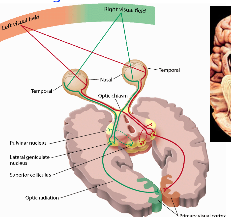
optic chiasm
the crossing of the nasal half fibers

LGN (lateral geniculate nucleus)
a 6 layered thalamic structure between retina and V1. It receives feedback from cortex and other modulatory inputs
What layers of LGN receive input from the contralateral eye
contralateral - opposite eye
layers 1, 4, 6

What layers receive input from the ipsilateral eye?
ipsilateral - same side
Layers 2, 3, 5

Magnocellular (M)
in layers 1-2
likes motion, rapid temporal canges, coarse spatial information
Parvocellular (P)
layers 3-6
likes fine detail and color
Fovea has many of these
smaller in size than magnocells
Koniocellular (K)
thin layers between the six main layers
Optic radiations
bundle of white matter axons connecting the LGN to the primary visual cortex
Meyer’s loop
travels through the temporal lobe and carries upper-visual-field information
the parietal toute carries loser-visual-field information

Lesion in the optic nerve causes
vision loss in one eye
Lesion in the optic chiasm causes
bitemporal hemianopia - loss of outer halves of both visual fields
lesion to the optic tract causes
contralateral homonymous hemianopia - damage to opposite side of brain, loss of vision in the same half of the visual field for both eyes
Lesion to the temporal optic radiations/Meyer’s loop
contralateral upper quadrantanopia - pie in the sky
Lesion to the parietal optic radiations
contralateral lower quadrantanopia
Lesion to V1
contralateral visual-field loss, extent depends on lesion
receptive field
region of space in which a stimulus changes a neuron’s response
V1
processes early features: orientation, contrast, spatial frequency, and location
retinotopic mapping
nearby locations in the visual field map to nearby locations in visual cortex
fovea and nearby central vision receive a disproportionately large V1 representation (cortical magnification)
fMRI mapping expanding ring stimulus and rotating wedge stimulus
outward ring mapping and angular position around fixation. What is on the retina corresponds with activity of fMRI mapping
orientation columns
neighboring neurons prefer similar orientations

ocular dominance columns
groups of neurons preferential to one eye
eye inputs remain separate in parts of layer 4 and combine in other cortical layers

hypercolumn
block of neurons in V1 that processes orientation, color, and both eye inputs for a small region of the visual field
not all neurons in a hypercolumn encode the same features

blobs
neurons that are color selective, not orientation
extrastriate cortex
region of the occipital cortex directly outside of striate cortex

Dorsal stream
where - spatial properties, motion-guided action (grasping for things)
projects toward parietal cortex
ventral stream
what - object identity and recognition
projects toward temporal cortex
supports recognition of objects, faces, and categories
MT/V5
Area V5 (also known as MT or the middle temporal area ) is a region of the extrastriate visual cortex specialized in visual motion perception (direction, speed, and depth)
Gets information from V1
Lateral Occipital Complex (LOC)
object shape and form, responses to complex objects
FFA (fusiform face areas)
face processing
PPA (Parahippocampal Place Area)
activates strongly to locations and buildings
receptive fields tend to become ___ larger in later stages of visual processing
larger
ventral areas emphasize
foveal vision and recognition-related features
Avoid assuming every neuron in a stream has identical properties
dorsal areas often emphasize
spatial properties relevant to action
Avoid assuming every neuron in a stream has identical properties
apperceptive agnosia
difficulty building an accurate visual representation of a form
ex: difficulty coping or matching an object
involved perceptual-form processing
associative agnosia
difficulty connecting a visual representation to a name or explain what the object does
ex: can copy an object but cannot identify it visually
involves recognition/association
prosopagnosia
difficulty recognizing familiar faces
optic ataxia
difficulty using vision to guide action/motion
ex: can see an object, but difficulty reaching for it
visual agnosia
impaired access to object identity/knowledge despite preserved basic vision
Tong et al. (1998) findings
during binocular rivalry between face and house images, fMRI activity in FFA and PPA tracked the participant’s reported percept more than the unchanging retinal input
supports a relationship between neural activity and conscious visual perception
Gauthier et al. (2000)
the FFA responded to trained visual categories and expertise effects
supporting the idea that experience can influence fusiform responses
study did not prove that FFA processes only faces
Chang and Tsao
monkey face-patch neurons represented dimensions of face appearance
findings support a population-based code rather than requiring a “grandma neuron” for every individual face
motion aftereffect
prolonged viewing of motion in one direction can make stationary objects move in the opposite direction
imaging work found MT activity related to the aftereffect, consistent with MT’s role in motion perception
blindsight
some people with V1 damage respond to visual stimuli without visual awareness
upper-left visual field loss poiniwer to
the right hemisphere, below the calcarine sulcus
lower-left visual-field loss points to
the right hemisphere, above the calcarine sulcus
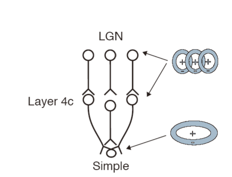
LGN neurons receptive field are combined in the simple cell in V1. LGN neurons project to simple

simple neurons activation timing of receptive field gives complex cells motion
quadranopsia
quadrant cannot be seen
hemianopsia
low vision of half visual field
scotoma
small part of visual field is lost
What causes Motion after effect?
perceptive system is designed to find changes
the illusion shows the adaptation
neurons and their preferences calibrate during an experience
[ Neurons preferring LEFT ] 🔄 Balanced Baseline 🔄 [ Neurons preferring RIGHT ] → Stare at continuous LEFT motion → [ Neurons preferring LEFT ] 📉 Become Fatigued 📉 [ Neurons preferring RIGHT ] → Look at a STATIONARY object → [ Neurons preferring LEFT ] (Reduced Output) < [ Neurons preferring RIGHT ] (Normal Baseline) → 🧠 Brain perceives RIGHT motion!
lesion to the parietal results in
struggling spatial task (are they in the same location)
fine in object task
lesion in temporal results in
struggling in object task (are they the same 3 objects)
fine in spatial task
ventral stream
receptive fields get larger as you move from V1 cortex to TE
receptive fields include more of foveal region than dorsal
neurons become sensitive to more complex objects further along stream
some neurons in TE response selectively to faces
some neurons in TE are invariant to changes in position, orientation, size, etx
mechanism of object recognition
lines and colors represented by V1 neurons are assembled by higher visual areas into recognizable objects
reccognizing objects is based on testing hypothesis. Looping in the brain. High level requires multiple brain areas to understand what you’re looking at
binding problem
how are the images put back together
How do you decide what is a part of the same object
binding criteria
nearness of lines
nearness of color
coherent motion
experience
hierarchical coding of an object
features (sides) → conjunction of features (corners) → component shapes → gnostic cell (the cell for recognition)
prosopagnosia
impairs a person's ability to recognize familiar faces, despite having normal vision
superior temporal sulcus (STS)
sees where people are looking and looks for biological motion
dorsal stream
receptive fields get larger as you move from V1 cortex to PPC
receptive fields include less of foveal region than ventral
neurons are sensitive to visual motion of a particular direction
neurons in dorsal stream are used to guide motor behaior
some neurons in PPC are invariant to changes stimulus features but code for position