Anp Unit 3
 Call Kai
 Learn
 Practice Test
 Spaced Repetition
 Match
 Flashcards
 Knowt Play
1/84

There's no tags or description
Looks like no tags are added yet.
85 Terms
Common CNS misconceptions
we only use 10% of brains
brain is supercomputer
listening to classical music increases intelligence
discrete limit to memories that can be stores
male and females process differently
CNS Operational
sense internal and external enviorment
specific and general controls of cell activites
control ranges from fast to slow
top down
nerurons with same process in same space
plasticity
chemically separate from blood and protected by bone and connective tissue
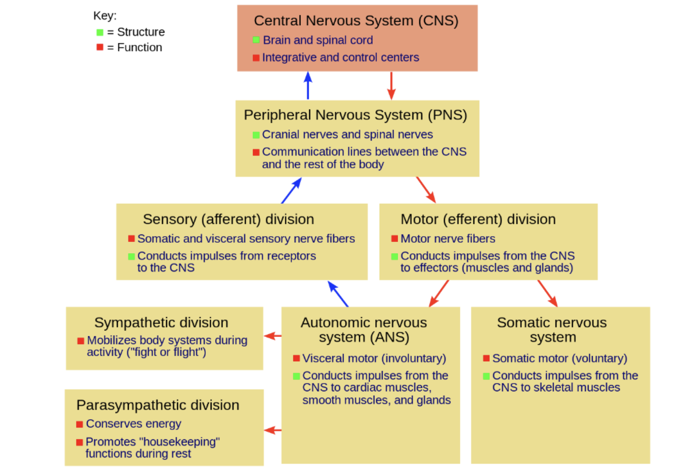
CNS
brain and spinal cord
physically separate from PNS from BBB
Afferent neurons
inward axonal projections
sensory
Efferent Neurons
outward axonal projections
motor
CNS compositions
10% neurons 50% volume
90% neuroglia 50% volume
nucleus
cluster of neuronal cell bodies in CNS
ganglion
Cluster of neuronal cell bodies in PNS
Grey matter
rich in neuronal cell bodies, dendrites, synapses
closer to surface
White matter
rich in myelinated axons
deep to grey matter
Circuts
anatomical and functional connections between neurons
Multipolar neurons
common CNS neuron with single axon and short dendrites
Pyramidal neurons
common
largest CNS neurons
multiple long axons and long dendrites
chart
CNS needs chemical isolation from rest of the body and from blood. Why?
needs stable environment because neurons are sensitive to ion changes. Blood also changes constantly.
Common ideas
Cranial bones provide protections against mechanical forces
CNS covered in meninges which are multiple layer connective tissue
isf - interstitial space fluid
csf - cerebral spinal fluid
bbb - blood brain barrier
Dura Mater
outermost
tough mother
made of 2 fibrous layer
space between has blood vessels, extracellular fluid, and lymphatic vessels
subdural space - between dura and arachnoid
dural venous sinus - drainage system allows CSF to re enter blood
Subdural Hematoma
Physical trauma leds to bleeds into brain surface
increased fluid pressure leads to neuronal cell damage
MRI
Arachnoid Mater
between layer
spiderlike mother
has epithelial layer that does not follow the sulci and gyri of brain
subarachnoid space - between dura and pia
Arachnoid villi move through venous sinuses allowing CSF to exit subarachnoid space
Pia Mater
Gentle mother
on surface of brain
accompanies branches of cerebral blood vessels
coverage and protection of CNS tissue. blood vessles
meningittis
Meningittis
viral or bacterial infection leading to pia matter inflammation
cause headaches
Vascularization
brain has 15-20% cardiac output
internal carotid and vertebral arteries supply along with internal jugular vein
anastomose
fusion of blood vessels
Circle of Willis
alternative path for blood flow
ensures blood supply in the face of blockage
Brain Interstitial System
Fluid filled space between neurons and capillaires
has ISF and EM
15-20% of brain volume
not ISF outside CNS
Originate from CSF and blood vessels
Immediate environment surrounding neurons
Blood Brain Barrier
Made of capillary endothelial cells in tight junctions with basal lamina, astrocytic endfeet, pericytes
is highly selectively permeable with limits paracellular path and control of ISS
Neurovascular Unit
has capillary endothelial cells, pericytes, astrocytes, microglia, neurons
Regulation of blood flow and BBB
Blood Brain Barrier Transport
Limited transport capacity
carrier mediated - selective and saturable, aa glucose nucleotides
Receptor mediated - binding and internalization; hormones growth factor
Hydrophobic molecules: diffusion
lipid soluble come through water soluble do not
adsorptive transcytosis - fuse with membranse come out other end in vessicle
CSF acess
Hydrophobic diffusion is very fast so hydrophilic drugs have limited acess and bacterial infections are rare
ISF vs Plasma
ISf has lower Ph, protien content, buffering, glucose, K, HCO3
CNS very sensitive to changes in plasma CO2
CO2 levels
decreased medullary extracellular PH and increased phrenic nerve activity
CSF
located around and within CNS
Source of water an dolsutes for ISF
Gives brain boancy, protection against trauma, ISF functional stability and removes waste
made via ependymal cells lining ventricles
Ependymal cells
line ventricles
form choroid plexus with capillaries
secretion and movement of CSF
CSF Pathway
Ventricle choroid plexus
exits 4th ventricle via paired lateral apparatus of lone median aperture
flows thorugh subarachnoid
into arachnoid vili
drains into dura venous sinuses
CSF composition
More like ISF than plasma
plasma has more protien and K+
500 ml per day
150 ml at one time
Sleep and CSF
Sleep triggers rhythmic alternating slow waves of blood and CSF
operational dynamic

Neuroglia
>50% volume and 90% cells in CNS
are the internal environment responsible for communication, guiding axons, making synapses, bbb, remove waste, make CSF and myelin, generate electrical currents, and take up NT and ions
Oligodendrocytes
myelin production
regulational axonal growth
cerebral palsy - loss of oligodendrocutes
Astrocytes
most abundant
multiple types
provides surface coverage, limits solute into ISF
Part of BBB
Regulation of CSF content
Microglia
specialized macrophages
removal of debris, cells, foreign
Radial Glia
scaffolding in which new neurons migrate
Circuitry
based on synaptic connections between cells
vary from simple to complex
made during fetal stage
emergent - consciousness
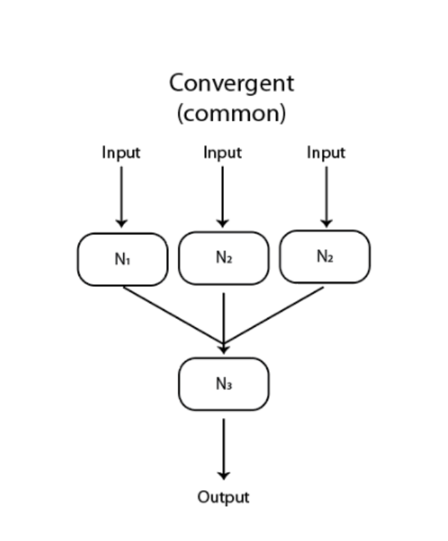
Convergent Circuit
many axons to one target neuron
intergate electrical info
trade sensitivity over spatial info
Divergent Circut
one axon project to many cells
skeletal muscle
coordinated activities of multiple cells

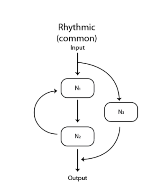
Rhythm Generating Circuit
central pattern generator with minimum of 2cells
complex movement with few cells like running breathing
Complex Circuits
emergent properties and unpredictable
whole is greater than parts
human consciousness
Circuit Formation
multiple levels
10^14 connections
each neuron has 7000 connections
Process of forming circuit
axon guided to right spot during fetal development
state function: right connection at the right time under right conditions
EM has attractive and repulsive proteins and cells
this is why repair of spinal cord is hard
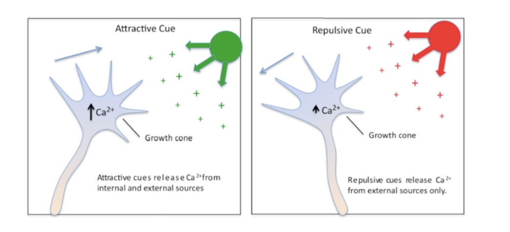
Growth cone helps be guided through chemotaxis of proteins and chemicals
Growth Cone Filopodia
membrane projection based on chemical cues
attract: both release Ca
repel: only external release Ca
Circuit Physiology
neuronal circuits develop during fetal and neuronatal growth
maintained by use
plasticity - changeable
wiring dictates functionality
complex behaviors are generated by simple circuits
Hemispheric Specialization
The left and right halves of your brain handle different jobs (like logic vs. spatial reasoning), but this specialization happens down at the cellular/circuit level rather than a major structural difference.
Uniform Signals, Diverse Functions
Even though every single neuron fires using the exact same basic electrical mechanism (ion fluxes across the membrane), complex groups of neurons can be assigned to completely different tasks depending on how they are wired.
Computational Power = Brain Volume
Harder tasks require more brain power, which is why larger areas of brain volume are dedicated to complex processing like vision.
Top-Down Control
the brain doesn't just react; it actively modifies and oversees activities happening down in the spinal cord.
SRMI
structural imaging
located differences in water content to outline shapes
examines shape not function
determine correct structure vs somthing wrong
Diffusion Tensor Imaging
structural
how water travels along axonal projections in brain brownian motion
info about neuron interconnectivity
Fmri
functional
past - association of brain trauma in region with change in physiology ie phinneas
now: measure small changes in blood flow different brain activities
EEG
functional
electrical brain activity with scalp electrodes
Phineas Gage
iron rod destroyed left frontal lobe
personality changes, loss of restraint
CNS common infections
need ct or mri that helps detect small bleeds
meningitis has high resolution and differentiates tissues
Brain mapping
normal surface structure
longitudinal fissures: left and right lobes
Latitudinal: top and bottom
has gyri/ridges sulci/valley and fissures
names for overlaying bones
Cerebrum
largest
cerebral cortex is outer layer
neocortex is largest part with 90% brain volume
higher level processing and cognition
Brainstem
autonomic and reflex center
Cerebellum
little brain
movement coordination
Diencephalon
homostatic control centers
Contralateral control
right side brain controls left side body and vice versa
Frontal lobe
motor speech memory formation personality and emotion
frontal cortex has : prefrontal, motor, and broccas area
Parietal lobe
somatosensory cortex
touch, taste, sight, smell
collection of info from multiple sources
proprioception: awareness of physical structures in #D space
Occipital lobe
visual processing and visual memories
primary visual cortex in back
visual association area to interpret info from cortex just before it
Temporal lobe
hearing speech language smell
passage of time - grey hair here
orginazation and language comprehension
memory formation and retreival
Primary motor cortex
back of frontal lobe
motorneurons betz cells that controls striated muscle
volume of area is indicative of control
middle finger neurons near pinky neurons
Myotome
PMC
representation of motoneurons controlling muscles on different regions of the body
Somatosensory cortex
sensory input from thermal, touch(mechanoreceptors), proprioception, pain
Hommunculus: visual representation of how much of cortex used by what
inputs from contralateral cord and thalmus
integration
Werinicke’s area
temporal lobe
speech comprehension
Wernicke’s aphasia
cant understand words and communicate
Brocas area
frontal lobe
helps speak
Primary language areas
broca
wernicke
angular gyrus which is superior edge of temporal lobe
arcutate fasiculus which is axonal tracts connecting broca and werinick allowing for coordination
Aphasia
cant comprehend or formulate languaue
many types
Brain Lateralization
left for language math reasoning logic
right for spatial, face recog, visual, music
caused by minute temporal delays in other hemispshere neuronal cells
CNS fibers
CNS made of interconnecting neurons
White matter
myeling coated axon projections
Projection
connects subcortical regions: telecenphalon, diencephalon, brain stem, spinal cord
commissural
connections between lobes
association
connections within lobes