Chapter 8 - Orthopedics/Skeletal System
 Call Kai
 Learn
Learn Practice Test
 Spaced Repetition
 Match
Match Flashcards
 Knowt Play
1/148

There's no tags or description
Looks like no tags are added yet.
Name | Mastery | Learn | Test | Matching | Spaced | Call with Kai | Chat |
|---|
No analytics yet
Send a link to your students to track their progress
149 Terms
orth/o
straight
ped/o
child
-ics
knowledge, practice
functions of the skeletal system
structurally support the body
protect soft tissues
protection for internal organs
storage site for calcium and phosphorus
Anatomy of the skeleton
divided into two major regions
Axial Skeleton
forms central frame work of the body
supports and protects vital organs
includes bones of the skull, vertebral column, and thoracic cage
Appendicular Skeleton
bones of the upper and lower limbs and what attaches them to the axial
includes bones of the hands, shoulders, arms, hips, legs, and feet
primarily functions to support movement and locomotion
Long Bone Structure
Major regions:
Diaphysis: shaft of the bone
Epiphyses: enlarged ends of the bone
Internal Structures:
Cortical Bone: Dense outer layer that provides strength
Cancellous bone: spongy internal bone with marrow-filled spaces
Medullary cavity: hollow space in the shaft containing primarily yellow marrow in adults

Osteoma
type of bone tumor
Benign growth
Osteosarcoma
type of bone tumor
malignant growth of osteoblasts
Ewing sarcoma
type of bone tumor
malignant growth
primarily affects children and young adults
more frequent in males than females
oste/o
bone
chondroma
benign tumor of the cartilage
chondr/o
cartilage
connective tissues of the bones include:
cartilage
ligaments
joints
What does red and yellow marrow do?
Red marrow produces blood
yellow marrow primarily stores fat
chondromalacia patellae
softening and deterioration of the cartilage on the underside of the patella
may cause pain in the front of the knee
especially when climbing stairs, squatting or bending the knee
can result from repetitive stress improper patellar alignment or injury
-malacia
softening
-blast
developing cell
sarc/o
connective issue
FX/Fx
Fracture
break in bone caused by injury, repetitive stress, or an underlying disease
types of fractures
stress fracture
a small crack in the bone caused by repetitive loading or overuse
Pathologic fracture
break in the bone weakened by disease such as osteoporosis
Malunion
a FX that heals in an abnormal position or alignment
por/o
pores; small openings
Ways to describe a fracture
Is the skin broken
open fx: Broken bone with an external wound
closed fx: skin remains intact over the fracture
Are the bones aligned
nondisplaced: bone fragments emain aligned
displaced: bone fragments have shifted out of alignment
comminut/o
break into small pieces
comminuted fracture
broken into many pieces

Transverse fracture
broken cleanly across

colles fracture
a common break at the wrist, typically happens because of trying to catch oneself when one falls

oblique fracture
breaks at an angle
spiral fracture
bone becomes twisted

greenstick fracture
where one side breaks but the other stays intact

hairline fractures
common fracture in older people, small crack like
compression fracture
pressure causes fracturing, common in the vertebrae
depressed fracture
a fracture that presses down

depress/o
press down
osteomalacia
softening of the bones
caused by inadequate minderalization
most commonly due to Vitamin D deficiency
causes bone wekness, pain, and increased risk of fractures
a similar disease in children is called rickets
inadequate sunlight can contribute to Vitamin D deficiency
Rickets
an osteomalacia like disease seen in children
Osteomyelitis
infection and inflammation of bone tissue and bone marrow, most commonly caused by bacteria
bacteria may reach bones through the bloodstream
can cause bone pain, swelling, fever
Osteoporosis
condition in which bone breakdown exceeds bone formations, results in reduced bone density and increased risk of fractures. Thinning of the bone structure.
creates many large porous areas in the bone

Thoracic cage anatomy
Protects the heart and lungs and consists of ribs, sternum, and thoracic vertabrae
sternum
Manbrium
body
xiphoid process
12 pairs of ribs
ribs 1-7 are referred to as “true” ribs
directly attached
ribs 8-10 are referred to as “false” ribs
indirectly attached
11-12 are referred to as “floating”ribs
no anterior attachment

Anatomy of the vertebral column
consists of 5 regions which help support hte body and protect the spinal cord
cervical
7 vertabrae (C1-C7)
Neck
Thoracic
12 Vert (T1-T12)
thorax
Lumbar
5 vert (L1-L5)
Lower bacl
Sacrum
5 fused vertebrae
coccyx
usually 4 fused vertebrae
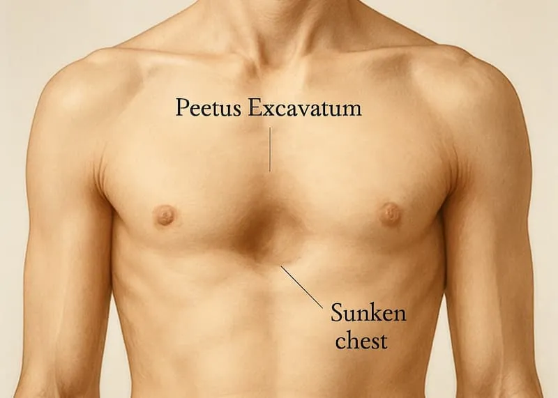
Pectus excavatum
congenital chest wall deformity in which the sternum is displaced inward
creates a sunken or funnel shaped appearance of the chest
often called funnel chest
severe cases may compress the heart and lungs, affecting breathing and exercise tolerance
Kyphosis
abnormal posterior curvature of the thoracic sipne
AKA humpback
kyph/o
bent; humpbacked
lordosis
abnormal anterior curvature of the lumbar spine
AKA swayback
scoliosis
abnormal c or s shaped lateral curvature of the spine
dexteroscoliosis = right sided
levoscoliosis = left sided
Ankylosing Spondylitis
chronic inflammation disease that primarily affects the spine, causing pain and stiffness
over time inflammation can lead to fusion of the vertebrae, reducing spinal flexibility and mobility
Spondylolisthesis
spinal condition in which one vertebrae slips forward over the vertebrae below it, causing misalignment of the spine
may result from degenerative changes, stress fractures or injury
can cause lower back pain, stiffness or nerve
-listhesis
slipping, displacement
anatomy of the leg bones
femur
thigh bone of the upper leg
tibia
shin bone of the lower leg
fibula
thin lateral bone of the lower leg
patella
kneecap
Bones of the foot
tarsals
7 bones forming the ankle and rearfoot
metatarsals
5 long bones forming the forefoot
phalanges
14 toe bones
2 in the great/big toe, 3 in the other toes
from ankle to toes is goes: Tarsals, Metatarsals, Phalanges

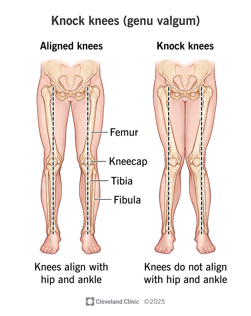
Genu Valgum
Genu = knee
Valgum = angled outward (referring to the distal section of a limb)
Called knock-knees
when knees touch together, causing the legs/feet to be angled outward
Genu Varum
Genu = Knee
Varum = angled inward (referring to the distal section of a limb)
Called Bowlegs
when knees angle outward, causing the legs/feet to be angled inward

Hallux valgus
deformity in which the great toe (the hallux) angles toward the other toes causing a bony prominence (bunion) at the base of the great toe
may cause pain swelling and difficulty wearing shoes
Talipes Equinovarus
AKA Clubfoot
congenital foot deformity in which the foot is turned inward and downward, often with the heel elevated
may affect one or both feet
the foot is typically stiff and cannot easily be moved into its normal position
early treatment usually with gental manipulation and serial casting can often correct the deformity without major surgery
talipes
foot/ankle deformity
equin/o
horse-like; heel elevated foot pointed downward
varus
turning inward
Anatomy of joints
joints (articulations) are locations where two or more bones meet
suture joints: immovable joints found between bones of the skull
symphysis joints: slightly movable joints connected by fibrocartilage, such as the pubic symphysis and joints between vertebral bodies
synovial joints: freely movable joints that allow a wide range of movements, such as the knee and hip
cartilage anatomy
Cartilage: connective tissue that provides cushioning and support within joints
Articular Cartilage: covers the ends of bones, reducing friction and absorbing shock during movement
Meniscus: a pad of fibrocartilage found in certain joints that helps distribute weight and absorb shock
Ligaments
strong bands of fibrous connective tissue that connect bone to bone, providing joint stability and limiting excessive movement
ligaments connect bone to bone, tendons connect muscle to bone
Arthalgia
pain in the joint due to injury inflammation, or infection from various causes
arthropathy
general term for disease of the joint from any cause
dislocation
complete displacement of a bone from its normal position within a joint, causing loss of contact between the articulating surfaces
causes
most commonly due to trauma, such as a fall or sports injury
congenital/development conditions: abnormal hip joint development in infants can result in hip dislocations
Gout
Form of inflammatory arthritis caused by the accumulation of uric acid crystals in joints
causes sudden episodes of severe pain, redness, and swelling
most commonly affects the base of the great toe
RA
Rheumatoid Arthritis
acute and chronic inflammatory disease of the connective tissue, particularly the joints
autoimmune condition in which the body’s own antibodies attack cartilage and tissues
joints are red and swollen with progressive deformity
OA
Osteoarthritis
chronic inflammation, pain, and stiffness in the joints as cartilage wears away and bone rubs on bone
sprain
overstretching or tearing of ligaments around a joint
term comes from middle english (spreynd) meaning to press out
subluxation
partial dislocation with slight displacement of bone ends from the joint
the difference between a dislocation and subluxation is the degree of separating of the articulating bones
subluxation is partial
dislocation is complete
torn meniscus
a tear in the cartilage pad in the knee joint because of an injury
causes pain swelling and difficultly moving the knee
hemarthrosis
condition of blood in the joint cavity, usually due to blunt trauma or penetrating injury
RF
Rheumatoid Factor
RF=autoantibody produced by the immune system
binds to a protein called immunoglobulin G (IgG)
This binding can lead to inflammation and damage to the joints
blood tests for RF are usually positive in patients w/ rheumatoid arthritis
rheumat/o
watery flow; associated w/ rheumatic conditions
Uric Acid
blood test that shows elevated level in patients w/ gout and gouty arth
X-ray
most common diagnostic procedure for the skeletal system
primary X-Ray means it is for diagnosing fractures, dislocations, and bone tumors
meniscus direct meaning/translation
crescent-shaped
greek for little moon
Bone density tests
measures bone mineral density (BMD) to diagnose osteoporosis
AKA bone densiometry
2 types:
DEXA
dual energy x-ray absorptiometry (uses 2 x-ray beams)
QCT
Quality Computerized tomography (x-ray+CT)
BMD
bone mineral density
Cast
a layer of plaster or fiberglass applied around a fractured bone to immobilize the bone as it heals
Closed Reduction
procedure in which bone ends in a displaced fracture are manually manipulated back into place
often followed with a cast
Goniometry
procedure that measures the angle of a joint and its range of motion
ROM
range of motion
orthosis
a orthopedic device like a brace or splint that is used to keep a body part straight and immobile
or to correct an orthopedic problem
PT
physical therapy
exercise that improves a patients ROM, joint mobility, strength, and balance
passive: exercises are done my the therapist
active: exercises are done by the patient
goni/o
angle
traction
procedure that uses weight to pull the bone ends of a fracture into correct alignment
skin traction uses wraps or straps
skeletal traction uses surgically inserted pins or wires
prosthesis
a orthopedic device like a leg or arm that is created for a patient following an amputation
greek for in addition, to place or put
prosth/o
artificial part
Analgesics
treats pain associated with bone injury or surgery
an- = without
alges/o=pain
-ic=pertaining to
bone resorption inhibitors
prevents osteoclasts from breaking down bone, helping to treat osteoporosis
corticosteroids
decreases severe inflammation
gold compound drugs
Inhibits immune respose
Used to treat RA
Contains real gold
NSAIDs
nonsteroidal anti-inflammatories
treat inflammation and pain from osteoarthritis and orthopedic injury
Amputation
removal of all or part of an extremity damaged by trauma or disease
BKA
Below the knee amputaion
performed at the level of the tibia and fibula
AKA
Above the Knee Amputation
performed at the level of the femur
Arthrocentesis
Removal of accumulated fluid from an injured joint using a needle inserted in the joint space
-centesis = procedure to puncture
arthrodesis
fusion of bones in a deteriorated, unstable joint
-desis
procedure to fuse together
arthroscopy
visualization of the inside of a joint using an arthroscope inserted into the joint
bone graft
replacement of bones severely damaged due to bone cancer
autograft = patients own bone
allograft = cadaver bone