Lecture 6 - Actin and Intermediate Filaments
 Call Kai
 Learn
Learn Practice Test
 Spaced Repetition
 Match
Match Flashcards
 Knowt Play
1/18

There's no tags or description
Looks like no tags are added yet.
Name | Mastery | Learn | Test | Matching | Spaced | Call with Kai | Chat |
|---|
No analytics yet
Send a link to your students to track their progress
19 Terms
Describe the structure, subunits, and function of Actin filaments (microfilaments)
Structure:
Strands in double helix
Subunits
Actin
Functions
maintain cell shape by resisting tension (pull)
move cells via muscle contraction or cell crawling
divide animal cells in two
move organelles and cytoplasm in plants, fungi, and animals

Describe the structure, subunits and functions of intermediate filaments
Structure:
Fibers wound into thicker cables
Subunits:
Keratin or vimentin or lamin or others
Functions
maintian cell shape by resisting tension (pull)
anchor nucleus and some other organelles

Describe the structure, subunits, and functions of microtubules
Structure:
Hollow tube
Subunits
a- and B-tubulin dimers
Functions:
maintain cell shape by resisting compression (push)
move cells via flagella or cilla
move chromosomes during cell division
assist formation of cell plate during plant cell division
move organelles
provide tracks for intracellular transport

What is the cortical cytoplasm?
It is just beneath the plasma membrane and helps to maintian cell shape
Dominated by an interconnected meshwork of microfilaments (acitn)
Large membranous organelles and other bulky structures are mostly excluded form this region
What is the Subcortical Cytoplasm (aka Endoplasm)
Major organelles of the cell reside in this region and are spatially organized and othen attached to cytoskeletal elements
What is the nuclear cytoskeleton?
Important in mitosis, allows the nucelus to aintain and change its shape
Nuclear lamins (intermediate filaments) give the nucleus form like a tennis ball
Describe the structure of actin (a microfilament). What is G-actin?
G actin: actin monomer
Globular
42 kDa protein (375 a.a.)
Non-symetrical
Pointed end (-)
Barbed end (+)
Binds ADP or ATP in a cleft
Binding ATP promotes polymerization
ATP eventually hydrolyzes

How are microfilaments formed? Mention nucleation and F actin

Nucleation: the formation of trimers, followed by more rapid growth
F-actin: Actin polymers
(-) ends only add to (+) ends, giving the chain polarity
this is reversible and depends on subunit availability
What is the microfilament tread-mill?
Occurs because the barbed end of a filament grows 5-10 times faster than the pointed end
G actin bound to ATP favorably associates with the positive end of the microfilament (where the barbed aspect of actin is exposed)
Binding of G actin will eventually lead to hydrolysis of ATP to ADP
The presence of ADP on an exposed end promotes depolymerization

Actin binding proteins are regulated via cell signaling. What are the different actin binding proteins
Profilin - stimulates ADP exchange for ATP
Formin - catalyzes nucleation and extension of a microfilament
Arp 2/3 complex - initiates the formation of branches
Capping proteins and tropomyosin - stabilize filaments
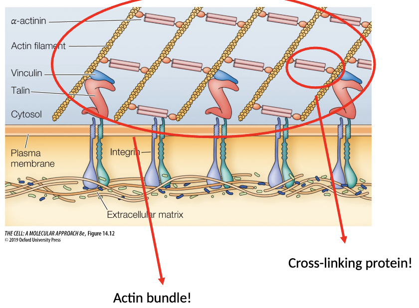
Cross-linking proteins: cross-link filaments into bundles and networks
Cofilin - severs acting filaments

Where are actin filaments located? What do they form?
Microfilaments (including actin filaments) are more concentrated in the periphery (creating the cell cortex)
Cortical cytoskeleton is made of actin and spectrin (anchoroed by ankyrin and protein 4.1)

What are focal adhesions and stress fibers?
Focal adhesions are areas of the cell that makes connections with the extracellular matrix
These areas of the membrane also tend to have a lot of actin attachments (stress fibers)
Differentiate microvilli and stereocilia
Microvilli
finger like extensions; abundant on cells involved in absorption formed by actin bundles
Stereocilia
finger like extensions; not involved in absorbtion, but involved with dtecting extracellular changes (auditory hair cells) formed by actin bundles
What are ‘podia?
These are cell dependent and allow for different variations of movement. They are a more transient structure (unlike microvilli)
What are the proposed steps in locomotion (true for all ‘podia)
Protrusion: The leading edge usually shows filopodia and lamelipodia. These extensions probe forward in search of a new substatum contact site
Substrate Adhesion: If and when the leading edge ecnounters and adhesive region or the substratum it will attach, stabilizing the forward extension. The mechanism of attachment involves integral membrane receptors and their coupling to internal cytoskeletal elements
Traction/Cell Body movement: Once a new forward contact has been made a mechanism must exist to move the bulk of the cell cytoplasm in that direction. Several activities are believed to contribute

What does this movement look like in a cell?
In most cases, cells move in response to cues from the environment
Rho family proteins are coupled to receptors and respond to envrionmental cues by activating Acting Binding Proteins
This stimulates actin remodleing and cell movement

List the actin associated proteins
Profilin
Formin
Arp 2/3 complex
Capping proteins
Tropomyosin
Cross-linking protiens
Cofilin
Spectrin
Ankyrin
Protein 4.1
Rho
Describe the structure and assembly of Intermediate filament proteins
All Intermediate filaments have a similar general structure
N and C terminal globular domains
Step 1 - Two polypeptides will dimerize connected by central rod domain (coiled-coil)
This process is in parallel (N-N/C-C)
Step 2 - Two dimers combine to form a tetramer
This process is in antiparallel (N-C/C-N)
Step 3 - Tetramers will stack on one another end to end to form a protofilament
Step 4 - 8 protofilaments will combine, twisting around each other to form a rope-like structure called a filament

Describe specifically the roles of intermediate filaments
Stablize structure and support within the cell
Intermediate filaments are more resistant to strech and bending forces than microtubules, but less resistant than actin
Positioning of the nucleus and certain organelles
support of the plasma membrane
support of the nuclear membrane (nuclear lamina)
Transmitting force between cells or to the extracellular matrix
Requires attachement to one or the other via peripheral and transmembrane proteins
Resisting and distributing externally generated stresses over multiple cell to prevent damage to individual cells
Desosomes - junctions between cells (cell to cell)
Hemidesmosomes - jucntions between cell and connective martrix (cell to substratum)
