Management of the Patient with Hematologic Disorders: lecture 4 exam 3
 Call Kai
 Learn
Learn Practice Test
 Spaced Repetition
 Match
Match Flashcards
 Knowt Play
1/77

There's no tags or description
Looks like no tags are added yet.
Name | Mastery | Learn | Test | Matching | Spaced | Call with Kai | Chat |
|---|
No analytics yet
Send a link to your students to track their progress
78 Terms
Blood
delivers oxygen and nutrients to all tissues, removes wastes, and transports gases, blood cells, immune cells, antibodies, and hormones throughout the body.
- Account for 7% to 9% of total blood volume
Plasma
fluid of blood; it contains various proteins, such as albumin, globulin, fibrinogen, and other factors necessary for clotting, as well as electrolytes, waste products, and nutrients.
- About 55% of the whole blood volume is plasma
blood review
Blood consists of three primary cell types (see Table 28-1): erythrocytes (red blood cells [RBCs], red cells), leukocytes (white blood cells [WBCs]), and thrombocytes (platelets)
Components of blood normally make up 40% to 45% of the blood volume; makes up approximately 7% to 9% of the normal body weight and amounts to 5 to 6 L of volume for men and 4 to 5 L of volume for women
Short lifespan
The body replenishes its supply of cells continuously; this process is termed hematopoiesis.
The primary site for hematopoiesis is the bone marrow.
![<p>Blood consists of three primary cell types (see Table 28-1): erythrocytes (red blood cells [RBCs], red cells), leukocytes (white blood cells [WBCs]), and thrombocytes (platelets)</p><p>Components of blood normally make up 40% to 45% of the blood volume; makes up approximately 7% to 9% of the normal body weight and amounts to 5 to 6 L of volume for men and 4 to 5 L of volume for women</p><p>Short lifespan</p><p>The body replenishes its supply of cells continuously; this process is termed hematopoiesis.</p><p>The primary site for hematopoiesis is the bone marrow.</p>](https://assets.knowt.com/user-attachments/20b12c87-1734-4527-a429-e61d876f51aa.png)
Excessive blood loss
Trauma can lead to excess blood loss
To prevent this, an intricate clotting mechanism is activated when necessary to seal any leak in the blood vessels. Increased clotting occurs to attempt to slow blood loss.
Fibrinolytic mechanism occurs
- The balance between these two systems—clot (thrombus) formation and clot dissolution or fibrinolysis—is called hemostasis
Low Hemoglobin (Hgb) & Hematocrit (Hct)
Low Mean Corpuscular Volume (MCV)
blood cells
blood cells

nursing assessment
nursing assessment

Major Disorders of the Hematologic System
Anemia
Polycythemia Vera
Leukocytosis
Hemophilia
Sickle Cell Disease
Thalassemia
Von Willebrand Disease
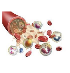
anemia
anemia

defective RBC production
iron deficiency (most common)
vitamin b12
folate deficient
decreased erythropoietin production (chronic kidney disease)
cancer/inflammation
RBC destruction
sickle cell disease, thalassemia
drug induced anemia
autoimmune anemia
symptoms of anemia
symptoms of anemia

Iron Deficiency Anemia
Most common type of anemia
Insufficient iron levels
Condition lacking adequate healthy red blood cells
Iron is required for the body to produce enough red blood cells to oxygenate the body
Low MCV (mean corpuscular volume), Low serum iron, monitor ferritin levels

Iron Deficiency Anemia: Underlying Cause
Insufficient dietary iron intake (vegetarian or vegan diets)
Chronic blood loss from GI bleeding
- Hemorrhage
- GI bleeding
- Surgery
Malabsorption of iron
- GI disorders (celiac and Crohn's disease)
- Medications (proton pump inhibitors, aspirin)
- Chronic inflammation
Increased iron requirements
- Rapid body growth
- Pregnancy, breastfeeding
- Menstruation
Iron Deficiency Anemia: Nursing
Monitor lab values
Head-to-toe assessment
Vital signs
Assess, document the presence of any clinical symptoms
Monitor intake and output
Assess response to prescribed treatments
Advise the patient
- Take iron supplements 1 hour before meals;
- Take liquid forms of iron via a straw to avoid staining teeth
- GI side effects
** take on empty stomach, take 1-2 hrs before meals, stools could turn black
Educate the patient
- Foods high in iron
- Vitamin C intake helps to facilitate iron absorption
Anemia of Inflammation
Associated with chronic diseases, including inflammation, infection, and malignancy
- Caused by an imbalance in iron metabolism, decreasing production of RBC
Inflammatory conditions
- Autoimmune diseases (rheumatoid arthritis, lupus)
- Infections (TB, HIV)
- Cancer
- Kidney disease
- Inflammatory bowel disease (Crohn's, ulcerative colitis)
The underlying condition can be identified so that it can be treated appropriately
Mild to moderate anemia- few or no symptoms
aplastic anemia
Rare disease caused by a decrease in or damage to bone marrow stem cells, damage to the microenvironment within the bone marrow, and replacement of marrow with fat.
Severe anemia; can be life-threatening
Significant neutropenia (lower-than-normal neutrophil count)
Thrombocytopenia (lower-than-normal platelet count)
Life-threatening condition associated with bone marrow failure evidenced by pancytopenia (i.e., anemia, neutropenia, and thrombocytopenia; lower-than-normal counts of erythrocytes, neutrophils, and platelets)
Most cases are idiopathic (i.e., without apparent cause)
May also be associated with certain medications, chemicals, or radiation damage, chemotherapy
Signs & Symptoms- infection, fatigue, pallor, dyspnea, bruising
Low Neutrophil, low hemoglobin, low platelet count

aplastic anemia
An immune-mediated condition in which T lymphocytes attack hematopoietic stem cells with subsequent reduced production of erythrocytes, leukocytes, and platelets.
Treatment
- Hematopoietic stem cell transplant (HSCT)
- Treatment with immunosuppressive therapy using antithymocyte globulin (ATG) and cyclosporin
- Transfusions with PRBCs and platelets are frequently required
- Prophylaxis against invasive fungal infection is needed for severely neutropenic patients
Megaloblastic Anemias
A type of anemia characterized by abnormally large and immature red blood cells, caused mainly by a deficiency in vitamin B12 or folate.
Folic Acid Deficiency
- Lack fruits and vegetables
- Can be depleted within months if dietary intake of folate is deficient
- High in alcohol abuse disorder and liver disease
- Small bowel diseases (celiac disease, Crohn's disease)
- High MCV, low folate
Vitamin B12 Deficiency
- Inadequate dietary intake, poor absorption
- High MCV, Low B12
- Pernicious anemia
Pernicious Anemia (B12 Deficiency)
A type of anemia caused by the body's inability to absorb vitamin B12 from the small intestine.
- decrease RBC occurs b/c the intestines cannot absorb vitamin B12
Associated with a lack of intrinsic factor; Intrinsic factor is normally secreted by cells in the gastric mucosa; it binds to vitamin B12 and transports it to the ileum, where it is absorbed.
Diseases of the pancreas and the ileum (Crohn's Disease) or a GI resection; bariatric surgery
Body can't absorb enough vitamin B-12.
Use of metformin for treatment of type 2 diabetes, chronic use of histamine blockers, antacids, and proton pump inhibitors to reduce gastric acid, can inhibit vitamin B12 absorption
Pernicious Anemia: Clinical Manifestations
Fatigue and weakness
Mood changes
Mental status changes
Tingling sensation
Pain
Diarrhea
Weight loss
Vision changes
Alterations in smell or taste
Glossitis (painful, smooth, red tongue)
Uncontrollable muscle movements

Pernicious Anemia: Assessment
Labs:
- Serum levels of both folic acid and vitamin B12
- Low B12, Low hemoglobin, presence of Intrinsic Factor Antibiotics (IFAb)
A positive intrinsic factor antibody test is more useful
- A positive test indicates that antibodies are present that interfere with the binding of the intrinsic factor-vitamin B12 complex to receptors in the ileum, preventing absorption.
Education: monthly vitamin B12 injections or daily oral vitamin B12 supplements
Hemolytic Anemias
Red blood cells are prematurely destroyed, leading to a shortage of healthy red blood cells in the body
Fewer erythrocytes result in decreased available oxygen, causing hypoxia, which in turn stimulates an increase in erythropoietin release from the kidneys.
Red cell destruction persists; the hemoglobin is broken down excessively; the majority of the heme is converted to bilirubin, conjugated in the liver, and excreted in the bile
Inherited: Sickle Cell Disease (SCD) and Thalassemia
Gerontologic Considerations
Most common causes of anemia in the elderly are chronic disease and iron deficiency.
Vitamin B12 deficiency, folate deficiency, gastrointestinal bleeding and myelodysplastic syndrome are some other causes of anemia in the aging population.

Sickle Cell Disease (SCD)
Autosomal recessive disorder caused by inheritance of the sickle hemoglobin (HbS) gene.
A defective hemoglobin molecule that causes the erythrocyte to change shape when exposed to low oxygen tension.
These sickle cells cause occlusion of small blood vessels, ischemia, severe pain episodes, delayed growth and development, and damage to many organs.
Sickle cells have a shorter than normal life span, causing premature destruction, resulting in a shortage of red cells (anemia), sticky and stiff, sickle-shaped

Sickle Cell Disease: Signs & Symptoms
Anemia signs and symptoms (Sickle Cell Anemia)
- SOB, fatigue, delayed growth in children
Symptoms and complications are the result of chronic hemolysis and thrombosis.
The bone marrow expands early in life to compensate for the resulting anemia, sometimes causing enlargement of the bones in the face and skull.
Jaundice is often present.
Chronic anemia is often associated with tachycardia, cardiac murmurs, and cardiomegaly.
Hypoxia, infections (pneumonia and osteomyelitis)
Arrhythmias and heart failure may also occur
Sickle Cell Crisis
Acute vaso-occlusive crisis (most common)
Blood flow is severely reduced, ischemia or infarction of the tissue can cause severe pain, swelling, and fever
Requires a multidisciplinary approach to manage painful crisis episodes, prevent complications, decrease emotional problems, and promote a healthy lifestyle.

acute management: sickle cell crisis
priority intervention: oxygen FIRST and then pain
acute management: sickle cell crisis
priority intervention: oxygen FIRST and then pain

Why is hydration critical in managing sickle cell disease?
Adequate hydration helps maintain blood viscosity and reduces the likelihood of sickling.
Hydration improves circulation and prevents blockages in small blood vessels.
Dehydration can trigger or worsen vaso-occlusive crises by increasing blood thickness.
recommended plan for pain management in sickle cell disease
recommended plan for pain management in sickle cell disease

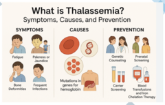
Thalassemia
Genetic blood disorder
Characterized by the abnormal production of hemoglobin, the protein in red blood cells that carries oxygen
Decrease in the hemoglobin content of erythrocytes,
Extreme microcytosis (smaller than normal-sized erythrocytes), hemolysis, and variable degrees of anemia.
Impaired hemoglobin synthesis
Thalassemia Major
- Severe anemia, profound hemolysis, ineffective erythropoiesis.
- May require numerous blood transfusions.
- Greater risk for iron overload, requiring frequent chelation therapy.
- Long-term complications: cognitive dysfunction, peripheral neuropathy, cerebrovascular disease.
Thalassemia Intermediate
- Anemias, jaundice, splenomegaly, and hemosiderosis resulting from increased intestinal iron absorption.
Thalassemia Minor
- Mild anemias, often asymptomatic.

Thalassemia: Alpha
Alph-globin genes mutated
Mediterranean or Southeast Asian descent
Less severe
Erythrocytes are microcytic
Anemia is mild
Thalassemia: Beta
Beta-globin genes mutated
African descent
More severe
Microcytosis and severe anemia
Thalassemia Major (Cooley's anemia)
- Blood transfusions
- Iron chelation therapy
beta thalassemia major features
beta thalassemia major features

thalassemia
thalassemia
Polycythemia
An increased volume (too many) RBCs leads to thickened blood, increasing the risk for clots
The term is used when the hematocrit is elevated (more than 55% in males and 50% in females).
Dehydration can cause elevated hematocrit, but not usually to the level seen with polycythemia.
Polycythemia is classified as either primary or secondary.
- Primary polycythemia, also known as polycythemia vera, is a myeloproliferative neoplasm

Polycythemia Vera
Increased hemoglobin above normal values
Elevated Hemoglobin and RBCs with microcytosis
Elevated histamine levels
Elevated WBCs with basophilia;
Bone marrow biopsy (excessive RBCs)
Signs & Symptoms
- Sweating, dyspnea, weakness, headaches, double vision, pain and swelling in joints (big toe), redness of skin, muscle pain, hypertension, tinnitus
Monitor for peripheral and cerebral thrombosis

Neutropenia
Neutrophil count less than 2,000/mm3; decreased production of neutrophils or increased destruction of cells
Are essential in preventing and limiting bacterial infection
High risk for infection, hypersplenism, viral diseases (infections, hepatitis, mononucleosis)
Caused by aplastic anemia, chemotherapy, metastatic cancer, lymphoma, leukemia, radiation
Lab- CBC w/ differential; low WBC count

bleeding disorders
A group of conditions that prevent the body from forming clots properly, leading to excessive or prolonged bleeding.
Categorized based upon: (1) deficiency of platelets or a defect in the platelets, or (2) inherited or acquired coagulation factor abnormality, or (3) based upon a defect in the vasculature.
A decrease in platelets results in petechia that often clusters
Coagulation defects
- Bleeding occurs deeper within the body (sub-Q, IM hematomas, joints)
Signs and symptoms vary according to the defect
- Thrombocytopenia
- Hemophilia A & B
- von Willebrand Diseases
- Liver Disease
- Vitamin K Deficiency
- Heparin-Induced Thrombocytopenia
- Disseminated Intravascular Coagulation (DIC)
bleeding disorders cont
Because platelets are primarily responsible for stopping bleeding from small vessels, patients with decreased platelets develop petechiae, often in clusters
Avoid activities that increase the risk for bleeding, such as contact sports.
Skin assessment: evidence of bleeding (petechiae and ecchymoses (bruises), nose and gums)
When bleeding disorders are severe, patients are monitored for bleeding by testing all drainage and excreta (feces, urine, emesis, and gastric drainage) for obvious and occult blood.
Assess for medications to increase bleeding: aspirin, NSAIDs, alcohol, and anticoagulants
Thrombocytopenia
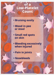
Deficiency of platelets in the blood; causes bleeding into the tissues, bruising, and slow blood clotting after injury.
Low platelet level
- Can result from a variety of factors, including reduced production of platelets in the bone marrow, increased destruction of platelets, or increased consumption of platelets (ex., the use of platelets for clot formation).
Platelet counts fall to 20,000/mm3 or less, and petechiae may occur.
- Nasal bleeding, gingival bleeding, excessive menstrual bleeding
Bone marrow aspiration and biopsy are used to identify platelet deficiency
Genetic causes of thrombocytopenia- Autosomal dominant, autosomal recessive, and X-linked mutations
Screening for hepatitis B or C
Hemophilia
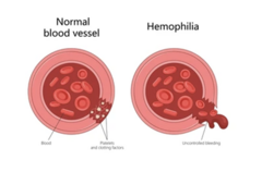
A rare, inherited bleeding disorder that affects the body's ability to form blood clots.
Two forms: hemophilia A and hemophilia B
- Determined by laboratory tests
- Both are inherited as X-linked traits, making both conditions more common in males than in females
Hemophilia A is caused by a genetic defect that results in deficient or defective factor VIII. Relatively common.
Hemophilia B, known as Christmas disease, is due to a genetic defect that causes a deficiency or defect in factor IX.
Often dx in early childhood, usually in the toddler
Manifested by hemorrhages into various parts of the body
75% of bleeding occurs in the joints
Hemophilia Management
Factor Replacement Therapy:
- Administering the missing clotting factor (factor VIII for hemophilia A or factor IX for hemophilia B) through intravenous injections.
- It can be used both prophylactically (to prevent bleeding) and on demand (to treat bleeds).
Emicizumab (Hemlibra):
- A monoclonal antibody that mimics factor VIII and can prevent bleeding in hemophilia A without the need for factor replacement.
Patients at risk for significant complications (bleeding into the respiratory tract or CNS)
Significant pain requiring analgesics for hematomas and joint hemorrhage.
Warm baths help relieve pain, promote relaxation, and improving mobility.
von Willebrand Diseases (vWD)
A bleeding disorder caused by a deficiency or dysfunction of von Willebrand factor (VWF), a protein that plays a crucial role in blood clotting.
Inherited condition that can be passed down from parents to children
Type 1: The most common type, characterized by a reduced amount of functional VWF; 70-80% of cases
Type 2: Several subtypes exist, where the VWF is present but does not function correctly.
Type 3: A rare and severe type where there is almost no VWF produced.
Signs & Symptoms:
- Bleeding of the mucous membranes, nosebleeds, heavy menses, easy bruising, and prolonged bleeding from cuts and surgical sites
The goal of treatment is to replace the deficient protein (e.g., vWF or factor VIII)
Acquired Bleeding Disorders
Liver Disease
- Liver dysfunction (due to cirrhosis, hepatitis, and tumor) can lead to reduced amounts of the factors needed for coagulation and hemostasis
Vitamin K Deficiency
- Vitamin K is an essential element for the synthesis of many coagulation factors
- Patients who are malnourished
Heparin-Induced Thrombocytopenia
- Serious complications of heparin-based therapy
- Decrease in the platelet count is the hallmark sign; it often develops 5 to 10 days after heparin therapy is initiated
- Increased risk for thrombosis, either venous, arterial, or both, deep vein thrombosis (DVT), acute coronary syndrome (ACS), stroke, or thrombosis to major vessels in an extremity
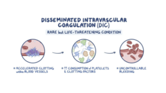
Disseminated Intravascular Coagulation (DIC)
The main cause is an underlying disease process, with severe infections (sepsis) being the most common cause, followed by cancer and major trauma.
- Conditions trigger widespread clotting, which then consumes platelets and clotting factors, leading to a dangerous state of both clotting and bleeding throughout the body.
- Systemic syndrome characterized by microthromboses and bleeding or excessive bleeding
Precipitated by sepsis, trauma, cancer, shock, abruptio placentae, allergic reactions, and other conditions.
Most cases are associated with infection or malignancy
Potentially life-threatening
Symptoms may include: Uncontrollable bleeding from several areas of the body; Bruising; Confusion, memory loss, or change of behavior; Difficulty breathing (dyspnea); Fever that's more than 103
Disseminated Intravascular Coagulation (DIC): Assessment
Providing treatment for the underlying disorders, such as the administration of antibiotics or surgical drainage in patients with infectious diseases and anticancer drugs or surgery in patients with malignant diseases.
Prolonged prothrombin time (PT), Prolonged activated partial thromboplastin time (aPTT), Low fibrinogen levels, and increased D-dimer; Elevated fibrin degradation products (FDPs)
Supportive care: Increase tissue oxygenation, replace fluids, correct electrolyte abnormalities, and administer vasopressor medications (maintain perfusion and blood pressure)

Human Immunodeficiency Virus (HIV)
A virus that targets the immune system
Over time, the immune system begins to fail and is known as immunodeficiency, which increases the risk of infections and tumors that a healthy immune system could fight off
- Results in complications and is referred to as acquired immunodeficiency syndrome (AIDS)
- HIV-1 and HIV-2; HIV-1 is the most common
HIV targets CD4+ cells
CD4 molecule
Helps T-help, macrophage, and dendritic cells attach to and communicate with other immune cells
Very important for the immune system, especially for HIV
HIV targets and attaches to the CD4 molecule via a protein

rna retrovirus
Single-stranded RNA Retrovirus

rna retrovirus
The "retro" part of the retrovirus refers to it needing to use an enzyme called reverse transcriptase
Transcribes it to a complementary double-stranded piece of "proviral" DNA, which is ready to be integrated into the host's DNA

CD4 molecule
HIV binds to CD4 molecules and co-receptors to get inside cells (T-cells)
Co-receptors
- CXCR4
-- T-Cells
- CCR5T-Cells
-- Macrophages
-- Monocytes
-- Dendritic cells

Inside the Cell
Infection---> Cell Activation
- Transcribing and translating proteins for the immune response
- Then translate new HIV viruses, which bud off from the cell membrane to infect more cells
- Makes errors when replicating and can alter the strain

HIV: Acute and Chronic
Flu-like symptoms in the acute phase of infection
In response, the immune system counterattacks and starts to control the amount of viral replication, and the viral load lowers
12 weeks enter the chronic or clinical latent phase (last b/w 2 to 10 years)

HIV: Low T-Cells
200 - 500 T-cells/mm3
- Swollen lymph nodes (lymphadenopathy)
- Hairy Leukoplakia (white patch on tongue, caused by Epstein-Barr virus)
- Oral Candidiasis (yeast infection in the mouth)
Less than 200 T-cells/mm3
- Immune system severely compromised
- HIV --> AIDS
-- Persistent fever, fatigue, diarrhea, weight loss
-- HIV count in the blood is higher in the blood
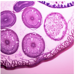
HIV
Virus that infects and damages the immune system
Transmitted through body fluids (blood, semen, vaginal, sharing needles, breast milk)
Can remain dormant in the body for years with no symptoms
HIV antibody test (chart 32-7)
- Enzyme immunoassay (EIA)
- Enzyme-linked immunosorbent assay (ELISA)
If left untreated, causes AIDS
Viral Load- Viral suppression through antiviral therapy decreases the amount of HIV in the blood
AIDS
Advanced stage of HIV infection
Occurs when HIV severely weakens the immune system
Characterized by opportunistic infections
CD4 falls below 200 cells/mm3
32 million people worldwide morbidity

Classification of HIV & Risk Factors
One of five HIV infection stages (0, 1, 2, 3, or unknown).
Stage 0 indicates early HIV infection, lab testing
Stages 1, 2, and 3 are based on the CD4+ T-lymphocyte count; viral load testing
Two markers
- Level of CDG + T-cell
- Plasma HIV RNA (viral load)
Risk Factors
- Sharing infected needles
- Sexual relations with an infected person
- Infants born to mothers with HIV
- Organ transplants, HIV infected blood, blood products (1978-1985)

Stage 1: Acute HIV Infection
Within 2 to 4 weeks after infection with HIV, about two-thirds of people will have a flu-like illness. Symptoms can last from days to weeks.
Fever and/or chills
Rash
Night sweats
Muscle aches
Sore throat
Fatigue
Swollen lymph nodes
Mouth ulcers
Stage 2: Clinical Latency
The virus still multiplies, but at very low levels.
Patients may not feel sick or have any symptoms.
This stage is also called chronic HIV infection.
No treatment- can live 10 to 15 years, but some patients may move quicker through this phase
With antiviral treatment, live long and healthy lives, keeps viral load low or undetected

Stage 3: AIDS
HIV and not on HIV treatment, eventually the virus will weaken the body's immune system, and progress to AIDS (acquired immunodeficiency syndrome).
Late stage of the infection
Symptoms- rapid weight loss, recurring fever, profuse night sweats, extreme fatigue, swelling of the lymph glands, diarrhea, sores in mouth, anus, genitals, pneumonia, neurologic changes, and red, brown, pink, or purple blotches under skin
Opportunistic infections
Stage-3-Defining Opportunistic Illnesses in HIV Infection
Stage-3-Defining Opportunistic Illnesses in HIV Infection

Treatment of HIV
Diagnosis
(1) antibody test (antibodies against HIV)
(2) antibody/antigen test (antibodies against HIV and HIV itself- antigens) ++ Recommended
(3) RNA/DNA test (viral RNA and copies of viral RNA in DNA)
Antiretroviral treatment-Guidelines for Adults and Adolescents
The CD4+ count serves as the major laboratory indicator of immune function and prophylaxis for opportunistic infections, and is the strongest predictor of subsequent disease progression and survival
Goals of ART treatment include:
(1) reduce HIV-associated morbidity and prolong the duration and quality of survival,
(2) restore and preserve immunologic function,
(3) maximally and durably suppress plasma HIV viral load,
(4) prevent HIV transmission
FDA Approved HIV Medications
Called Antiviral therapy (ART)
Combination or treatment regimen
Usually at least 3 HIV medications (2 from different drug classes)
Different oral medicines may be combined in a single pill or capsule
Newer long-acting injectable medicines every 2 months

antiretroviral therapy
antiretroviral therapy

approved HIV meds
approved HIV meds

Clinical Manifestations
Respiratory
Pneumocystis pneumonia (PCP)
- Associated w/ CD4 T-cell count less than 2300 cells/mm
- Progressive dyspnea, fever, nonproductive cough
Mycobacterium Avium Complex (MAC)
- Common infection associated w/ CD4 T-cell count less than 50 cells/mm
- Fever, night sweats, weight loss, fatigue, diarrhea, and abdominal pain
Tuberculosis (TB)
- Can occur with any CB4 T-cell count
- High risk with untreated HIV; high risk for latent TB
- Can develop in the lungs, CNS, bone, pericardium, stomach, peritoneum, and scrotum
- Advanced AIDS is rapid progression and fatal
- PRODUCTIVE GREEN SPUTUM
Gastrointestinal
- Loss of appetite, nausea, vomiting, oral and esophageal candidiasis, and chronic diarrhea
- Symptoms may be related to the direct inflammatory effect of HIV on the cells lining the intestines.
Candidiasis
- Oropharyngeal or esophageal fungal infections
- Creamy white, plague-like lesions
- Women-vulvovaginitis
HIV Wasting Syndrome
- Involuntary weight loss
- More than 10% of body weight
- Diarrhea or weakness > 30 days
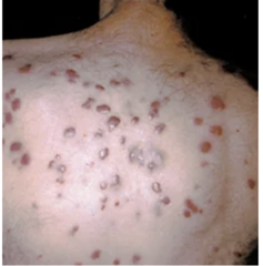
Kaposi Sarcoma (KS)
is caused by human herpesvirus-8 (HHV-8)
Affects eight times more men than women and may spread through sexual contact.
Type of cancer that forms in the skin, lymph nodes, or internal organs; usually involves the epithelial layer of blood and lymphatic vessels.
AIDS-related KS exhibits a variable and aggressive course, ranging from localized cutaneous lesions to disseminated disease involving multiple organ systems.
- Requires frequent skin assessments for infections
-- Provide skin care
- Assess for purple, red, brown lesions on the skin or mouth
Neurologic Manifestations
HIV-related brain changes have profound effects on cognition, including motor function, executive function, attention, visual memory, and visuospatial function
Direct effects of HIV on nervous system tissue, opportunistic infections, primary or metastatic neoplasm, cerebrovascular changes, metabolic encephalopathies, or complications secondary to therapy.
- Subcortical Neurodegenerative Disease
- Peripheral Neuropathy
- HIV Encephalopathy (AIDS dementia)
- Progressive Multifocal Leukoencephalopathy
Prep, or pre-exposure prophylaxis (PrEP)
is a medicine people at risk for HIV take to prevent getting HIV from sex or injection drug use.
PrEP can stop HIV from taking hold and spreading throughout the body.
Reduces the risk of getting HIV from sex by about 99% when taken as prescribed.
2 oral medications approved by FDA
- Truvada (emtricitabine/tenofovir disoproxil fumarate) and
- Descovy (emtricitabine/tenofovir alafenamide)
1 injectable long-acting approved by FDA
- Apretude (cabotegravir)
HIV: Gerontologic Considerations
91,127 persons between the ages of 60 and 64 years are living with HIV infection, and 58,526 persons are living with AIDS (CDC 2019a).
65 years and older, 82,046 persons are living with HIV infection, and an additional 52,995 persons are living with AIDS (CDC, 2019a)
Comorbidities in older persons with HIV/AIDS include type 2 diabetes, non-AIDS cancerous diseases, cardiovascular disease, osteoporosis, and depression
Loneliness has been associated with increased mortality in the general population.
Health Promotion
Behavioral interventions have been effective in reducing the risk of acquiring or transmitting HIV by ensuring that people have the information, motivation, and skills necessary to reduce their risk
HIV testing, because most people change their behaviors to protect their partners if they know they are infected with HIV
Treatment and care enable individuals with HIV to live longer
Correct and consistent use of condoms
Voluntary medical male circumcision (VMMC) is 50% to 60% effective at preventing the acquisition of HIV infection
Reducing the Risk of Transmission to Health Care Providers
The use of standard precautions to prevent exposure, using post-exposure prophylaxis
Appropriate hand hygiene measures
To reduce the risk of exposure of health care workers to HIV, the CDC developed standard precautions
Standard precautions are used when working with all patients in all health care settings, regardless of their diagnosis or presumed infectious status.
Post-exposure prophylaxis (PEP)
- 72 hours after exposure
- Three different drugs
- Urgent medical concern and should be managed immediately after possible exposure—the sooner the better; every hour counts
Supportive Care
Nutritional support may be as simple as assisting in obtaining or preparing meals.
For patients with more advanced nutritional impairment resulting from decreased intake, wasting syndrome, or gastrointestinal malabsorption associated with diarrhea, parenteral feedings may be required.
Imbalances that result from nausea, vomiting, and profuse diarrhea often necessitate IV fluid and electrolyte replacement.
Management of skin breakdown associated with KS, perianal skin excoriation, or immobility entails thorough and meticulous skin care that involves regular turning, cleansing, and applications of medicated ointments and dressings.
Address pain associated with skin breakdown, abdominal cramping, peripheral neuropathy, or KS
Pulmonary symptoms - dyspnea and shortness of breath, may be related to opportunistic infections, KS, or fatigue.
Oxygen therapy, relaxation training, and energy conservation techniques may be effective
Emotional and Ethical Concerns
Health care professionals
- Fear of infection, responsibility for giving care, values clarification, confidentiality, developmental stages of patients and caregivers, and poor prognostic outcomes
- May still are afraid and have anxiety about disease transmission despite education concerning infection control and the low incidence of transmission
ANA's Code of Ethics for Nurses (ANA, 2015)
- "The nurse practices with compassion and respect for the inherent dignity, worth, and unique attributes of every person" (p. 1).
What are the key nursing interventions in managing a newly dx HIV patient?
Establish a therapeutic relationship and provide emotional support.
Educate the patient about HIV, disease progression, and treatment options.
MD initiates ART promptly and monitors for side effects.
Encourage adherence to ART and lifestyle modifications.
Prevent opportunistic infections through vaccinations and prophylactic medications.
Collaborate with interdisciplinary teams for comprehensive care.
How can nurses support patients in adhering to ART therapy?
Educate patients about the importance of adherence to prevent viral resistance.
Simplify medication regimens and provide reminders (e.g., pill organizers, phone alerts).
Address barriers to adherence, such as financial constraints or side effects.
Offer counseling and support groups to encourage adherence.
Monitor for side effects and adjust therapy as needed in collaboration with the healthcare team.
What role does patient education play in preventing HIV transmission?
Educate patients about safe sex practices, including condom use and pre-exposure prophylaxis (PrEP) for partners.
Emphasize the importance of viral load suppression in reducing transmission risk.
Discuss the risks of needle sharing and promote harm reduction strategies.
Encourage routine partner testing and disclosure in a safe environment.
Provide resources for community support and prevention programs.
How can nurses address stigma and mental health concerns associated with HIV?
Provide a nonjudgmental, confidential, and supportive care environment.
Educate patients and families to reduce misinformation and stigma.
Refer patients to mental health professionals for counseling if needed.
Encourage participation in peer support groups for social support.