Liver Cirrhosis
 Call Kai
 Learn
 Practice Test
 Spaced Repetition
 Match
 Flashcards
 Knowt Play
1/76

There's no tags or description
Looks like no tags are added yet.
77 Terms
What is cirrhosis?
End-stage disease of any chronic liver disease
Histology of cirrhosis
Regenerative nodules surrounded by diffuse fibrous tissue with disruption of the architecture of the entire liver.
Why is focal liver fibrosis not considered cirrhosis?
Cirrhosis involves diffuse fibrosis and architectural disruption of the entire liver; focal fibrosis, such as healing of a liver abscess, does not.
How can cirrhosis be clinically classified?
As either compensated or decompensated cirrhosis.
What is the progression from chronic liver disease to cirrhosis?
Liver injury → inflammation → fibrosis and hepatocyte regeneration → cirrhosis.
What are the main causes of cirrhosis in the Western world?
Alcoholic liver disease (60-70%)
Viral hepatitis (10%)
Biliary diseases(5-10%) e.g. primary biliary cirrhosis
Primary Haemochromatosis (5%)
Wilson Disease
Alpha-1 antitrypsin deficiency
Cryptogenic cirrhosis (10-15%)
What are the basic pathological events in the pathogenesis of cirrhosis?
Liver damage followed by replacement of liver tissue by fibrous tissue (fibrosis).
What type of cells are responsible for liver fibrosis?
Stellate cells.
What is the key cellular event in hepatic fibrosis?
Activation of stellate cells.
What is the normal function of hepatic stellate cells?
In the normal liver, stellate cells store vitamin A.
What 4 things happen to hepatic stellate cells when they become activated?
They lose vitamin A, proliferate, differentiate into myofibroblasts and become fibrogenic.
Effects of activated Kupffer cells in liver fibrosis

What happens to protein synthesis when hepatic stellate cells are activated?
Protein synthesis increases.
What happens to collagen synthesis when hepatic stellate cells are activated?
Collagen synthesis increases.
What does activated stellate cell activity lead to (with ECM)?
Formation and deposition of extracellular matrix (ECM).
What is ECM composed of
Collagens
Glycoproteins
Proteoglycans
Which types of collagen are produced in hepatic fibrosis?
Type I and type III collagen.
What glycoproteins are involved in the extracellular matrix of hepatic fibrosis?
Laminin and fibronectin.
What proteoglycans are involved in the extracellular matrix of hepatic fibrosis?
Dermatan and chondroitin sulphate.
What is the consequence of extracellular matrix deposition in the liver?
Formation of a fibrotic scar.
How does a normal liver appear histologically compared with a cirrhotic liver?
A normal liver has conserved architecture, whereas a cirrhotic liver has regenerative nodules surrounded by fibrous tissue.
How does a cirrhotic liver appear grossly?
It has an irregular surface with nodules
What are the major consequences of cirrhosis?
Progressive liver failure
Hepatocellular carcinoma
Disruption of the hepatic microvasculature → Portal hypertension.
Approximately how much hepatic capacity must be lost before hepatic failure develops?
80–90% of hepatic capacity must be eroded.
What can precipitate decompensation in a patient with chronic liver disease?
Intercurrent conditions such as GIT bleeding, sepsis, electrolyte disturbances or major surgery.
What are the three major morphological patterns associated with hepatic failure?
Acute liver failure/massive hepatic necrosis
Chronic liver disease
Hepatic dysfunction without overt necrosis.
Signs of hepatic failure
Jaundice
Coagulopathy
Oedema
Infection
Hepatic encephalopathy
Hepatorenal syndrome
Fetor hepaticus
Hyperestrogenism
What happens to catabolic activity in hepatic failure & what is the outcome of this
Catabolic activity becomes impaired → ↓breakdown of by-products of metabolism & ↓conjugation & excretion of bilirubin → jaundice
What happens to hepatic synthetic activity in hepatic failure & what is the outcome of this
Synthetic activity is impaired → ↓ synthesis of molecules (esp. proteins) → ↓ clotting factor production → coagulopathy
What is hypoproteinaemia in hepatic failure?
A reduction in circulating plasma proteins, including albumin and globulins.
Effect of hypoalbuminaemia in hepatic failure
↓ Albumin → ↓ Plasma oncotic pressure → fluid moves out of blood vessels → oedema.
What is the consequence of low serum globulin levels in hepatic failure?
Reduced immunity and increased susceptibility to infection.
What causes hepatic encephalopathy in liver failure?
Impaired metabolism of ammonia.
What clinical features can result from hyperestrogenism in hepatic failure?
Spider naevi, palmar erythema, hypogonadism and gynaecomastia.
What major systemic complication can occur in severe hepatic failure?
Susceptibility to multi-organ failure.
What is portal hypertension?
Increased pressure within the portal venous system, associated with disruption of the hepatic microvasculature in cirrhosis.
What does the normal liver offer in terms of resistance to portal blood flow?
The normal liver offers almost no resistance to flow.
Why can the normal liver accommodate increased portal blood flow without a major increase in portal pressure?
The normal portal venous system is a low-pressure system and the normal liver can withstand significant increases in flow without a corresponding increase in portal pressure.
Which vessels drain into the portal venous system?
Vessels draining intra-abdominal organs, including the coronary vein & splenic vein, drain into the portal venous system.

What are the four major clinical consequences of portal hypertension?
Ascites
Formation of portosystemic venous shunts
Congestive splenomegaly
Hepatic encephalopathy.
How does portal hypertension contribute to ascites?
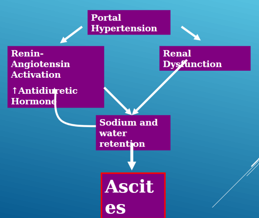
What are portosystemic shunts?
Abnormal venous connections that allow portal blood to bypass the liver and enter the systemic circulation.
What happens to the spleen in portal hypertension?
Congestive splenomegaly can develop.
What are the 3 main categories of primary liver tumours?
Benign tumours, tumour-like lesions and malignant tumours.
Name 5 benign primary liver tumours
Haemangioma
Bile duct adenoma
Von Meyenburg complex
Hepatic adenoma
Hepatobiliary cystadenoma.
Name 2 tumour-like liver lesions
Focal nodular hyperplasia
Angiomyolipoma.
Name 4 malignant primary liver tumours
Hepatocellular carcinoma
Cholangiocarcinoma
Angiosarcoma
Neuroendocrine tumours.
What is a hepatic haemangioma?
A benign tumour of blood vessels and the commonest benign tumour in the liver.
What are the histological features of a hepatic haemangioma?
Proliferated thin-walled blood vessels lined with flat endothelial cells.

How are hepatic haemangiomas commonly detected?
Incidentally on imaging.
Do hepatic haemangiomas usually require intervention?
No. They rarely require intervention.
What is a bile duct adenoma - describe using histology
A well-circumscribed benign lesion composed of closely packed tubules and acini in fibrous stroma, without atypia.

What is another name for bile duct adenoma?
Peribiliary gland hamartoma.
What is a Von Meyenburg complex?
A biliary microhamartoma caused by a developmental malformation involving persistence of the embryonic ductal plate.
What are the histological features of a Von Meyenburg complex?
Nodular lesion
Proliferated ductular structures containing bile
Loose fibrous stroma

Can Von Meyenburg complexes be multiple? If so, when
Yes. They can be multiple.
Associated with polycystic liver and kidney disease
What is focal nodular hyperplasia (FNH)?
A benign, non-neoplastic, tumour-like focal lesion of the liver.
What is the gross/histological appearence of focal nodular hyperplasia?
A central stellate scar with radiating fibrous septae.
Bile duct proliferation
Lymphocytes

What vascular feature is characteristic of focal nodular hyperplasia?
Thick-walled arteries.
What staining pattern is characteristic of focal nodular hyperplasia?
A characteristic geographical staining pattern with glutamine synthetase.
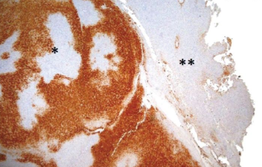
Why can focal nodular hyperplasia cause diagnostic difficulty?
It can mimic a tumour on imaging and can mimic cirrhosis on core biopsy.
What is hepatocellular carcinoma (HCC)?
A malignant primary liver tumour composed of neoplastic cells resembling hepatocytes.
In what setting do liver cancers generally arise?
In general, liver cancers arise in the setting of cirrhosis.
What is the most common cause of liver cancer worldwide
Viral hepatitis, particularly hepatitis B virus (HBV).
What is the most common cause of liver cancer in the United States?
Chronic alcoholism.
What tumour marker is associated with hepatocellular carcinoma?
Raised alpha-fetoprotein (AFP)
What is a characteristic architectural feature of HCC?
Thickened hepatocyte cell plates containing more than 3 cells.
What is the role of reticulin staining in HCC?
Reticulin highlights the cell plates and can demonstrate their abnormal thickening.
What hepatocyte marker can be expressed by HCC?
Hep Par 1 (hepatocyte marker).
Does HCC still produce bile?
Yes. HCC forms small bile canaliculi & can produce bile.
What features on histology can support a diagnosis of well-differentiated HCC?
Neoplastic cells resembling hepatocytes, thickened cell plates, abnormal reticulin architecture, Hep Par 1 positivity and formation of bile canaliculi.

What can CD10/CEA staining demonstrate in HCC?
It can demonstrate canalicular staining.
What is cholangiocarcinoma?
A malignant tumour of the intrahepatic or extrahepatic bile ducts.
What type of carcinoma is cholangiocarcinoma?
An adenocarcinoma.
Is cholangiocarcinoma associated with cirrhosis?
No
Are metastatic tumours to the liver common compared with primary liver cancers?
Yes. Metastases to the liver are far more common than primary liver cancer.
Name 2 metastatic cancers that can → liver
Metastatic breast carcinoma
Metastatic colorectal adenocarcinoma