blood sampling in small mammals and reptiles
 Call Kai
 Learn
Learn Practice Test
 Spaced Repetition
 Match
Match Flashcards
 Knowt Play
1/90

There's no tags or description
Looks like no tags are added yet.
Name | Mastery | Learn | Test | Matching | Spaced | Call with Kai | Chat |
|---|
No analytics yet
Send a link to your students to track their progress
91 Terms
what is the circ vol of rabbits
55-70ml/kg
what is safe vol to collect from rabbit
10% of circ vol
5.5-7ml/kg
what does rabbit blood tend to do
clot quickly
size of v, flow into syringe is slow
what tubes should be used for rabbits and what else should do
EDTA for haematology
Serum or Lithium Heparin for biochemistry
Fluoride Oxalate for glucose
bc serum tube lowers glucose
Two fresh blood smears
what v for rabbits
Jugular → more common
Cephalic
Saphenous
Marginal ear vein (not middle artery)→ more common

where is v and a

why not use central ear a
Best to avoid using artery to avoid pinna necrosis→ would create a larger area of necrosis
higher pressure so risk haematoma
why do you still need to care with marginal ear v
•Pinna necrosis if poor/rough technique, but smaller area effected compared to artery.

difficulties of jug v in rab
Dewlap can get in the way
Restraint may inhibit respiration
Poor/rough technique may cause blindness in the ipsilateral eye

why care with restraint in saphenous v in rab
kick out and damage back

Saphenous vein restraint
blood vol of g pig
§70-75ml/kg (on average)
safe vol to sample in g pig
1% BW rule (or 10% blood volume)
when should you limit blood sample size in small mamals and exotics
if anaemic or in circulatory shock.
why is it better to use a % of total blood vol rather than bw to know how much to sample
using 10% of blood vol is a lower vol than using bw
so safer

what v use in g pig
Cephalic vein (situated more lateral)
Cranial vena cava most useful for large blood draws
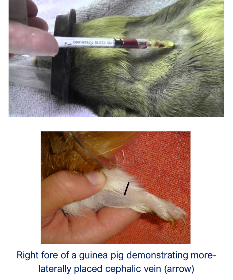
when can you only do cr vena cava in g pig
under sed or ga
risk of using cr vena cava in g pig
blind
risk puncture heart
ga sed risk
blood vol rat, mouse hamster gerbil
§60-70ml/kg (on average).
safe vol to take in rat, mouse hamster gerbil
10% blood volume
locations for rat mouse gerbil hamster
Lateral tail vein - warm tail first. Not suitable in gerbils → tail slip
Lateral saphenous vein
Cranial vena cava

locations for blood sample in ferret
Jugular vein (more lateral compared to cat)
Cranial vena cava
why is less risk to do cr vena cava in ferret
heart sits further back so less risk cardiac puncture
considerations in blood sample ferret
Consider chemical restraint
Skin can be very thick especially in mature hobs
blood vol of ferret
50-70ml/kg
locations in hedgehog
Cranial vena cava
Jugular vein (hard to visualise due to thick skin)
Femoral vein (inside hind leg)

considerations when using cranial vena cava method
Potential for severe complications if performed incorrectly
In some species (e.g. guinea pig, hedgehog) the heart lies very close to sample site.
Thoracic pathology may change normal anatomy (radiograph first helps)
Anaesthesia is essential
cr vena cava method
Patient in dorsal recumbency, head extended, and forelimbs retracted caudally
25G, 0.5" needle (or similar)
Needle placed at the notch where first rib meets the manubrium (bone at top of sternum)
Advance towards the contra-lateral hindlimb
Approximate 30o angle until blood flash


what does this highlight
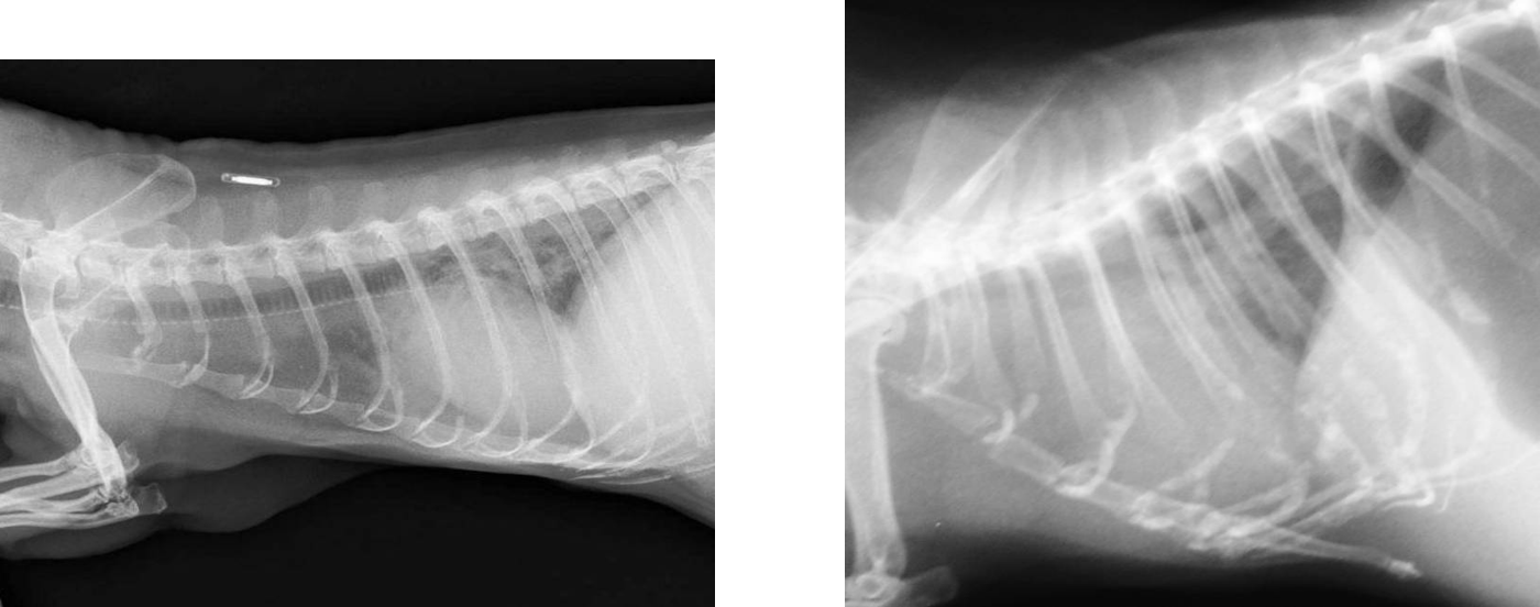
ferret l
g pig r
Note different in the heart’s position → risk less in ferret
why do we use landmarks in reptiles
Most veins are not visible = use landmarks and feel
why may we contam blood samples in reptiles
The lymphatic system is closely associated with blood vessels which may lead to contamination of blood samples
other issue with reptile venipuncture
Blood pressure low and blood draws slowly
why not use edta in reptiles
EDTA tends to haemolyse reptile red blood cells.
what anticoagulant in reptiles
Lithium Heparin is anticoagulant of choice
what else should be made with reptiles
Make two good quality fresh smears
how do we perform haematology in reptiles
Haematology is usually performed manually since automated techniques are made difficult due to nucleated red blood cells
why is good skin prep in reptiles important
Skin is often heavily contaminated so ensure thorough skin preparation.
eg salmonella
what is approx circ vol of reptiles, what is safe to take
Approximately 5-8% of body weight
May collect up to 10% (i.e. 5-8ml/kg)
BUT many reptiles are presented in poor to very poor health and are often anaemic
Therefore, often take half this amount
Rarely require more than 2ml
samp site in lizard
ventral coccygeal v
ventral abd v
jugular
how do ventral coccygeal v in lizard
Entry approximately 1/3rd down the tail to avoid hemipenes and anal glands
Ventral or lateral approach may be used

risk of ventral coccygeal v
necrosis (esp b dragons, iguanas)
tail autotomy (geckos) → ga can help reduce

why not use ventral abd v
Difficult to prevent haematoma

benefit of ventral abd v
§May be visualised in some species
when better to use jug v
Useful in large, longer-necked species such as Monitor lizards

risk of using jug v
Blind approach along imaginary line from dorsal ear to shoulder
but Often quite superficial
sample sites in snakes
ventral coccygeal v
heart
palatine v (can be cannulated)
when is harder to use ventral coccygeal v in snake
§Difficult in small snakes

why can we sample from heart in snake
low bp and flow in heart
so less risk of losing blood from the hole we have created
how sample from heart in snake
Dorsal recumbency
Heart located visually, with doppler or ultrasound
Stabilise heart between finger and thumb
Needle entry ventral midline at a 30-degree angle in a cranial direction (aim for caudal point of ventricle)
always under ga

risk of palatine v
Prone to haematoma formation
how do palatine v
Large, anaesthetised snakes only

sample sites in chelonians
jugular v
subcarapacial v
dorsal coccygeal v
features of using jug v in chelonians
Along line from dorsal ear scale to shoulder
Tends to be superficial
Visible in some species
Lymph contamination least likely
Carotid artery deeper and more ventral


how do subcarapacial v/sinus in chelonians
Entry point craniodorsal midline where skin joins carapace
Aim for point at junction of 1st & 2nd vertebral scute on midline
May need to bend needle slightly
Head may be retracted or extended

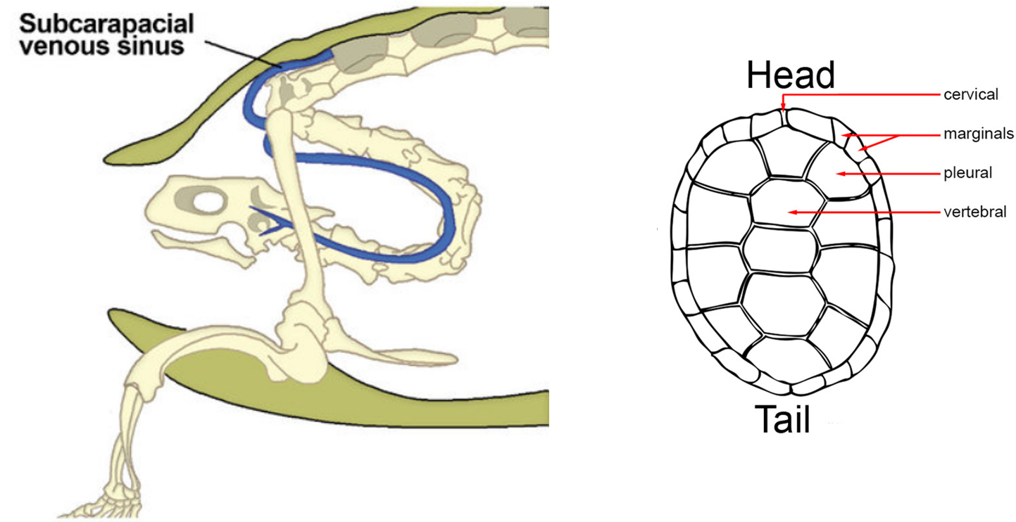
how do dorsal coccygeal v
Often superficial
Aim as far cranial as possible

risk of using subcarapacial v/ sinus
Often lymph contaminated
risk of using dorsal coccygeal v
Often lymph contaminated
anisocytosis
= RBCs that are unequal in size
polychromasia
high number of immature red blood cells
features of rabbit rbc
Anisocytosis and polychromasia is a common, normal finding
May be due to shorter erythrocyte life span (57-67 days)

rabbit heterophils
Heterophils have granular cytoplasm and may be mistaken for eosinophils.
het should have rod shaped granules (eos more round)
rabbit basophils
Basophils are present quite commonly.
rabbits lymphocytes, eos, monocytes
§Lymphocytes, eosinophils and monocytes similar to dog/cat.

Representative rabbit blood cells
(A) RBCs and platelets.
(B) Heterophil
(C) Lymphocyte
(D) Eosinophil
(E) Basophil
(F) Monocyte
what happens to rabbit wbc in infection/ stress
Typically, the heterophils (neutrophil) to lymphocyte ratio is 1:1 (45%:45%).
The total white cell count rarely increases, instead the neutrophil : lymphocyte ratio changes in response to stress or disease.
Relative neutrophilia and lymphopaenia occurs with infection and chronic stress

rabbit
heterophil
what is kurloff cell in g pig
produced by the thymus under oestrogen stimulation. Resembles lymphocyte with round to oval inclusions
is still present in male g pigs

function of kurloff cell
protect fetus from maternal immune functions
do g pigs have neutrophils or heterophils
g pigs have heterophil
ferret neutrophil
contain small, pale-red granules

ferret eosinophil
granules rounded and bright red

how are ferrets erythron diff
have higher HCT, HB, and RBC counts
how are ferrets wbc diff
White cell count generally quite low (3-4 x10⁹)
blood groups in ferrets
no
features of blood cells in reptiles
Both cell types are nucleated
what is the pcv like in reptiles
§PCV 20-40% in most species
probs due to lifespan
erythrocyte lifespan in reptiles
Erythrocyte life span up to 800d in some species
why do you need to care when blood sampling reptiles (or of know blood has been lost)
May take 4 months for losses to be replenished
what is Polychromasia and reticulocytosis like in reptiles
Polychromasia and reticulocytosis low (<1%)
describe heterophils in reptiles
Predominant cell type
Equivalent to mammalian neutrophils
Intracytoplasmic fusiform granules
Increase in summer, decrease during hibernation

what happens to hetrophils in infection, inflam and stress in reptiles
Heterophilia
what happens to heterophils in chronic stress/ disease, severe infection
Heteropaenia
probs due to slow replenishment
‘Toxic’ heterophils in severe inflammation

features of reptile lymphocytes
Morphology very similar to mammals
Lymphocytes generally lowest in winter
what can cause Lymphopaenia ( low)
malnutrition, stress immunosuppression and poor husbandry
what can cause lymphocytosis (high)
= inflammation, parasitic and viral disease, wound healing, ecdysis
features of reptile eosinophils
Eosinophils generally lower in summer
Eosinophilia = parasites and inflammation
features of reptile basophils
Basophils minimal seasonal change
Basophilia = parasitic and viral disease
what is azurophil in reptiles
Unique to reptiles
Like monocytes in mammals (but still have mono too)
Red-purple cytoplasm
More common in snakes

reptile

monocyte
what happens in reptiles to monocytes and azurophils in inflam and granulomatous disease
Monocytosis & Azurophilia