Axial Skeleton Bones, Skull Bones, Muscles of Head Neck and Trunk
 Call Kai
 Learn
 Practice Test
 Spaced Repetition
 Match
 Flashcards
 Knowt Play
1/130

There's no tags or description
Looks like no tags are added yet.
131 Terms
Vertebral column
the spine, formed of a number of individual bones called vertebrae and two composite bones (sacrum and coccyx)

Body of Vertebra
big circular part, is weight bearing

vertebral foramen
canal through which spinal cord passes

pedicle of vertebra
attached to and extends posteriorly on either side of the body

Lamina of vertebra
supporting bone between the spikes of the vertebra

spinous process
Posterior projection of a vertebra...usually points down

Transverse process of vertebra
forms joint with ribs in thoracic region, site for muscle attachment in lumbar region

superior articular process
bony process that extends upward from the vertebral arch of a vertebra that articulates with the superior articular process of the next upper vertebra

inferior articular process
bony process that extends downward from the vertebral arch of a vertebra that articulates with the superior articular process of the next lower vertebra

superior articular facet
the part of the superior articular process that articulates with the superior bone to it

inferior articular process
the part of the inferior articular process that articulates with the inferior bone to it

Intervertebral disks
23 total, disks in the spine

cervical vertebrae
C1-C7, have transverse foreamen, has arteries

transverse foramen
holds arteries in cervical vertebra

Atlas
C1, the first vertebra, fits into C2

transverse ligament
This ligament attaches to the lateral masses of C1 to hold the dens in place

Axis C2
has bump where C1 fits into (shake head)

Dens (odontoid process)
a bony projection that extends upward from the vertebral body where C1 rotates on

Thoracic Vertebrae (T1-T12)
second set of 12 vertebrae; they articulate with the 12 pairs of ribs to form the outward curve of the spine

costal facets of thoracic vertebrae
where head of rib connects

Lumbar Vertebrae (L1-L5)
third set of five larger vertebrae, which forms the inward curve of the spine, lack facets and foreamer

Sacrum (S1-S5)
Shapes the posterior wall of pelvis
Formed from 5 fused vertebrae
Superior surface articulates with L5
Inferiorly articulates with coccyx
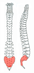
anterior sacral foramina
holes on the front of the sacrum

Posterior sacral foramina
holes on back of sacrum

sacral canal
continuation of vertebral canal

sacral hiatus
inferior opening of the sacral canal

ala of sacrum
The wing-like projections on either side of the sacrum.

auricular surface
where pelvic bone articulates with sacrum

superior articular process of sacrum
paired processes that extend upward from the sacrum to articulate (join) with the inferior articular processes from the L5 vertebra

superior articular facet of sacrum
where smooth surface is on process

Coccyx
Tailbone

Sternum
Breastbone

manubrium
upper portion of the sternum

clavicular notch
where sternum connects to clavicles

sternal angle
horizontal ridge across front of sternum where manubrium and body meet

body of sternum
large middle portion of the sternum

costal notches of sternum
indentations on sternum where costal cartilages from the ribs articulate
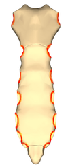
xiphoid process
lower, narrow portion of the sternum

True ribs (1-7)
Cartilage leads to sternum

false ribs (8-12)
ribs that do not have a direct attachment to the sternum

floating ribs
last two pairs of ribs; do not attach to sternum

head of rib
Articulates with the costal facet of a thoracic vertebral body
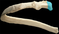
superior articular facet of rib
articulates with inferior costal demifacet of the thoracic vertebral body one numeric segment superior

inferior articulating facet of rib
articular surface at bottom of rib
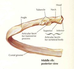
neck of rib
between head and tubercle

tubercle of rib
bump on head (lower) points down

Angle of rib
where rib bends

Costal groove of rib
groove on the inferior side of the rib shaft

hyoid bone
a U-shaped bone in the neck that supports the tongue.

body of hyoid bone
middle of bone

greater cornu/horns of hyoid bone
long projections on back

lesser cornu/horns of hyoid bone
teeth like projections

Cranial Bones
skull bones: ethmoid, frontal, occipital, parietal, sphenoid, and temporal

frontal bone
forehead

supraorbital foramen
opening above each orbit allowing blood vessels and nerves to pass

parietal bones
Bones that form the sides and top of the cranium.

temporal bone
bone that forms parts of the side of the skull and floor of the cranial activity. There is a right and left temporal bone. By the ear

Zygomatic arch of temporal bone
cheek bone

zygomatic process of temporal bone
extension from the temporal bone that forms the posterior portion of the zygomatic arch

mandibular fossa of temporal bone
Articulates with the head of the mandible to form the temporomandibular joint. The mandible (lower jaw) joins with the skull at this site.
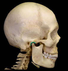
External auditory/acoustic meatus of temporal bone
opening in the temporal bone extending to the inner ear

sphenoid bone
forms part of the base of the skull and parts of the floor and sides of the orbit...looks like a butterfly

lesser wing of sphenoid bone
anterior and superior to greater wing on the interior of the skull

optic foramen of sphenoid bone
opening to the optic canal

greater wing of sphenoid bone
posterior and inferior to lesser wing on the interior of the skull, also visible in the posterior orbit and lateral side of the skull

sella turcica of sphenoid bone
houses the pituitary gland

lateral pterygoid plate of sphenoid bone
sharp process that laterally and inferiorly projects from the sphenoid

medial pterygoid plate of sphenoid bone
Location: Sphenoid bone (inferior surface)
Thin plate of bone attached to the pterygoid process, longer and more narrow than lateral pterygoid plat, lower end forms hook-like pterygoid hamulus, lateral surface forms wall of pterygoid fossa, medial surface forms lateral boundary of corresponding posterior nasal aperature
Tendon of tensor veli palatine muscle deflects around the pterygoid hamulus, pterygomandibular raphe is attached to pterygoid hamulus

ethmoid bone
forms part of the posterior portion of the nose, the orbit, and the floor of the cranium

perpendicular plate
forms superior part of nasal septum

Cribiform plate of ethmoid
forms the roof of the nasal cavity and has holes through which axons of the olfactory nerves pass.

crista galli of ethmoid bone
superior projection in the middle of the cribriform plate of the ethmoid bone; providing a point of attachment for the dura mater, helping to secure the brain within the skull

Vomer bone
forms the base for the nasal septum

Maxillary bone (maxilla)
Carries the upper teeth and forms the boundary of three cavities: the roof of the mouth (palate), the floor of the nose, and the floor of the orbits.

infraorbital foramen of maxilla
opening under the orbit that forms a passageway for the infraorbital artery and nerve

palatine process of maxilla
roof of the mouth

palatine bone
either of two irregularly shaped bones that form the back of the hard palate and helps to form the nasal cavity and the floor of the orbits

greater palatine foramen
the largest of three openings located in the posterior region of the hard palate

Zygomatic bone
the arch of bone beneath the eye that forms the prominence of the cheek

zygomatic arch
front part of arch that meets the occipital bone

nasal bone
bridge of nose

lacrimal bone
small fragile bone making up part of the front inner walls of each eye socket and providing room for the passage of the lacrimal ducts
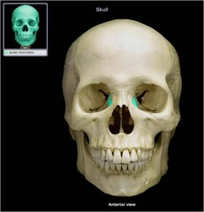
Mandible
lower jaw bone

body of mandible
the horizontal portion of the lower jaw

mental foramen of mandible
one of two holes located on the anterior surface of the mandible. It permits passage of the mental nerve and vessels.

angle of mandible
a bony angle formed by the junction of the posterior edge of the ramus of the mandible and the inferior surface of the body of the mandible; marks widest part of lower 1/3 of face.

ramus of mandible
vertical part of mandible

coronoid process of mandible
flattened upward projection from the anterior margin of the mandibular ramus

condyloid process of mandible
Portion of the mandible that fits into the mandibular fossa and forms the TMJ joint

Mandibular condyles
articulate with mandibular fossae to form temporomandibular joints

mandibular notch
Notch between condyles in mandible

mandibular foramen
hole in back of jaw where novocaine goes

Levator scapulae m.
elevates scapula

(anterior, middle, posterior) scalene muscles
•Work with sternocleidomastoid to flex neck
•Pull cervical vertebrae toward 1st and 2nd ribs (lateral flexion of neck)
•Elevate 1st and 2nd ribs during forced inhalation

Sternocleidomastoid m.
Bilaterally, flexes cervical portion of vertebral column
Unilaterally, rotates head to the side opposite the contracting muscle

Sternohyoid m.
larynx and hyoid depression

Sternothyroid m.
thyroid cartilage and hyoid depression

Digastric m.
open mouth and depress mandible

Mylohyoid m.
hyoid elevation

Frontalis m.
draws scalp anteriorly, raises eyebrows, wrinkles forehead
