3.2
 Call Kai
 Learn
 Practice Test
 Spaced Repetition
 Match
 Flashcards
 Knowt Play
1/45

There's no tags or description
Looks like no tags are added yet.
46 Terms
subdivisions of somites

recall that when cells are born out of the tailbud region, they alr know their
R/C axis from differential Mesp2 expression and A/P from differential Hox gene expression
the first couple somites contribute to the base of the skull, so we are ignoring them for this class
somites form
R—>C
EMT is used to make
sclerotome
medial dermamyotome becomes the
primaxial/epaxial skeletal muscles (muscles at the base of the skull)
intercostal muscles
intevertebral muscles of the back

central dermamyotome becomes
dermis of the back
brown fat
muscle stem cells (satellite cells)

lateral dermamyotome becomes
abaxial/hypaxial skeletal muscles (ventral skeletal muscle of the body wall)
diaphragm
limb musculature
tongue

sclerotome becomes
cartilage
bone
tendons of axial skeleton
somitocoel cells become
outer part of intervertebral disks
satellite cells
3 parts of dermamyotome
medial
central
lateral

brown fat
regulates/maintains high temp by decoupling ATP in oxidative phsophorylation
satellite cells
help make more muscle
central dermamyotome does not contribute to any
skeletal muscle (unlike medial and lateral dermamyotome)
after they form, somites are patterned along their
D/V and medial/lateral (M/L) axes
somites are not born with these axes
Exps that showed us that DV and ML axes are acquired after formation of somites
somite explantation and rotation
if flipped 180º, somites form per normal if from a younger somite (0) but NOT if in an older somite (S3/4), which already specified its DV axes
if do same thing in young cell, no change, but flips ML axis if S5/6
so something happens between these somite stages
ML is acquired after DV axis
someite explantation Exps: S1-3 express Pax3 but older somites 4-5 express Pax1 in sclerotome but Pax3 in dermamyotome (so dorsal is normal and ventral is induced state)

medial dermamyotime expresses skeletal muscles earlier than
lateral
so ML is a function of time
ML is a consequence of
BMP gradient
BMP coming from lateral and inhibitors more medially

D/V patterning
ventral has high Shh from notochord and ventral part of the neural tube
Wnt is high dorsally

high concentrations of ______ from the notochord induce the V side
Shh
grafted notochord to dorsal location, and ventralized scelrotome forms dorsally

EMT helps form
sclerotome
dermamyotome is more
dorsal
myotome formation (skeletal muscle cells)
earliest marker of commitment to this lineage is MyoD or Myf5 (TFs that lead to skeletal muscles)
myoblasts do EMT to underneath the dermamyotome to form dermamyotome
stop dividing and fuse to form multinucleated skeletal muscle fibers

in mice, Myf5 turns on first and activates MyoD
in chx, the reverse occurs: MyoD turns on first and activates Myf5
primaxial/epaxial muscles
epaxial comes from DML (dorsal medial lip) of the dermamyotome and forms axial skeletal muscles
muscles at base of skull, intercostal muscles, most intevertebral muscles of the back
stuff from DML commits sooner to its given lineage than things from the VLL

abaxial/hypaxial skeletal muscles
hypaxial form from the VLL (ventro-lateral lip of the dermamyotome)
migrate away and become skeletal muscles of the ventral body wall (abs), diaphragm, limb, tongue
so determined after they get the the correct location

2 types of somite-derived skeletal muscle
primaxial/epaxial
abaxial/hypaxial
DML gives rise to
epaxial/primaxial myotome THEN epaxial/primaxial skeletal muscle
VLL becomes
abaxial/hypaxial myotome THEN abaxial/hypaxial skeletal muscle
primaxial/epaxial
comes from DML
primaxial vs abaxial
primaxial: from medial part of dermamyotome
abaxial: from lateral part of dermamyotome

epaxial vs hypaxial
epaxial: innervated by dorsal ramus
hypaxial: innervated by ventral ramus$
based on how these muscles are later innervated
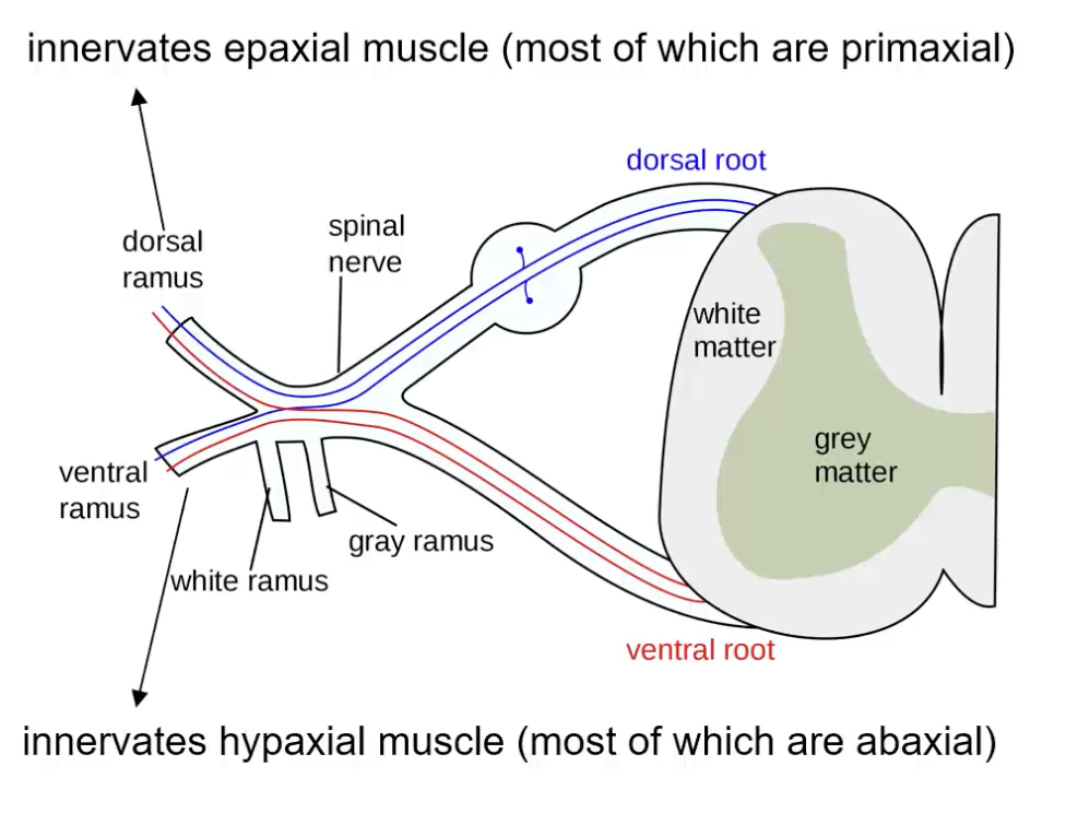
although epaxial vs hypaxial and primaxial vs abaxial have diff meanings,
we just say that primaxial is epaxial and abaxial is hypaxial
they are not quite synonyms, but are just saying that they are

initiation of epaxial skeletal muscle
little bit of Shh from notochord and ventral part of the neural tube, which primes these cells to later respond to Wnt from dorsal neural tube and ectoderm
Myf5 or MyoD activated first and activates the other
later decr in BMP prepares myoblasts for EMT (migrate down)
DML also produces Noggin (activated by Wnt), which —| BMP (to get BMP lower and initiate the EMT)
to actually move down, must be coordinated with movement of neural crest cells as well (reciprocal interactions between the two)

lateral part of dermamyotome is too far from neural tube and notochord to receive
much Shh

myotome precursors cells must undergo EMT to form myotome at same time as
neural crest cells that left dorsal part of neural tube get into the same area
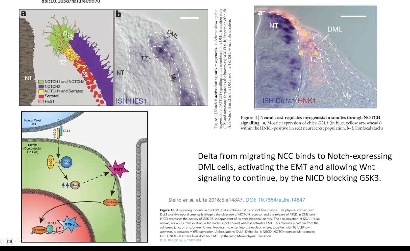
coordinated via a Notch-Delta high-five interaction
Delta is on neural crest cells
DML (dorsal medial lip) cells express Notch
they touch each other and undergo EMT and continue along their journey
so their contact is what initiates their EMT to continue migrating
so cross-talk between somite-derived and neural tube-derived cells
abaxial/hypaxial muscles differentiation does not occur until
these cells are out in the periphery
delayed by BMP4 from LPM so that the cells migrate to their location before differentiating
lots of BMP4 laterally
once they get laterally, they get Wnt and Shh that initiate myogenesis in the limb bud area

central dermamyotome becomes
dermis
responding to later signals of ectoderm and NT

cartilage vs tendon differentiation
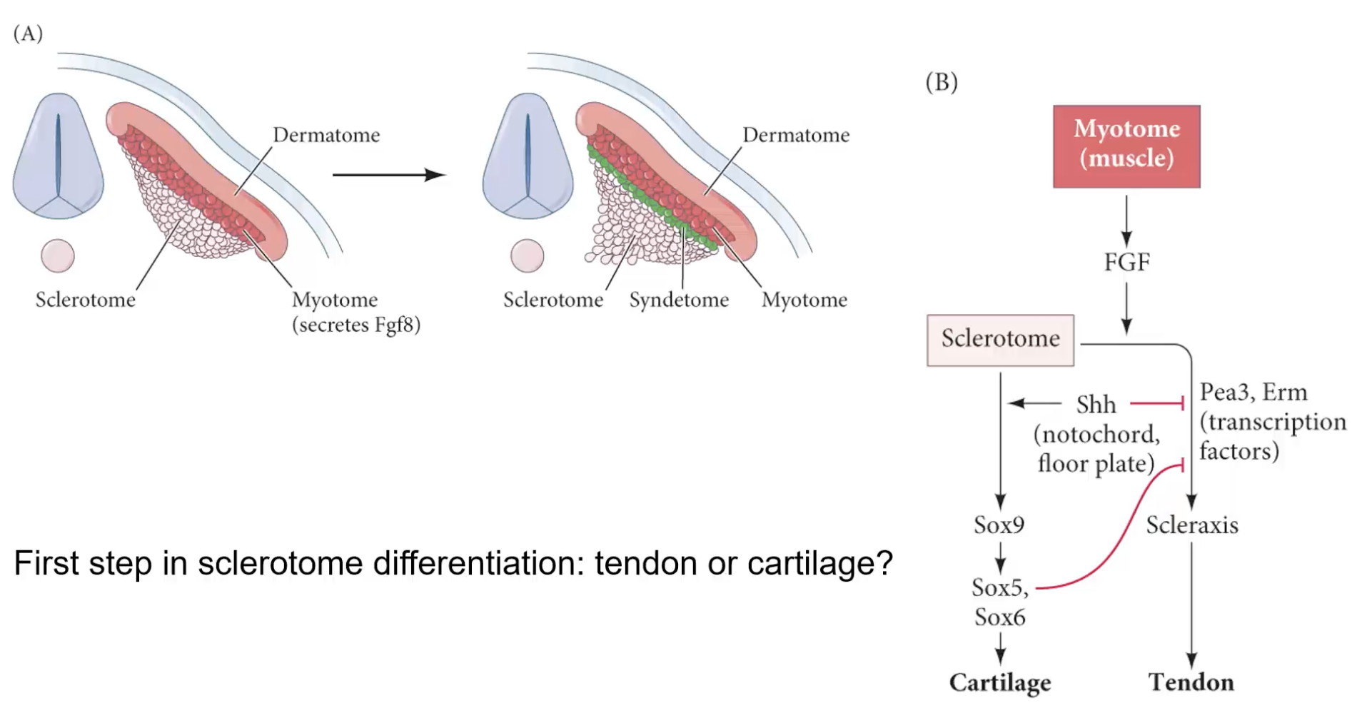
sclerotome becomes the cartilage and tendon
early FGF goes sclerotome from myotome
myotome laterally secretes Fgf8, and shifts dorsal-most sclerotome from cartilage path to a tendon path
other sclerotome cells that didn’t get Fgf8 go cartilage path
vertebrae are mostly derived from sclerotome, and sme from
notochord itself
inner part of each intervetebral disk (nucleus pulposus) is from notochord
dermamyotome makes
skeletal muscle

how to offset muscle vs bone to form vertebrate organization the way it is in adults
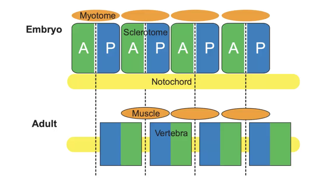
must offset sclerotome and myotome derivatives so that bone and muscle alternate (resegmentation)
sclerotome moves while dermamyotome stays put
sclerotome moves differently if from A or P side of embryo
vertebrate formation/resegmentation
sclerotome cells precursor split A/P sides themselves
each vertebrate comes from two diff somites
rostral half of one somite and the caudal half of another fuse to form a single vertebrate (resegmentation)
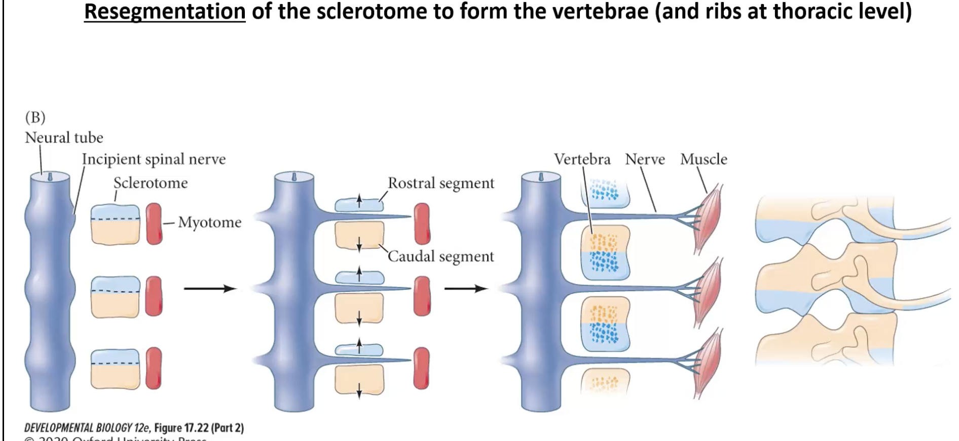
segmentation
makes somites
resegmentation
splitting sclerotome into R/C or A/P halves to form the vertebrae
vertebrate derivatives
sclerotome—> vertebral body
notochord—> central part of intervertebral cushions (nucleus pulposus)
somitocoel OR sclerotome—> anulus fiberosus (outer part of intervertebral disks)
