SBMS1431: Cell-Cell Communication and Signaling
 Call Kai
 Learn
Learn Practice Test
 Spaced Repetition
 Match
Match Flashcards
 Knowt Play
1/45

There's no tags or description
Looks like no tags are added yet.
Name | Mastery | Learn | Test | Matching | Spaced | Call with Kai | Chat |
|---|
No analytics yet
Send a link to your students to track their progress
46 Terms
Different Forms of Cell-Cell Communication
Local communication
Long-distance Communication
Local communication
Gap junctions
Contact-dependent signals (e.g., juxtacrine signals)
Autocrine signals
Paracrine signals
Long-distance Communication
Endocrine signals (Hormones)
Neurotransmitters (of the nervous system)
Neurohormones (of the nervous system)
Gap Junctions
Gap junctions link adjacent cells and are formed by connexons.
Each connexon is made of connexins that form a channel.
Their open and closed states are regulated.
When open, they allow ions and small molecules to pass directly between adjacent cells down the concentration gradient.
Flow of chemical messengers via gap junction can be bidirectional.

How do signaling molecules (ligands) differ based on their chemical nature, and how do they work?
1. Lipophilic / small (e.g., steroids)
Bind to intracellular receptors.
Produce genomic action: activation or repression of gene expression → changes in protein synthesis.
2. Lipophobic / hydrophilic / polar / large (e.g., amines, proteins)
Bind to cell surface membrane receptors.
Produce nongenomic action: transient changes in the cytoplasm.
For surface receptor binding:
Uses signal transduction (a signaling cascade).
Leads to second messengers or changes in enzyme/biochemical activity.
Results in amplification and termination of signals.
Chemical Classification of Messengers
Class | Chemical property | Location of receptors on target cell | Functional classification |
|---|---|---|---|
Amino acids | Lipophobic | Plasma membrane | Neurotransmitters |
Amines* | Lipophobic | Plasma membrane | Paracrines, neurotransmitters, hormones |
Peptides/proteins | Lipophobic | Plasma membrane | Paracrines, neurotransmitters, hormones |
Steroids | Lipophilic | Cytosol† | Hormones |
Eicosanoids | Lipophilic | Cytosol | Paracrines |
* One exception is the thyroid hormones, which, although amines, are lipophilic and have receptors in the nucleus of target cells.
† A few steroid hormones have receptors on the plasma membrane.
Signal Transduction Mechanisms for Selected Chemical Messengers
Messenger | Functional class | Chemical class | Signal transduction mechanism |
|---|---|---|---|
Epinephrine | Hormone, neurotransmitter | Amine | G protein–coupled receptors |
Thyroid hormones | Hormone | Amine (lipophilic) | Altered transcription of mRNA |
Vasopressin (ADH) | Hormone, neurotransmitter | Peptide | G protein–coupled receptors |
Insulin | Hormone, neurotransmitter | Peptide | Enzyme-linked receptors |
Estrogen | Hormone | Steroid | Altered transcription of mRNA |
Glutamate | Neurotransmitter | Amino acid | Channel-linked receptor, G protein–coupled receptors |
Serotonin | Neurotransmitter, paracrine | Amine | Channel-linked receptor, G protein–coupled receptors |
Prostaglandins | Paracrine | Eicosanoid | G proteins, unknown for many |
Interleukins | Paracrine | Peptide | Enzyme-linked receptors |
GABA | Neurotransmitter | Amino acid | Channel-linked receptor |
Choline derivative
A neurotransmitter made from choline. Example: Acetylcholine (used in memory, muscle movement, and autonomic function).
Biogenic amines
Small molecules derived from amino acids, often involved in mood, arousal, and alertness.
Examples: Dopamine, epinephrine, norepinephrine, serotonin, histamine.
Amino acids
The building blocks of proteins, but also act as fast neurotransmitters.
Examples: Glutamate (excitatory), GABA and glycine (inhibitory), aspartate.
Purines
Molecules normally involved in energy metabolism (like ATP), but also act as signaling molecules. Examples: ATP, ADP, adenosine.
What are the two factors that determine whether chemical messengers bind intracellular receptors or membrane receptors, and how do they dictate receptor location?
1. Chemical nature (water soluble/lipophobic or lipid soluble/lipophilic; polar or nonpolar)
2. Molecular size
Target-cell receptors may be found in the nucleus, in the cytosol, or on the cell membrane.
Lipophilic Messengers / Signal Molecules
What they are: Chemical messengers that are lipid-soluble (lipophilic) and small in molecular size — e.g., steroid hormones (cortisol, estrogen, testosterone), thyroid hormone, vitamin D.
Why they can go inside: Because they are fat-soluble, they diffuse freely through the phospholipid bilayer of the cell membrane — no door needed.
Where they bind: Intracellular receptors — located in the cytosol or nucleus of target cells. These receptors are readily accessible to them.
What they do: Produce genomic action — activation or repression of gene expression → changes in protein synthesis.
Speed: Relatively slow — cell response may not be noticeable for an hour or longer (because it takes time to make new proteins).
Step-by-step mechanism of genomic action for lipophilic messengers
Hormone diffuses through the plasma membrane into the cell.
(1a) If the receptor is in the nucleus → hormone diffuses into the nucleus and binds → forms a hormone-receptor complex.
(1b) If the receptor is in the cytosol → hormone binds there → the complex then enters the nucleus.
Inside the nucleus, the hormone-receptor complex acts as a transcription factor → binds to a region of DNA called the hormone response element (HRE), located at the beginning of a specific gene.
Binding to the HRE activates or deactivates the gene → affects transcription of mRNA → ultimately increases or decreases synthesis of the protein coded by that gene.
mRNA moves into the cytosol.
mRNA is translated by ribosomes → yields new proteins.
Lipophobic Messengers / Signal Molecules
What they are: Chemical messengers that are polar (water-soluble/lipophobic) or large in molecular size — e.g., peptides, proteins, catecholamines (epinephrine, norepinephrine), serotonin.
Why they stay outside: They cannot pass through the cell membrane (lipid bilayer blocks them).
Where they bind: They remain in the extracellular fluid and bind to receptors on the cell membrane.
What they do: Exert effects through signal transduction / second messenger systems → alters activity of enzymes inside the cell → non-genomic action.
Speed: Very rapid — responses within milliseconds to minutes
The signal transduction mechanism of lipophobic messengers, including the roles of first and second messengers.
Signal transduction: An extracellular signal molecule activates a membrane receptor → which in turn alters intracellular molecules → creating a response.
First messenger: The extracellular signal molecule itself (e.g., epinephrine, insulin).
Second messenger system: The intracellular molecules that relay and amplify the signal inside the cell (e.g., cAMP, IP₃, Ca²⁺).
Result: Alters the activity of enzymes inside the cell → non-genomic action (no direct gene transcription).
Response time: Very rapid — milliseconds to minutes.
4 Categories of membrane receptors
1. Receptor-channel (is a channel)
Ligand binding opens or closes the channel.
2. Receptor-enzyme (is a receptor)
Ligand binding to a receptor-enzyme activates an intracellular enzyme.
3. G protein-coupled receptor (is a receptor)
Ligand binding to a G protein-coupled receptor opens an ion channel or alters enzyme activity.
4. Integrin receptor (is an integrin)
Ligand binding to integrin receptors alters the cytoskeleton.

Lipophobic Messenger vs. Lipophilic Messenger
Property | Lipophobic messenger (hydrophilic) | Lipophilic messenger (hydrophobic) |
|---|---|---|
Chemical classes | Amino acids, amines, peptides | Steroids, eicosanoids, thyroid hormones* |
Storage in secretory cell | Secretory vesicles | None |
Mechanism of secretion | Exocytosis | Diffusion |
Transport in blood† | Dissolved | Bound to carrier protein |
Location of receptor | Plasma membrane | Cytosol or nucleus |
Signal transduction mechanism | Open/close ion channels‡ | Alter transcription of mRNA (alter protein synthesis) |
Relative time to onset of response | Fast | Slow |
Relative duration of response | Short | Long |
Relative half-life | Short | Long |
What is a signaling cascade, and what two things happen to the original signal during it?
Definition: A series of sequential steps that progressively increase in magnitude (like tiny brooks joining to become a major river).
Two things happen to the signal:
It is transformed → signal transduction
It is amplified → signal amplification
Describe the step-by-step sequence of a signaling cascade.
A stimulus (the signal molecule) converts inactive molecule A (the receptor) into its active form.
Active A then converts inactive molecule B into active B.
Active B in turn converts inactive molecule C into active C, and so on.
At the final step, a substrate is converted into a product.
What is an example of an extracellular signaling cascade, and what happens if one factor is deficient?
Example: Blood clotting (coagulation cascade)
If one clotting factor is deficient: Leads to coagulation disorders, e.g., hemophilia
Coagulation disorders are medical conditions where the body's ability to form blood clots is impaired (too little clotting) or excessive (too much clotting)
How does an amplifier enzyme contribute to signal amplification in a cascade?
The receptor-ligand complex turns on an amplifier enzyme.
The amplifier enzyme activates several molecules, which in turn each activate several more molecules as the cascade proceeds.
This creates exponential amplification of the original signal (e.g., one messenger molecule can lead to millions of product molecules).
What is signal transduction, and what are the first and second messengers?
Signal transduction: The process by which an extracellular signal molecule activates a membrane receptor that in turn alters intracellular molecules to create a response.
First messenger: The extracellular signal molecule.
Second messenger system: The intracellular molecules that relay the signal.
What are the two main things membrane receptors and their associated proteins do?
Activate protein kinases — enzymes that transfer a phosphate group from ATP to a protein.
Activate amplifier enzymes — which create intracellular second messengers.
What are the three things second messenger molecules do?
Alter the gating of ion channels — opening/closing ion channels creates electrical signals by altering the cell's membrane potential.
Increase intracellular calcium — calcium binding to proteins changes their function, creating a cellular response.
Change enzyme activity — especially protein kinase (adds phosphate) or protein phosphatase (removes phosphate). Phosphorylation/dephosphorylation changes protein configuration and creates a response (e.g., increased/decreased enzyme activity, opening/closing of gated ion channels)
The proteins modified by calcium binding and phosphorylation control one or more of the following
a) metabolic enzymes
b) motor proteins for muscle contraction and cytoskeletal movement
c) proteins that regulate gene activity and protein synthesis
d) membrane transport and receptor proteins
What are ion channel-linked receptors, and what are the two categories?
Definition: A type of ligand-gated channel in which the ligand is a messenger that binds to a receptor.
Two categories:
Fast channels — the receptor and channel are the same protein
Slow channels — the receptor and channel are separate proteins but are coupled together by a G protein
Describe fast channels (ionotropic receptors), including their mechanism, speed, and typical role.
Structure: Receptor and channel are the same protein.
Mechanism: Messenger binding to receptors only opens the channel → increases permeability of the target cell for the specific ion.
Speed: Rapid — response within a millisecond.
Role: Mostly for neurotransmitters — their fast response time matches the requirement for rapid neurotransmission.
Describe slow channels (metabotropic receptors), including their mechanism, speed, and typical role.
Structure: Receptor and channel are separate proteins, coupled together by a G protein.
Mechanism: G protein-linked ion channels can be either opened or closed by messenger binding to the receptor.
Speed: Slow to open or close in response to receptor binding, and stay open or closed for longer periods (often minutes).
Role: Involved in nervous control, e.g., controlling heart rate by vagal stimulation through acetylcholine.
What is the overall sequence of signal transduction using ion channels?
Receptor-channels open or close in response to signal molecule binding.
Change in membrane permeability to Na⁺, K⁺, Cl⁻.
Creates an electrical signal.
Voltage-sensitive protein responds.
Cellular response occurs.
Receptor vs. Messenger vs. Channel
Term | What It Is | Simple Analogy |
|---|---|---|
Messenger | The signal molecule itself — the chemical that carries the message | The letter being delivered |
Receptor | The protein that binds the messenger — it "receives" the signal | The mailbox where the letter is placed |
Channel | A pore/tunnel through the membrane that lets ions pass through | The door that opens to let people in |
How They Work Together
Messenger (e.g., acetylcholine) arrives at the cell.
Messenger binds to the receptor (the mailbox).
Binding causes the channel to open or close (the door swings open).
Ions flow through the channel → creates an electrical signal → cellular response.
What happens when a messenger binds to a fast channel, and how do ions move?
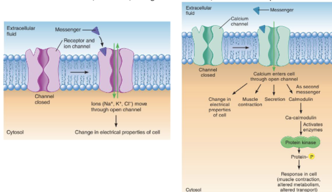
Binding of a messenger to the receptor/ion channel causes the channel to open, increasing the membrane's permeability for that specific ion.
Open ion channels allow a specific ion to move across the cell membrane down its electrochemical gradient.
What are the two different effects ion movement can have on a target cell?
Changing ion permeability (e.g., Na⁺, K⁺, Cl⁻) rapidly changes the cell's membrane potential → creates an electrical signal that alters voltage-sensitive proteins.
Entering ions (e.g., Ca²⁺) can interact with proteins inside the cell (e.g., calmodulin & troponin) → induces responses such as muscle contraction, secretion, change in metabolism, or altered transport of a substance.
How can Ca²⁺ enter the cell, and what does it bind to inside the cell?
Entry routes for Ca²⁺:
Ligand-gated channels
Voltage-gated channels
Mechanically-gated channels
Released from intracellular compartments (e.g., sarcoplasmic reticulum)
Binds to cytosolic proteins: calmodulin and troponin
What are the three effects of Ca²⁺ binding to cytosolic proteins?
Ca²⁺-calmodulin complex = an activated "switch" that turns on/off enzymes, transporters, and ion channels to create a cellular response.
Ca²⁺-calmodulin complex → alters enzyme or transporter activity or the gating of ion channels.
Ca²⁺ binding to troponin → initiates muscle contraction in a skeletal muscle cell.
Ca²⁺ binds to regulatory proteins → triggers exocytosis of secretory vesicles (e.g., release of insulin from pancreatic beta cells).
What are G proteins, how are they structured, and why are they called "G" proteins?
What they are: A diverse group of proteins.
Structure: Formed from at least 20 different alpha (α) subunits, 5 beta (β) subunits, and 13 gamma (γ) subunits.
Why "G": They get their name from their ability to bind guanosine nucleotides (GDP/GTP)
When G proteins are activated, what are the two things they can do?
Open an ion channel in the membrane (slow ligand-gated channel), OR
Alter enzyme activity on the cytoplasmic side of the membrane.
Most common amplifier enzymes for G protein-coupled receptors:
Adenylyl cyclase
Phospholipase C
Describe the step-by-step action of a G protein on a slow ligand-gated ion channel.
Binding of the messenger to the receptor activates the G protein.
The alpha (α) subunit moves to an ion channel in the membrane.
The ion channel opens or closes, changing the permeability of the membrane to a specific ion.
Movement of ions across the plasma membrane is altered → changes the electrical properties of the cell.

Describe the G protein–coupled adenylyl cyclase–cAMP signalling pathway and how it amplifies a signal.
This pathway transmits signals from many protein hormones:
A signal molecule binds to a G protein–coupled receptor (GPCR), activating a G protein.
The G protein activates adenylyl cyclase, an amplifier enzyme.
Adenylyl cyclase converts ATP into many cyclic AMP (cAMP) molecules, amplifying the signal.
cAMP acts as a second messenger, activating protein kinase A (PKA).
PKA phosphorylates other intracellular proteins, continuing the signalling cascade and producing a cellular response.
Describe the G protein–coupled phospholipase C pathway and the roles of its second messengers
A signal molecule binds to a GPCR, activating its associated G protein.
The G protein activates phospholipase C (PLC), an amplifier enzyme.
PLC splits the membrane phospholipid PIP₂ (phosphatidylinositol bisphosphate) into two second messengers:
DAG (diacylglycerol): Remains in the membrane and activates protein kinase C (PKC), which phosphorylates proteins to produce a cellular response.
IP₃ (inositol trisphosphate): Diffuses through the cytoplasm and triggers Ca²⁺ release from the endoplasmic reticulum.
Released Ca²⁺ can:
Act on proteins to stimulate contraction or secretion.
Act as a second messenger, binding to calmodulin and activating protein kinases that phosphorylate target proteins, producing a cellular response.
How do enzyme-linked receptors work, and what are their main types and effects?
A ligand binds to the receptor, changing its shape and activating its enzyme. Two examples:
Tyrosine kinase: Transfers a phosphate group from ATP to tyrosine residues on intracellular proteins, producing ADP. This phosphorylation changes protein activity through covalent regulation, causing responses such as altered metabolism or protein synthesis.
Example: insulin receptor
MAP kinase pathway: Promotes cell growth, proliferation and gene expression.
PI3K pathway: Promotes lipid, protein and glycogen synthesis; cell survival and proliferation; and movement of GLUT4 transporters to the cell membrane, increasing glucose uptake.
Guanylyl cyclase: An amplifier enzyme that converts GTP into cGMP. cGMP activates a protein kinase, which phosphorylates target proteins to produce a cellular response.
Covalent regulation means changing a protein’s activity by adding or removing a chemical group through a covalent bond.
What are the main second messengers, how are they produced, and what are their actions and effects?
Second messenger | Production and pathway | Actions and effects |
|---|---|---|
cAMP (nucleotide) | ATP → cAMP by membrane adenylyl cyclase, linked to a GPCR. | Activates PKA, leading to protein phosphorylation; also binds ion channels and alters their opening. |
cGMP (nucleotide) | GTP → cGMP by guanylyl cyclase: membrane enzyme linked to an enzyme-linked receptor, or cytosolic enzyme activated by nitric oxide (NO). | Activates PKG, leading to protein phosphorylation; also binds ion channels and alters their opening. |
IP₃ (lipid-derived) | Membrane PIP₂ is split by phospholipase C (PLC), linked to a GPCR. | Releases Ca²⁺ from intracellular stores, triggering calcium-dependent responses. |
DAG (lipid-derived) | Produced alongside IP₃ when PLC splits PIP₂. | Activates PKC, leading to protein phosphorylation. |
Ca²⁺ (ion) | Released from intracellular stores, including in response to IP₃. | Binds calmodulin and other proteins, altering enzyme activity and controlling exocytosis, muscle contraction, cytoskeletal movement and ion-channel opening. |
How does cholera toxin disrupt G protein signalling and cause severe diarrhoea?
Cholera is caused by Vibrio cholerae, which releases a toxin into the gut.
The toxin binds to a membrane ganglioside on small-intestinal secretory cells.
Its A subunit enters the cell and prevents the stimulatory G protein from switching off.
The persistently active G protein overstimulates adenylyl cyclase, continuously converting ATP into cAMP.
Increased cAMP activates protein kinase A (PKA), which phosphorylates proteins and increases chloride ion (Cl⁻) secretion into the intestinal lumen.
Na⁺ follows, and water follows by osmosis, causing massive watery diarrhoea and potentially 15–20 litres of fluid loss per day in severe cases.
What are the ways of terminating a signalling event?
Eliminate the signal: Remove or break down signalling molecules, such as neurotransmitters.
Down-regulate receptors: Decrease the number of receptors on the cell membrane by removing them through endocytosis, reducing sensitivity to the signal.
Remove protein phosphorylation: Phosphatases remove phosphate groups from tyrosine, serine or threonine residues, reversing phosphorylation-dependent effects.
Break down second messengers: Phosphodiesterases (PDEs) hydrolyse the cyclic phosphodiester bond in cAMP and cGMP, converting them into AMP and GMP and stopping their signalling activity.
Negative feedback: A downstream response inhibits an earlier step in the pathway, reducing or stopping further signalling.
How are signal molecules eliminated to terminate signalling?
Signals are eliminated by one or more of three mechanisms:
Removal or uptake: Moves signals away from their receptors or targets.
Ca²⁺ is pumped from the cytosol back into the endoplasmic reticulum or out into the extracellular fluid.
Neurotransmitters undergo reuptake into the presynaptic neuron or uptake into postsynaptic cells or neighbouring glial cells.
Enzymatic degradation: Specific enzymes break down signalling molecules.
Example: acetylcholinesterase breaks down acetylcholine.
Diffusion away: Signal molecules diffuse away from receptors and out of the synaptic cleft, becoming diluted in surrounding fluid and the general circulation.

How does receptor internalisation down-regulate receptor number, and how are receptors recycled?
Receptor internalisation means bringing a receptor from the cell membrane into the cell.
A ligand binds to a membrane receptor, and the receptor–ligand complex moves into a clathrin-coated pit.
Endocytosis brings the complex into the cell in a vesicle, reducing the number of surface receptors and decreasing the cell’s sensitivity to the signal.
The vesicle loses its clathrin coat, and the receptor and ligand separate in an endosome.
The ligand is sent to a lysosome or Golgi complex for processing.
Receptors enter transport vesicles, which fuse with the cell membrane, returning the receptors to the surface by exocytosis.