Biochemistry week 1-6
 Call Kai
 Learn
 Practice Test
 Spaced Repetition
 Match
 Flashcards
 Knowt Play
1/168

There's no tags or description
Looks like no tags are added yet.
Name | Mastery | Learn | Test | Matching | Spaced | Call with Kai | Chat |
|---|
No analytics yet
Send a link to your students to track their progress
169 Terms
What is the difference between elements, compounds and molecules?
element - simplest substance made up from one type of atom
molecule - any group of 2 or more atoms bonded together (oxygen 02)
compound - specific molecule made from 2 or more different elements which are chemically bonded (water H20)
What is the arrangement of an atom?
elements are composed of atoms which all have the same number of protons in their nucleus.
an atom contains
protons (nucleus)
neutrons (nucleus)
electrons (orbital shells)
What is the atomic number and the relative atomic mass?
atomic number - number of protons
relative atomic mass - number of protons and neutrons
What are isotopes?
atoms of the same element can have different numbers of neutrons. For examples chlorine has various isotopes

What are moles?
unit for counting tiny particles, like atoms and molecules as these molecules are too small to individually count
A mole of different atoms weight different amounts because they have different amounts of protons and neutrons
A mole of atoms = 6x10^23 atoms
1 mole of hydrogen atoms
Each hydrogen atom has about 1 proton
Very light
Mass ≈ 1 g
1 mole of carbon atoms
Each carbon atom has 6 protons and 6 neutrons
Much heavier
Mass ≈ 12 g
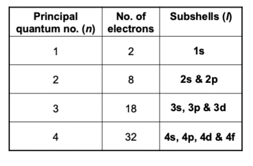
What is the maximum number of electrons each shell can hold?
1= 2 electrons
2 = 8 electrons
3 = 18 electrons

What are subshells?
each energy level or shells are split into smaller subshells. each shell contains the same number of subshells as the original shell number.
there are 4 different subshells
S,P,D,F
What are atomic orbitals?
this is the region around an atom in which there is a high probability of finding an electron or a pair of electrons.
each atomic orbital can hold max 2 electrons
(speedy, people, drink, fast)
Subshell | Number of orbitals | Maximum electrons |
s | 1 | 2 |
p | 3 | 6 |
d | 5 | 10 |
f | 7 | 14 |
What are the principles considered in electron configurations?
pauli exclusion - An orbital can contain a maximum of 2 electrons and those 2 electrons must have opposite spins
Aufbau principle - Electrons occupy the available orbitals of the lowest energy first before filling higher energy orbitals
hands rule - Electrons fill empty orbitals one at a time before they start pairing up

What is the difference between monatomic and diatomic and why are they different?
Monatomic = one atom (he, Ne, AR)
Diatomic = two atoms (H2, O2, N2)
these tend to not form bonds with other elements as thier outer shell is already full and stable
What is the Lewis Octet rule?
Atoms tend to form bonds so they have 8 electrons in their outer shell, having these 8 shell electrons makes an atom more stable (noble gases)
What is ionic bonding?
ionic bonding - electrostatic attraction that forms between oppositely charged ions. This occurs as electrons are transferred from one atom to another in order to gain a full outer shell
Metal (group 1 and 2) --> looses electrons --> positive ions
Non metal (group 6 and 7) --> gains electrons --> negative ion

What is covalent bonding?
covalent bonds happen when 2 atoms share a pair of electrons so they both achieve a full outer shell
Example Cl2 - each chlorine atom has 7 valence electrons, they each need one more to reach 8, therefore they share one pair of electrons
normally between non metals

What is the difference between cations and anions?
cations are positively charged ions. It forms when an atom loses electrons.
anion are negatively charged ions negative charge (−). It forms when an atom gains electrons.
What is an ionic compound?
This is a substance made up of positive and negative ions held together by strong electrostatic forces of attraction
Example - sodium chloride NaCl
Sodium has one electron on its outer shell electron and chlorine has 7
Sodium donates one electron to chlorine in order for them to both achieve a full valence shell, this leaves sodium as a cation as it has lost a electron ( a negative charge) and chlorine as an anion as it has gained a negative charge.
These are then both attracted to each other and form a strong ionic bond due to the electrostatic forces of attractions

What electronegatively?
how strong an atom attracts the shared pair of electrons in a covalent bond. if one atom has a higher electronegativity, it pulls the shared electrons closer itself.
Example: H–Cl
Chlorine is more electronegative than hydrogen:
H δ⁺ — Cl δ⁻
The electrons are still shared, but they're pulled more towards Cl.
So:
Cl gets a slight negative charge (δ⁻)
H gets a slight positive charge (δ⁺)
These are called partial charges
Rules of electronegativity?
Electronegativity generally increases across a period, this is because the nucleus tends to have more protons and therefore a higher nuclear charge, so it attracts bonding electrons more strongly
Electronegativity increases up a group, this is because up a group the smaller atomic radius and less electron shells due to the smaller atomic number, therefore the protons in the nucleus are closer to the electrons and are therefore more strongly attracted
What is the Pauling scale of electronegativity?
if the difference value is greater than 1.7 the bond is likely to be more ionic
if the difference in values is less than 1.7 the bond is more to be covalent

What is the drop and swap method?
drop and swap of the charges

What is a dative covalent bond
A dative covalent bond is a covalent bond where both electrons in the shared pair come from the same atom

What is the structure of an amino acid?
Amino acids differ in their r group, there are 20 different types of amino acids
R group
Carboxyl group
Amine group
Hydrogen group
an amino acid differ in their R groups, there are 20 amino acids

What is the protein structure?
Amino acid monomers are joined via peptide bonds, the sequence of these amino acids joined via peptide bonds is the primary structure
During the secondary structure the polypeptide folds into either a alpha helices and beta pleated sheet
The polypeptide then folds into a unique 3d shape called the tertiary structure
Some proteins consist of one or more polypeptide chain which Is the quaternary structure
What is the alpha carbon bonded to in the amino acid?
the alpha carbon is bound to a hydrogen atom and the side chain group (R)
What is chirality?
this is when the alpha carbon is surrounded by 4 different substituents
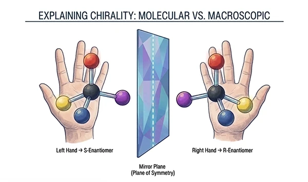
What is enantiomers?
Chirality creates two possible arrangements, called enantiomers. They are mirror images of each other

What is pH?
pH is a measure of the concentration of hydrogen ions in a solution.
the Lower the pH the higher the concentration of H+
the higher the pH the lower the concentration of H+
What is pKa?
The specific pH at which each functional group is 50% protonated and 50% deprotonated. Each ionizable group has its own pKa. This just tells you how easily an acid gives away protons.
Low pKa --> stronger the acid --> gives up hydrogen ions more easily
High pKa --> weaker the acid --> holds onto H+ more strongly
What is the general rule of pKa?
If the pH is higher than the pKa the molecule will lose a proton more easily
If the pH is lower than the pKa the molecule will bind a proton
What is a zwittern?
molecule that contains a positive and a negative charge on different functional groups, this makes the net overall charge 0
At neutral pH of 7 free amino acids exist predominantly as Zwittern
Carboxyl group looses a proton as the pH (7) is higher than the pKa -3.1
Amino group gains a proton as the pH (7) is lower than the pKa -8
The pKa tells you the pH at which a group is 50% protonated and 50% deprotonated
Explain the pH titration of free amino acids?
Starting in a strong acid there is a low pH, both carboxyl and amine groups are fully protonated, this gives the amino acid a net charge of +1 (this is because the pH is lower than both groups pKa)
The charge is +1 as the amino group (NH2) grabs an extra H+ ion from the acidic solution to become NH3+
The carboxyl group (COO-) also grabs an H+ ion to become COOH. This is now neutral so carries a charge of 0
Therefore + 1 + 0 = 1
As the pH rises and gets to 7 the carboxyl group gives up its proton first (because the pH is higher than the pKa) now you have one positive end NH3+ and one negative end COO- this balanced double charged state is called a zwitterion (0 charge)
At very high pH, the pH is now higher than the pKa of both groups. Protons are so scarce that the amino group is also forced to give up its proton too, this makes the overall charge -1
What does aliphatic mean?
carbon atoms can join together to make chains, these chains can be straight or branched.
aliphatic - carbon based structure that isn’t aromatic, it can be:
straight
branched
non aromatic ring
What does aromatic ring mean?
sometimes carbon atoms join together to form a closed loop. this is called a ring and there is different types
aromatic - this is a special type of ring containing a delocalised π electron system making it unusually stable
benzene is a classic example of an aromatic molecule
What does imidazole ring mean?
It has 5 atoms in a ring:
3 carbon (C) atoms
2 nitrogen (N) atoms
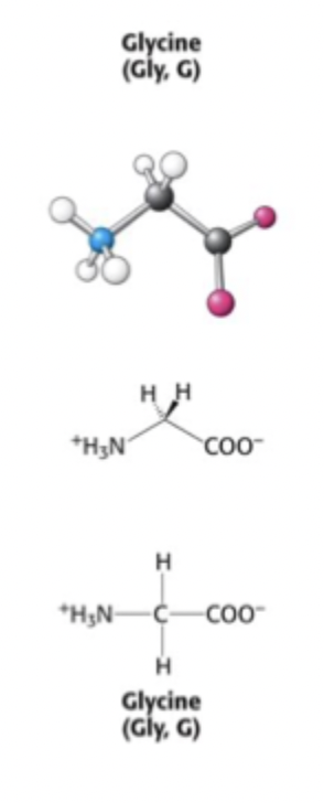
Glycine
R group is a single H atom
Only amino acid which lacks chirality this is because its central carbon is bonded to 2 identical hydrogen atoms (normally a carbon is bonded to 4 entirely different groups)
side chain = extreme flexibility
Alanine
R group - CH3
Aliphatic side chain
Small size
Non polar
Hydrophobic

Serine
R group CH2-OH
Hydroxyl functional group
Polar (oxygen more electronegative than hydrogen therefore forming a hydrogen bond)
Hydrophilic (water loving) therefore readily attracts water
Can partake in hydrogen bonding interactions
Biological role
Often found in active sites of enzymes, this is because it is a nucleophilic (reactive). It can form temporary covalent bonds to a substrate during an enzyme catalysed reaction

Propine
Unusual amino acid where the side chain loops around and bonds directly with the nitrogen atom of its amino group forming a rigid pyrrolidine ring
Aliphatic
Non polar
Hydrophobic
Common in proteins such as collagen and at sharp turns in globular proteins
Because of its fixed ring shape it introduces tight conformational constraints (kinks or bends) into protein chains


Histidine
R group - an imidazole ring (5 membered aromatic ring containing nitrogen
Side chain has a pKa of around 7
Can switch between neutral and positive charged depending on subtle changes in its local microenvironment
Frequently found in the active site of enzymes
Because of its pKa is around 6.7 the imidazole readily accepts or donates protons during chemical reactions acting as a flexible proton shuttle

Cysteine
R group CH2SH - thiol (sulphur and hydrogen atom binded) or sulfhydryl functional group
Hydrophobic
Side chain has a pKa of 8.4 and is frequently found in enzyme active sites
Thiol group (SH) is more reactive than the hydroxyl group
When a long protein folds up into its 3D shape, the 2 cysteine residues sit right next to each other, this causes both cysteine side chains to loose their hydrogen atoms
The 2 bare sulphurs snap together and from a strong covalent bond (S-S) called a disulphide bridge
The linked pair of cysteines is then called cystine
Oxidation - (loss of hydrogens / electrons)
When exposed to oxygen or an oxidising environment, cysteines lose hydrogen atoms and form disulphide bridges
Reduction - (gain of hydrogens / electrons)
In a reducing environment hydrogens are forced back onto sulphurs, this breaks the bridge back into 2 separate cysteines

Phenylalanine, Tyrosine and Tryptophan
All 3 have methylene linked CH2 aromatic ring as their side chain.
Phenylalanine - r group is a functional phenyl group
Highly hydrophobic (benzene ring)
Tyrosine - r group a phenol group
hydroxyl group means it is less hydrophobic and more reactive
Tyrptophan - r group an indole functional group
Hydrophobic and large

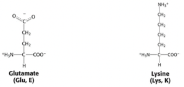
Glutamate and Lysine
Glutamate r group - CH2CH2COO
Contains acidic carboxyl functional group
Negatively charged -4.1 pKa
Hydrophilic
Partake in hydrogen bonding and ionic interactions
Lysine r group - CH2CH2CH2CH2NH3+
Contains basic amine functional group
Positively charged pKa 10.8
Hydrophilic
Partakes in hydrogen bonding and ionic interactions
What are Angstroms (A˚) and Daltons (Da), and what are their conversions and biological uses?
Angstrom (Å)
Definition: unit of length equal to 10⁻¹⁰ meters (1 A˚=10−10 m=10−8 cm=10−4 μm=0.1 nm)
Use: Expressing chemical bond lengths and atomic scale distances in proteins.
Dalton (Da):
Definition: A unit of mass equal to one atomic mass unit (amu), which is roughly the mass of a single hydrogen atom.
Use: Expressing molecular weight on the atomic scale, specifically for proteins (1 kDa=1,000 Da)
Why does the peptide bond have less flexibility, and how does its bond length compare to standard single and double bonds?
different bond (double or single peptide bonds) are different lengths
Partial Double Bond Character: The peptide bond exhibits resonance, giving it a partial double bond character that makes it stronger and restricts rotation (less flexibility).
Bond Length Comparison:
Standard C-N single bond: 1.49 A˚
Peptide bond C-N: 1.32 A˚ (shorter and stronger than a standard single bond due to resonance)
Standard C=N double bond: 1.27 A˚

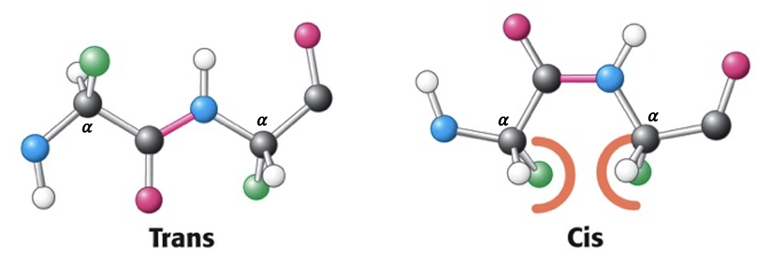
What is the configuration of a peptide bond?
Why is the peptide bond planar? Because of resonance, the peptide bond (C-N) shares electrons with the carbonyl group (C=O), giving it partial double-bond character. Double bonds cannot rotate freely, which locks the six participating atoms into a flat, rigid 2D plane.
The Two Configurations:
Trans-configuration: The two adjacent α-carbons (and their attached side chains/R-groups) are on opposite directions of the peptide bond to avoid steric clashes
Cis-configuration: The two adjacent α-carbons are on the same direction of the peptide bond.
praline can be a cis configuration
What are the Phi (ϕ) and Psi (ψ) dihedral angles, which bonds do they rotate around, and why are they important?
Why do these angles exist? Unlike the rigid, flat peptide bond (C-N), the single bonds connected directly to the α-carbon atom can rotate freely. This flexibility allows the protein backbone to adjust its shape and fold into 3D structures.
The Two Rotational Angles:
Phi (ϕ): The angle of rotation around the single bond between the nitrogen atom (N) and the α-carbon (Cα).
Psi (ψ): The angle of rotation around the single bond between the α-carbon (Cα) and the carbonyl carbon (C).

What is a Ramachandran Plot, and why are certain combinations of ϕ and ψ angles forbidden?
A Ramachandran Plot is a 2D graph that visualizes the allowed torsion angles (ϕ and ψ) for amino acid residues in a protein. Many combinations of ϕ and ψ are forbidden because they cause steric collisions (atomic overlaps) between atoms in the protein backbone and side chains.

Describe the secondary structure - alpha helix?
Physical Structure: An α-helix is a rigid, rod-like structure where the tightly coiled polypeptide backbone forms the inner core of the cylinder, while the amino acid side chains (R-groups) project outwards in a helical array.
Dihedral Angles (ϕ and ψ): In a standard right-handed α-helix (the common form found in proteins), both ϕ and ψangles are negative (typically around ϕ≈−57∘ and ψ≈−47∘).
Handedness Comparison
Right-handed helix: Very common; clusters in the lower-left quadrant of the Ramachandran plot (negative ϕ, negative ψ).
Left-handed helix: Very rare; clusters in the upper-right region (positive ϕ, positive ψ).

Alpha helix properties
3.6 residues per turn
right handed helix
5.4 A pitch
Hydrogen bonding stabilises structure. H-bond at nearly optimal length (2.8 Å).
𝛼-helices vary from 4 to >40 residues: Average length in globular proteins is ~10 residues.

Explain alpha helix hydrogen bonding?
Hydrogen bonding is arranged such that the peptide C=O group of residue n points along the helix towards the peptide N-H group of the n + 4 residue.
There are three amino acid residues between the two groups that make up a H-bond.
the H bond formed is at a nearly optimal length of 2.8 Å

Explain the alpha helical wheel and what are the 3 types of helices
this is just looking down the middle of the alpha helix
a full circle is 360 degrees
there are 3.6 amino acids per turn
therefore each amino acid sits 100 degrees apart
CH - all residues on the wheel are non polar
mixed - one face of the wheel is hydrophobic and the opposite face is polar
CP - all the residues on the wheel are charged / polar

Protien rich in alpha helices?
The 𝛼-helical content of proteins ranges widely, from none to almost 100%.
About 25% of all soluble proteins are composed of 𝛼-helices connected by loops and turns of the polypeptide chain.
Example: iron storage protein, ferritin, with 75% of the residues in 𝛼-helices.

Explain the secondary structure - Beta pleated sheet?
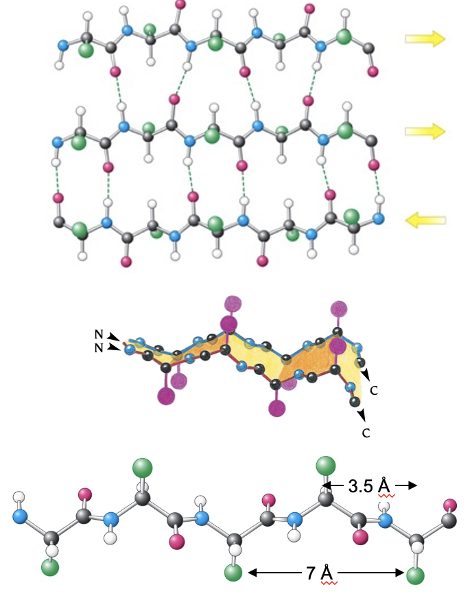
The polypeptide chain in a 𝛽–sheet, called a 𝛽-strand, is almost fully extended. (many beta strands come together to form a beta pleated sheet)
phi angle negative
psi angle positive
Properties of a beta pleated sheet?
Made from at least two (linked) polypeptide chains. Typically, 4 or 5 but, 10 or more 𝛽-strands can come together in 𝛽-sheets.
Strands can be parallel, anti-parallel or mixed.
They have rippled or pleated appearance.
Distance between adjacent amino acid residues is 3.5 Å.
Hydrogen bonding stabilises sheet.
𝛽-strands are typically between 3 and 10 residues long.
𝛽–sheet: hydrogen bonding?
Hydrogen bonding occurs between C=O of one strand to N-H of adjacent strand.
Pattern varies depending on whether adjacent strands are parallel or anti-parallel.
image 1 - antiparralel strands
image 2 - parallel strands
describe the hairpin turn?
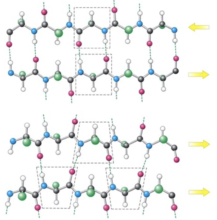
Hydrogen bonding stabilises abrupt changes in direction of the polypeptide chains in 𝛽-sheets.
The C=O group of residue i of the polypeptide is H-bonded to the N-H group of residue i + 3.

describe the structure of omega loops?
Unlike 𝛼-helices and 𝛽-sheets, 𝛺-loops do not have a regular periodic structure.
Loop structures are often rigid and well defined which join alpha helices and beta strands
Turns and loops invariably lie on the surfaces of proteins and often participate in interactions with other proteins or molecules.

Explain structural motifs?
secondary structure elements link together into common recognisable combinations.
mini domains - form structural units or motifs which process a degree of stability
helix turn helix motif - Two α-helices connected by a short loop region. Very common in DNA-binding proteins
hairpin motif - Two adjacent antiparallel β-strands linked by a sharp tur
B-a-B motif - Two parallel β-strands joined by an intervening α-helix link
greek key motif - Four antiparallel β-strands folded over into a compact shape that resembles a traditional Greek decorative key pattern.

Describe the tertiary structure?
refers to the complete fully folded 3D arrangement of a single polypeptide chain. it is build from any combination of secondary structural elements such as alpha helices, beta sheets, turns and loops packed together
Active form of protein/enzyme may require post-translational modifications and/or binding of co-factors.
Explain the properties of myoglobin?
first protein structure to be determined in 1958 by John Kendrew
it is a single polypeptide chain of 153 amino acids
binds a prosthetic Haem group (non protein molecule containing fe+ which allows oxygen to bind)
overall dimensions 45 Å× 35 Å × 25 Å
about 70% of the main chain is folded into an 8 alpha helices (held together by hydrogen bonds) and the rest forms turns and loops

How are hydrophobic and charged amino acids distributed between the surface and core of an all α-helix protein like Myoglobin?
Charged (hydrophilic) amino acids are located on the surface to interact with the surrounding water, while hydrophobic amino acids are packed inside the core (cross-section) to hide away from water and stabilize the protein's structure.

What is Concanavalin A (ConA), and what defines its tertiary structure?
Concanavalin A is a lectin (a carbohydrate-binding protein). Its tertiary structure is an all β-sheet protein composed of 14 β-strands

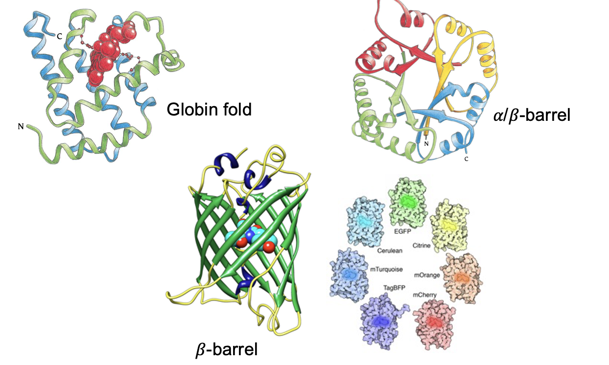
What are the common tertiary structural folds with 𝛼-helices and 𝛽-sheets?
What is a protein domain, and what are its key characteristics regarding folding, frequency, and size?
Definition: A distinct section of a protein that folds independently.
Frequency: Every protein folds into at least 1 domain, but some can have many (>10).
Size: Typically ranges from 30 to 400 amino acid residues in size

What is quaternary structure, and what is the difference between homomultimers and heteromultimers?
Quaternary Structure: The arrangement formed when two or more individual protein chains (subunits) assemble into a functional protein complex.
Homomultimers: Proteins formed by two or more identical copies of the same protein monomer.
Heteromultimers: Proteins formed by several different protein chains coming together to make the mature protein

What is the quaternary subunit composition of haemoglobin (α2β2), and how many oxygen molecules can it transport?
Composition: Haemoglobin is a tetramer (four individual protein subunits bound together) made up of two pairs of slightly dissimilar subunit monomers, designated as α2β2.
Each monomer binds one haem group, and each of these haem groups binds one oxygen molecule, allowing a single mature haemoglobin molecule to transport four O2 molecules in total

What are the key vital statistics regarding protein size, molecular weight, and scale?
Amino Acid Range: Most proteins contain between 50 and 2,000 amino acid residues (smaller chains are called oligopeptides or peptides).
Largest Protein: Titin is the largest single polypeptide known, containing over 34,000 amino acids.
Molecular Weight: Most proteins range between 5,500 and 220,000 Da (Titin is 3,816,030 Da).
Physical Dimensions: Proteins are on the nanometre scale:
Width: Typically ranges between 2 to 100 nm (20 to 1000 A˚).
Length/Stretch: Titin can stretch from 0.9 to 1.5 μm (900 to 1500 A˚).
What are the three major types of protein visualization models, and what does each highlight?
Space-filling model: Shows all individual atoms as spheres packed together; best for seeing overall 3D shape, volume, and surface features.
Ribbon or cartoon diagram: Traces the backbone path; best for clearly visualizing secondary structures like α-helices and β-sheets.
Wireframe or ball and stick diagram: Shows individual bonds and atomic connections; best for examining precise chemical interactions and side-chain arrangements

What causes cystic fibrosis at a molecular level?
It is caused by the misfolding of a specific protein, which leads to a lack of the protein needed to transport chloride (Cl−) ions across cell membranes
How do many neurodegenerative disorders form toxic aggregates?
They occur when partly folded or misfolded polypeptides (or polypeptide fragments) associate with similar protein chains to form toxic aggregates
What structural change occurs when a normal prion protein transitions into a diseased prion protein?
A prion (short for proteinaceous infectious particle) is a misfolded, infectious protein that causes severe brain and nervous system diseases
The structural composition shifts—amino acids in the normal alpha-helix conformation convert into beta-sheet structures in the diseased prion

What holds proteins together in their folded shape, and how stable are they?
Proteins are held together by an accumulation of weak forces and are only marginally stable.
How does pH affect protein stability, and what is the typical stable pH range for most proteins?
Extremes of pH cause proteins to denature. Most proteins are only stable within a pH range of 5–9
What effect does high temperature have on protein stability?
High temperatures cause proteins to denature
Why are proteins usually unstable in organic solvents?
Organic solvents strip away the essential water surrounding the protein, leading to instability
Why is protein folding described as an equilibrium process?
Proteins exist in a dynamic balance (is a state where two opposing processes occur at the exact same rate) between unfolded and folded states, naturally shifting toward their energetically most favorable conformation
To what conformation do proteins naturally fold?
Proteins fold to their energetically most favorable conformation
What role does the solvent play in protein folding?
Interaction with the solvent plays a major role in driving the folding process
How does solvent contact compare between the unfolded state and the folded state of a protein?
Contact with the surrounding solvent is at its maximum when the protein is in the unfolded state
What are the five key forces that stabilize the 3D structure of proteins?
Hydrophobic interactions
Ionic interactions
Van der Waals interactions (short range attractions between non polar molecules)
Hydrogen bonds
Disulfide bonds
Which stabilizing force in protein tertiary structure is unique because it involves covalent interactions?
Disulfide bonds—these are the only covalent interactions among the stabilizing forces which form between 2 sulphur atoms
What type of bonding interactions stabilize protein structure non-covalently?
Hydrophobic interactions, ionic interactions, Van der Waals interactions, and hydrogen bonds
where do hydrophobic amino acid side chains position themselves during the folding of globular proteins and why and what chemical properties do hydrophobic amino acid side chains share?
They position themselves on the inside (core) of globular proteins to move away from the surrounding water.
They have non-polar side chains (alanine, isoleucine, tryptophan, phenylalanine)
What thermodynamic equation governs the free energy change (ΔG) associated with the hydrophobic effect?
ΔG=ΔH−TΔS

What happens when separate non-polar molecules aggregate together in an aqueous solution?
They cluster together to reduce their exposed surface area, which releases ordered water molecules back into bulk solution, increasing entropy (ΔS)

What causes an ionic interaction to form between amino acid residues?
An electrostatic attraction between oppositely charged amino acid side chains
example = positively charged lysine forming an ionic bond with negatively charged glutamate

What step is required before two oppositely charged amino acid residues can form an ionic bond?
The removal of their surrounding solvation shell (dynamic layer of solvent molecules that surrounds and interacts with dissolved ion or solute molecules) is required

What causes van der Waals forces and why are they important?
weak attractions between nearby atoms caused by differences in electron distribution, Many together help stabilise the protein’s 3D structure

How strong are van der Waals forces compared to ionic bonds?
they are much weaker individually, but they matter because there are so many of them in a folded protein
What two opposing forces balance each other to determine the van der Waals distance between two atoms and what is the optimal distance between the 2 atoms interacting via van der Waals forces called?
Attractive forces (proportional to r−6) and repulsive forces (proportional to r−12).
The van der Waals distance

What happens to the net energy when two atoms get closer than their optimal van der Waals distance?
Repulsive forces increase sharply (r−12), this is because electron shells are closer and potentially overlapping, causing the energy to rise rapidly and making the interaction unfavorable
What mathematical expression describes the net energy of van der Waals interactions (Lennard-Jones Potential)?
energy=A/r12−B/r6
What are the 3 types of van der waals shown?
Permanent dipole: two molecules already have partial charges (δ+ and δ−) that attract.
Dipole-induced dipole: a permanent dipole causes a nearby molecule to develop a temporary dipole.
London dispersion forces: temporary fluctuations in electrons create temporary dipoles.
What does optimal packing do?
Brings atoms close together, maximising van der Waals interactions.
What is a hydrogen bond in terms of electrostatic interactions?
An electrostatic attraction between a weakly acidic donor group (D−H) and an acceptor atom (A) that carries a lone pair of electrons
Because atoms like oxygen (O) and nitrogen (N) are highly electronegative, they pull shared electrons away from the hydrogen atom attached to them. This leaves the hydrogen atom with a partial positive charge (δ+), making it a donor(D−H). The acceptor atom (A) is another electronegative atom with a non-bonding lone pair of electrons (δ−). The electrostatic force is simply the partial positive charge on hydrogen attracting the partial negative lone pair on the acceptor.
How is the hydrogen atom positioned in a hydrogen bond?
The hydrogen atom is "sandwiched" between two electronegative atoms—typically oxygen or nitrogen (e.g., O−H⋯Oor N−H⋯O).
What is the ideal bond length for a hydrogen bond in protein structures?
2.7−3.1A˚

Explain why hydrogen bonds formed between solute molecules are relatively weak when dissolved in water?

What type of chemical reaction converts two cysteine residues into a cystine residue, and what byproduct is produced?
The formation of a disulphide bridge requires an oxidizing environment where hydrogens are removed from the two thiol (−SH) side chains. During this oxidative cross-linking ([O]), the two hydrogen atoms combine with oxygen to form a molecule of water (H2O), leaving behind a covalent −CH2−S−S−CH2− linkage that firmly holds the protein structure together

How do the five main stabilizing forces differ between the unfolded state and the folded state of a protein?
Force | UNFOLDED | FOLDED |
Hydrophobic | Exposed to solvent | Shielded from solvent |
Ionic | Charged groups are solvated | Ionic bonds formed in protein |
Van der Waals | Protein with water | Protein with protein |
Hydrogen bonds | Protein with water | Protein with protein |
Disulphide | Not present | Present |
What determines how a protein folds into its final 3D structure?
A protein's primary structure (its specific sequence of amino acids).