Cornea (Alex's quizlet)
 Call Kai
 Learn
 Practice Test
 Spaced Repetition
 Match
 Flashcards
 Knowt Play
1/268

There's no tags or description
Looks like no tags are added yet.
269 Terms
t or f: the epithelium is able to regenerate
true
t or f: the stroma is able to regenerate
false
what is the average corneal thickness?
540 um
hallmark findings for punctate epithelial erosions (PEEs)
areas of epithelial loss that stain with fluorescein

superior PEEs are associated with which conditions?
SLK, VKC, floppy eyelid syndrome
inferior PEEs are associated with which conditions?
exposure keratopathy, chronic blepharitis
interpalpebral PEEs are associated with which conditions?
dry eye
diffuse PEEs are associated with which conditions?
HSK, HZO
central PEEs are associated with which conditions?
contact lens wear
hallmark findings for punctate epithelial keratitis (PEK)
areas of swollen epithelial cells that appear grey or opalescent
stain with Lissamine Green, variable staining with fluorescence

hallmark findings for superficial punctate keratitis (SPK)
areas of epithelial loss similar to PEE
stain with fluorescence

what is the difference between PEEs and SPK?
look the same under SLE
SPK is associated with inflammation, while PEEs are not
hallmark findings for subepithelial infiltrates
non-staining inflammatory cells that are below the epithelium
appear grey with indistinct margins
associated with sectoral redness
PEK is associated with which conditions?
Thygeson superficial punctate keratitis, viral infections
subepithelial infiltrates are associated with which conditions?
marginal keratitis, HZO, EKC, contact lens overwear
hallmark findings for deep infiltrates
non-staining inflammatory cells, cellular debris & tissue necrosis located in the anterior stroma
appear grey to white with indistinct margins
associated with larger area of conj hyperemia

deep infiltrates are associated with which conditions?
PUK, microbial keratitis, HSK
hallmark findings for ulceration
a deep infiltrate with overlaying staining and tissue loss

in order to be called an ulcer, what must be present?
deep infiltrate
ulcerations are associated with which conditions?
microbial keratitis, PUK
hallmark findings for corneal melting
epithelial defect with stromal loss
precursor to corneal perforation

corneal melting is associated with which conditions?
severe corneal infections, prolong topical NSAID use
hallmark findings for descemetocele
protrusion of Descemet membrane into anterior layer of the cornea
seen with severe corneal ulceration

descemetocele is associated with which conditions?
severe corneal infections
hallmark findings for epithelial edema
swelling of the epithelium seen as cyst
will be seen when the endothelium is compromised

epithelial edema is associated with which conditions?
Fuch's endothelial dystrophy
hallmark findings of corneal neovascularization
blood vessel growth into the cornea at the limbus

corneal neovascularization is associated with which conditions?
contact lens overwear, blepharitis
hallmark findings of pannus
blood vessel growth into the cornea with overlaying scar tissue

pannus is associated with which conditions?
chronic inflammation
hallmark findings for folds in Descemet's membrane
dark crisscross lines that appear in deep corneal layers
will have corneal edema

folds in Descemet's is associated with which conditions?
post-cataract surgery
*usually self-limiting
hallmark findings for breaks in Descemet's membrane
deep linear opacities that run vertically

breaks in Descemet's membrane is associated with which conditions?
hydrops associated with keratoconus
infantile glaucoma
traumatic birth
common etiologies for bacterial keratitis
Staph spp, Pseudomonas aeruginosa
common etiologies for fungal keratitis
Fusarium spp, Candida spp, Aspergillus
common etiologies for viral keratitis
herpes simplex-1, Varicella zoster
t or f: microbial keratitis is most commonly viral
false, bacterial is most common
risk factors for bacterial keratitis
CL wear (especially if non-compliant), topical steroid use, ocular surface disease, ocular trauma, previous infection
clinical findings for bacterial keratitis
deep corneal infiltrates
epithelial defect over the infiltrate and typically the size of the infiltrate
diffuse conj hyperemia
cells & flare in ant chamber

complications of bacterial keratitis
corneal scarring, irregular astigmatism, corneal neovascularization, corneal perforation
Tx for bacterial keratitis
topical antibiotic solution/ointment (broad spectrum is used until cultures are returned)
topical cycloplegics (pain control & prevent post synechia formation)
topical steroids after 2-3 days of improvement (re-epithelialization)
oral antibiotics for gonococcal
Is Candida a yeast or filamentous fungi?
yeast
Is Fusarium a yeast or filamentous fungi?
filamentous
Is Aspergillus a yeast or filamentous fungi?
filamentous
risk factors for fungal keratitis
ocular surface disease, long term topical steroid use, trauma with vegatative matter, CL wear, immunocompromised
clinical findings for yeast fungal keratitis
conj hyperemia
endothelial plaques (collection of inflamm/immune cells)
hypopyon in ant chamber
round "button" infiltrate
**NO feathery borders or satellite lesions

complications for yeast fungal keratitis
corneal scarring, corneal perforation, endophthalmitis
Tx for yeast fungal keratitis
debridement, topical antifungal drop, topical antibiotic
clinical findings for filamentous fungal keratitis
conj hyperemia
endothelial plaques (collection of inflamm/immune cells)
hypopyon in ant chamber
feathery ulcer border
satellite lesions
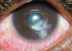
complications of filamentous fungal keratitis
corneal scarring, corneal perforation, endophthalmitis
Tx for filamentous fungal keratitis
debridement, topical antifungal drop, topical antibiotic
what are the 2 life stages of Acanthamoeba keratitis?
trophozoite- active, reproducing
cyst- dormant, resistant
risk factors for Acanthamoeba keratitis
CL wear with water exposure
clinical findings of Acanthamoeba keratitis (early, intermediate, and late stages)
pain that does not match SLE findings (reported pain is worse than expected)
cells & flare in ant chamber
conj hyperemia
early: PEEs, diffuse microcystic epithelial changes, pseudodendrites
intermediate: radial perineuritis, multifocal stromal infiltrates
late: ring infiltrate

complications from Acanthamoeba keratitis
scleritis, hypopyon, corneal scarring/melting, secondary glaucoma, retinal inflammation
Tx for Acanthamoeba keratitis
refer out
topical antimicrobials (pool cleaners- PHMB, Chlorhexidine, Brolene)
t or f: dendritic keratitis has live virus replication
true
clinical findings for dendritic (epithelial) herpes keratitis
day 1-3: swollen epithelial cells in a punctate or stellate pattern
day 4: dendritic epithelial lesion
follicular conjunctivitis
cells & flare
reduced corneal sensitivity
elevated IOP (from trabeculitis)
possible eyelid vesicles

complications of dendritic (epithelial) herpes keratitis
corneal scarring, geographic ulcer, neurotrophic keratitis
Tx for dendritic (epithelial) herpes keratitis
topical antiviral drop or gel
oral antiviral medication (cheaper than topical)
topical cycloplegic
topical steroid (after epithelium heals)
what is a geographic ulcer?
enlargement of the dendritic ulcer after the use of topical steroids

what is neurotrophic keratitis (or keratopathy)?
cornea loses trigeminal nerve innervation
there is no stimulus of pain/feeling and no signal to re-epithelialize after injury
which conditions can have neurotropic keratitis?
dendritic (epithelial) herpes keratitis, herpes zoster, ocular surface disease, diabetes, topical medication toxicity
describe the 3 stages of neurotrophic keratitis
1- epithelial irregularities and PEEs
2- persistent epithelial defects with stromal involvement
3- corneal ulceration with stromal melting (EMERGENCY)
complications of neurotrophic keratitis
corneal perforation (EMERGENCY)
Tx for stage 1 neurotrophic keratitis
preservative-free AT
autologous serum
punctal occlusion
moisture chamber goggles
Tx for stage 2 neurotrophic keratitis
cenegermin (Oxervate)- contains human growth factor
amniotic membrane
scleral or bandage CL
tarsorrhaphy (join upper & lower lids)
Tx for stage 3 neurotrophic keratitis
amniotic membrane
advanced corneal surgery
what are the 2 types of stromal herpes simplex keratitis?
immune (interstitial) & necrotizing
t or f: immune stromal herpes simplex keratitis has live virus
false
t or f: necrotizing herpes simplex keratitis has live virus
true
which is more aggressive: immune or necrotizing stromal herpes keratitis?
necrotizing
clinical findings for immune (interstitial) stromal herpes simplex keratitis
deep stromal infiltrates
intact epi & endo
cells & flare
increased IOP

complications from immune stromal herpes simplex keratitis
corneal neovas, corneal scarring
clinical findings for necrotizing stromal herpes simplex keratitis
deep stromal infiltrate
epithelial defect
significant cells & flare with hypopon developement
increased IOP
thinning of cornea

Tx for immune stromal HSK
**prescribe all at the same time
topical steroid drop
oral antiviral
topical cycloplegic
Tx for necrotizing stromal HSK
**prescribe all at the same time
topical antiviral
oral antiviral
topical cycloplegic
if perforation: penetrating keratoplasty
t or f: there is live virus with herpes simplex endotheliitis (disciform) keratitis
false
clinical findings for disciform keratitis
intact epi
disc-shaped stromal edema (from endotheliiitis)
Wessely ring (circular, immune-mediated stromal infiltrate)
granulomatous cells & flare
elevated IOP

t or f: stromal defects stain with fluorescence
if the epithelium is intact, then no
if epithelium is not intact, then maybe
complications of disciform keratitis
corneal scarring, corneal neovas, secondary glaucoma
Tx for disciform keratitis
topical steroid + oral/topical antiviral + topical cycloplegic
etiology of herpes zoster ophthalmicus
Varicella zoster (virus)
demographics of herpes zoster ophthalmicus
70-80 years old
immunocompromised
Hx of chicken pox
what are the 5 phases of herpes zoster ophthalmicus?
1. prodromal phase
2. maculopapular rash
3. epithelial keratitis
4. nummular keratitis
5. stromal (interstitial) keratitis
hallmark findings for prodromal phase of HZO (including timing)
3-5 days before appearance of the rash
can mimic upper respiratory infection
tiredness, malaise, fatigue
dermatome has tingling, itching, or burning sensation
hallmark findings for maculopapular rash phase of HZO (including timing)
vesicles appear 24 hrs after onset of symptoms
respect midline
eyelid edema
Hutchinson sign

hallmark findings for epithelial keratitis of HZO (including timing)
2-3 days after rash appears
spontaneously resolves
pseudodentrite appearance (no staining on terminal bulbs)

t or f: herpes zoster has live virus
there is NO live virus on the cornea
there is live virus on the skin
hallmark findings for nummular keratitis of HZO (including timing)
occurs 10 days after rash appears
develops at site of pseudodendrites
appear as subepithelial granular deposits with stromal haze

hallmark findings for stromal (interstitial) keratitis of HZO (including timing)
occurs 3 weeks after rash appears
deep infiltrates with intact epithelium & endothelium

how does HZO affect the conj & sclera?
conjunctivitis with follicles & pseudomembranes/membranes
episcleritis
complications from HZO
mucus plaques, neurotropic keratitis, post-herpetic neuralgia
Tx for HZO maculopapular rash
oral antiviral
topical antibiotic ointment (use when rash begins to crust over)
Tx for HZO epithelial keratitis
artificial tears
spontaneously resolves
Tx for HZO nummular/stromal/disciform keratitis
topical steroid
Tx for HZO mucus plaques
topical steroid
Tx for neurotropic keratitis/keratopathy
artificial tears
bandage CL
amniotic membrane
Cenegermin (Oxalate)
why is it important to treat HZO as quickly as possible?
reduce risk of developing post-herpetic neuralgia