ANPS 1190- Exam 2 flashcards
 Call Kai
 Learn
Learn Practice Test
 Spaced Repetition
 Match
Match Flashcards
 Knowt Play
1/82

There's no tags or description
Looks like no tags are added yet.
Name | Mastery | Learn | Test | Matching | Spaced | Call with Kai | Chat |
|---|
No analytics yet
Send a link to your students to track their progress
83 Terms
Neuron doctrine
the neuron is the main structural and functional unit of the nervous system
Neurons
the main info processing cell in the NS, the ability of the NS to process info is dependent on rapid intercellular communication between neurons
Glial cells
carry out diverse functions, including providing support for neurons:
Astrocytes- diverse functions, regulation of extracellular space, vascular cells (make up blood vessles), only found in the CNS, analogous cell type, satellite cells, are found in the PNS, regulate the levels of chemicals (neurotransmitters), and ions (K+) in the environment surrounding neurons (at nodes of ranvier, synapses), surrounds blood vessels in the brain, helping to form the blood-brain-barrier
Microglia- immune cells of the NS, resident of immune cells of the CNS, but can also travel to the PNS following injury, through phagocytosis, microglia can remove foreign intruders (microbes), as well as debris after injury to the NS
Oligodendrocytes- myelinating cells of CNS, one of the 2 kinds of myelinating glia, myelination- dramatically increases the speed of electrical signaling in neurons, only found in CNS, processes reach out and wrap lipid-rich myelin around axons of multiple neurons
Schwann cells- myelinating cells of PNS, one of 2 kinds of myelinating glia, only found in the peripheral NS, wrap lipid-rich myelin around axons of a single neuron, one neuron has multiple schwann cells wrapping arounds its axon
Ependymal cells- produce cerebrospinal fluid
Astrocytes
diverse functions, regulation of extracellular space, vascular cells (make up blood vessles), only found in the CNS, analogous cell type, satellite cells, are found in the PNS, regulate the levels of chemicals (neurotransmitters), and ions (K+) in the environment surrounding neurons (at nodes of ranvier, synapses), surrounds blood vessels in the brain, helping to form the blood-brain-barrier
Microglia
immune cells of the NS, resident of immune cells of the CNS, but can also travel to the PNS following injury, through phagocytosis, microglia can remove foreign intruders (microbes), as well as debris after injury to the NS
Oligodendrocytes
myelinating cells of CNS, one of the 2 kinds of myelinating glia, myelination- dramatically increases the speed of electrical signaling in neurons, only found in CNS, processes reach out and wrap lipid-rich myelin around axons of multiple neurons
Schwann cells
myelinating cells of PNS, one of 2 kinds of myelinating glia, only found in the peripheral NS, wrap lipid-rich myelin around axons of a single neuron, one neuron has multiple schwann cells wrapping arounds its axon
Ependymal cells
produce cerebrospinal fluid
Dendrite
info receiving component of the neuron, dendritic spines- tiny extensions on dendrites that increase surface area of plasma membrane, enables cell to receive more inputs, more dendrites/spines=more input
Axon
info transmitting component of the neuron
Axon hillock
the initial segment of the axon that extends from the cell body, this is where an action potential starts
Cell body
biosynthetic centric center of the cell, contains nucleus and other organelles. Primary site of protein synthesis in the neuron, also called soma, contains cytosol and organelles surrounding the nucleus, primary cite of other cellular metabolic processes, there is often prominent rER
Axon terminal
contains neurotransmitters, after the action potential spreads down the axon and reaches the axon terminal, neurotransmitters will be released into the synapse
Synapse
includes a presynaptic neuron, the synaptic cleft and postsynaptic neuron
Neurons can be divided into four functional regions
Receptive region- dendrites and cell body receive signals
Impulse-generating region- action potential is generated at the axon hillock (aka trigger zone)
Conducting region- action potential travels down the axon
Secretory region- neurotransmitters are released from the axon terminal into the synapse
Morphology of neurons
multipolar, bipolar, pseudounipolar
The neuronal cytoskeleton is made up of
microfilaments (smallest), intermediate filaments (neurofilaments), microtubules (largest), these are important for axonal transport
Axonal transport
the cell body communicates with the axon terminal via this, neurons rely on bidirectional axonal transport, where cargo contained in a transport vesicle is shipped down a microtubule
Anteograde transport
movement of materials from cell body to axon terminals, neurotransmitters, new membrane, mitochondria, enzymes, vesicles
Retrograde transport
movement of materials from axon terminals to cell body, organelles for recycling, signaling molecules brought in at terminal, potentially hazardous molecules can be brought into terminals
Myelin
is a lipid-rich substance that insulates and protects the axon, myelination increases the speed of electrical impulses traveling along the axon, more myelin= faster electrical signaling (unmyelinated vs lightly myelinated vs highly myelinated), gives white matter its color
White matter
mostly myelinated axons
Grey matter
mostly cell bodies, dendrites, and unmyelinated axons
Differences between neurons and glia
Neurons: basic functional unit of the NS, excitable cells that transmit electrical signals, don’t divide after developmental period
Glia: “non-excitable”, undergo mitosis throughout life
Three general functions of the NS
collect sensory input, integrate info, produce motor output
Collect sensory input
collect sensory input, integrate info, produce motor output
Integrate info
interpret sensory input and determine proper response
Produce motor output
activate effectors including muscles, organs, and glands
The NS is divided into structural and functional divisions
info is sent to the CNS via the sensory division (afferent) of the PNS, the CNS system integrates info and stimulates a response by activating the motor division (efferent) of the PNS, motor responses can include control of voluntary movement via the somatic motor division or alteration of organ function via the autonomic (visceral motor) division
The autonomic division
is divided into the:
Sympathetic NS- stimulates “fight or flight” response (increase hr and respiratory rate)
Parasympathetic NS- stimulates the “rest and digest” response (ex. Decrease hr, increase digestive function)
Comparison chart for the nervous system

PNS structures: nerve and a dorsal root ganglion
nerve- cable like bundle of parallel axons with their glia in the peripheral NS, ganglion- cluster of neuron cell bodies in the peripheral NS
Action potential
when stimulated, excitable cells can produce an electrical signal called this, nerve and muscle cells are excitable cells Electricity: “a phenomenon associated with stationary or moving electrical charges”, in the case of electrical signaling in neurons, ions move across the cell membrane to carry electrical charge
Ions carry electrical charge
in a non-aqueous environment most ions will form ionic bonds and crystalize, in an aqueous environment (like the human body) most ions will dissociate, interacting with polar water molecules, ions are electrolytes and can carry electrical current in solution, sodium (Na+), potassium (K+), and chloride (Cl-) and calcium (Ca+2) are major ions relevant for physiology
Excitable membranes contain membrane proteins that determine a cell’s electrical behavior
transmembrane proteins are key to the electrical excitanility of neurons and muscle cells- leak ion channels, gated ion channels, receptor, ion pumps
Ion channel
an integral membrane protein that allows ions to pass through a pore between intracellular and extracellular environments (leakage ion channels, gated ion channels)
Receptor
an integral membrane protein that produces a physiological change in a cell after a logan (signaling molecule) bind
Selective permeability
ion channels have pores that are selective for specific ions, ions can’t cross the cell membrane, based on cell size and charge, can be leakage channels which are always open, or gated channels which open and close, ion flows across membrane will change the membrane potential
Leakage channels
ion channels that are always open– leakage channels are ungated meaning they are always open, leakage channels are important for establishing the resting membrane potential, leak channels are selective for a specific ion
Gated ion channels
open and close in response to specific stimuli, allow ion movement only when open, control permeability for an ion, open channels allow passive movement of ions down their chemical and electrical gradient while the channel is open, voltage gated and chemically (ligand) gated
Voltage gated
open in response to a change in membrane voltage, highly concentrated in muscle and nerve tissue, voltage gated sodium channel, voltage gated potassium channel, closed under normal physiological conditions, but open with change in membrane voltage around the protein: voltage-gated sodium channels, potassium, and calcium channels
Chemically (ligand) gated)
open in response to binding of an extracellular signal, many neurotransmitters will work via this mechanism, specific chemical binding, have selective binding site on extracellular side of plasma membrane, neurotransmitters are the ligands, receptor is specific for a particular NT, closed under normal physiological conditions, many drugs target ion channels
Ionotropic receptors
ligand-gated ion channels that open or close in response to an extracellular signal; open ion channels allow ions to move and carry charge across the membrane, when a neurotransmitter binds to this receptor, this opens an ion channel that allows specific ions to flow into or out of the cell, these types of receptors are called ligand-gated ion channels (chemically gated) and the flow of ions through the channel cause electrical changes in the neuron
Metabotrophic receptors
are membrane proteins that respond to an extracellular signal by altering the metabolism in the cell, many are G-protein coupled receptors, because when the neurotransmitter binds, it activates a G-protein, after the G-protein is activated, it can stimulate intracellular signaling pathways, when a neurotransmitter activates a metabotrophic receptor, the response is slower but more widespread and potentially longer lasting
Small molecule neurotransmitters/neuropeptides
most neurotransmitters fit into one of the two categories
Small molecule neurotransmitters
glutamate, GABA, glycine
Glutamate
generally excitatory, the principle excitatory NT in the brain
GABA
generally inhibitory, principle inhibitory neurotransmitter in the brain
Glycine
inhibitory, major inhibitory neurotransmitter in the spinal cord+brain stem
Neuropeptides
small chains of amino acids made in the rER/golgi at cell body, substance P, endorphins (Endogenous opiates)
Life cycle of neurotransmitter
synthesis → packaging into vesicles → release into the synapse → binding to a post-synaptic receptor → removal from the synapse
Acetylcholine
synthesized from choline, and acetyl coenzyme A by an enzyme in the presynaptic terminal and then loaded into a synaptic vesicle, released from all somatic motor neurons, many neurons of the ANS, and many CNS neurons, the impact of Ach depends on which type of receptor it binds to, mechanism of removal from synapse, broken downby the enzyme acetylcholinesterase, neurons typically syntehsize and release only one small molecule neurotransmitter and are named for the dominant transmitter released (ex. Cholinergic, serotonergic, dopaminergic)
Nicotinic acetylcholine receptor
ionotropic, effect of Ach binding: Na+ influx/depolarization
Muscarinic acetylcholine receptor (metabotropic)
effect of Ach binding: Activation of a G-protein stimulates intracellular signaling that leads to calciu, release from intracellular organelles
The neuronal membrane can be broken down into 4 functional segment
Receptive region: dendrites and cell body, impulse-generating region- axon hillock, conducting region- axon, secretory region- axon terminals
The different functional segments have different complements of ion channels

Membrane potential (Vm)
refers to the difference in electrical potential (voltage) across the cell membrane, membrane potential=Vinside-Voutside
The resting membrane potential (Rm)
is the difference in electrical potential (voltage) across the cell membrane at rest
All human cells are said to be polarized at rest
Neurons= -60 - -80mV, Muscle= -90mV, red blood cells= -7mV
The resting membrane potential (Rm) of a typical neuron is -70mV
because the charge on the inside of the membrane (negative) is the opposite charge of the outside of the membrane (positive), we say that the cell is electrically polarized
Polarize
to separate into uneven groups
The human body is_____ but the plasma membrane _______
electrically neutral, properties allow for the generation of a membrane potential
Phospholipids
phosphate head (polar)- hydrophilic, hydrocarbon tail (nonpolar)-hydrophobic
The phospholipid bilayer is impermeable to
ions allowing for storage of charge, functions as a capacitor- a device that can store electrical charge
The cell membrane is selectively permeable
only certain molecules can freely cross the cell membrane, ions and polar molecules cannot freely cross through the cell membrane, they require transmembrane proteins like ion channels and pumps to move into or out of the cell
What makes the resting potential ~ -70mV?
Unequal distribution of charge across the cell membrane due to the movement of ions
1. Sodium/potassium pump
2. Potassium efflux through leakage channels
3. Movement of other ions
4. Intracellular organic anions (A-)
What are ions
atoms that have an electrical charge resulting from gaining or loosing electrons, when they pass through the cell membrane they change the membrane potential
Cation
positively charged ion
Anion
negatively charged ion
Depolarization
if Na+ moves into the cell, the membrane potential becomes more less negative (more positive) (depolarizing current)
Hyperpolarization
if K+ moves out of the cell, the membrane potential becomes more negative (less positive) (hyperpolarizing current)
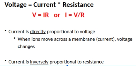
Currents (I)
the flow of electrical charge measured in amperes (A), in cells charge is carried by ion movement
The sodium potassium pump
uses ATP to move 3 sodium ions out of the cell and 2 potassium ions into the cell
By pumping 3 sodium ions extracellularly and 2 potassium ions intracellularly, this pump sets up the concentration gradient for sodium and potassium across the cell membrane, slight accumulation of a + charge extracellularly and - charge intracellularly
K+ efflux occurs through passive transport
they can be “pumped” in energetically unfavorable ways that require ATP, they can “flow” energetically favorable ways based on electrical and chemical driving force
Electrical driving force
the force that results from attraction between positive and negative charges
Chemical driving force
the force that results from movement of ions from high concentration to low concentration
How can you predict which way an ion will flow
Equilibrium potentials allow you to predict the direction of ion movement, the voltage of the cell membrane at which an ion is at electrochemical equilibrium. Tells you which direction and ion “wants” to flow, based on electrical and chemical driving forces. An ion will always flow in a qay that drives the membrane potential towards the equilibrium potential for that ion.
If the membrane is permeable to a particular ion
that ion will flow in a way that drives the membrane potential towards its equilibriu potential, which tells you how an ion wants to flow, based on electrochemical driving forces, the actual flow of an ion across the cell membrane depends on the permeability of the cell to that ion, which is determined by the presence of ion channels ion flow=driving force X permeability
The neuronal cell membrane displays selective ion pemeability
neurons are highly permeable to K+ relative to other ions due to abundant K+ leakage channels, high permeability to K+ allows K+ to leave the cell, driving the voltage of the cell towards the equilibrium potential for K+ (Kk= -80mV), as a result the resting membrane potential (-70mV) is very close to Ek
What are the most important determinants of the resting membarne potential
Na/K pump, and high permeability to potassium
Resistance (R)
hindrance to charge flow by substances through which current must pass measured in ohms (Ω ), in cells, resistance comes from basic property of the plasma membrane, resistance is the inverse of permeability, more permeability=less resistance
Voltage potential (𝛥V)
measure of potential energy generated by separated electrical charges measured in volts (V), separation is most often a physical barrier (in cells, this is the plasma membrane), in cells we refer to this as membrane potential
Bioelectricity is governed by Ohm’s law