combined 3200
 Call Kai
 Learn
Learn Practice Test
 Spaced Repetition
 Match
Match Flashcards
 Knowt Play
1/1114

There's no tags or description
Looks like no tags are added yet.
Name | Mastery | Learn | Test | Matching | Spaced | Call with Kai | Chat |
|---|
No analytics yet
Send a link to your students to track their progress
1115 Terms
What type of tissue is skeletal muscle, and where is it usually found?
Skeletal muscle is usually attached to bones and includes both obvious muscles (like the biceps, triceps, and quadriceps) and less obvious ones (like the tongue, diaphragm, and pharynx).
Fascicle
A bundle of muscle fibers grouped together within a skeletal muscle.
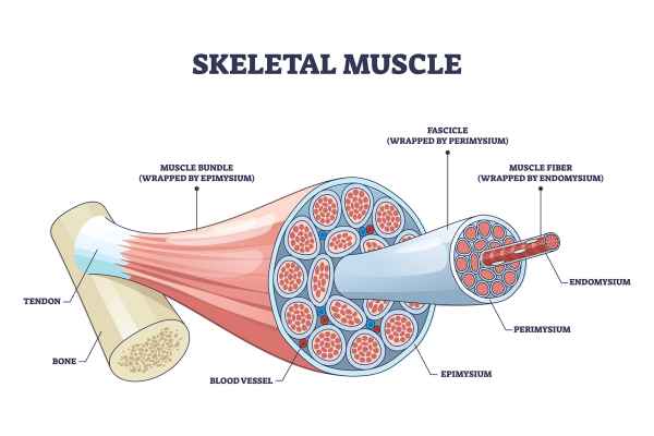
What connective tissue surrounds all fascicles and helps form tendons?
The epimysium.
Perimysium
The connective tissue that surrounds and holds together each fascicle within a skeletal muscle.
What are skeletal muscle fibers, and what is unusual about them?
They are large, fused cells (syncytia) with shared cytoplasm and multiple nuclei.
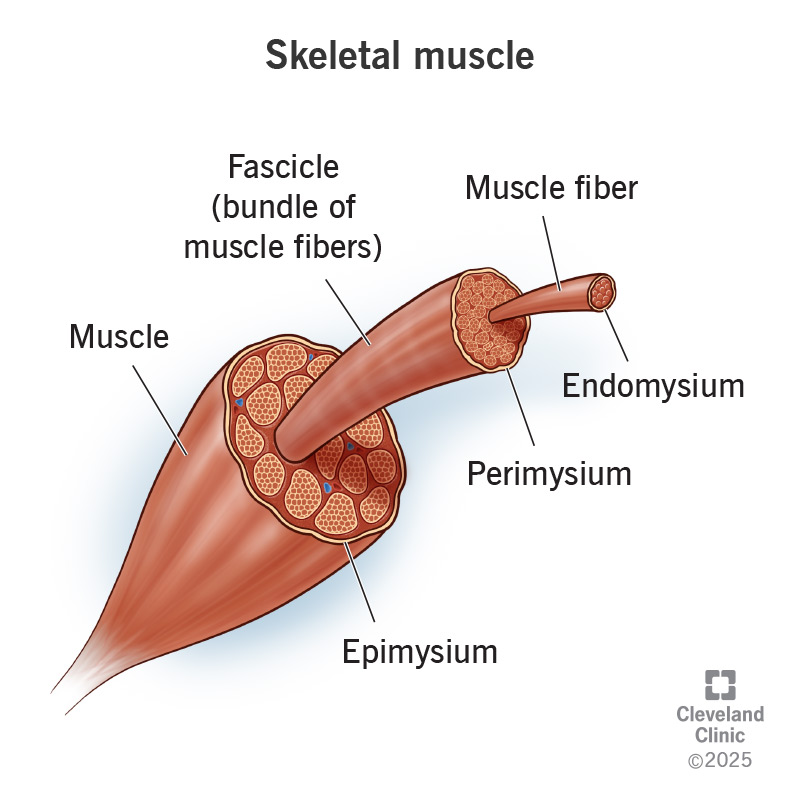
Syncytium
A single large cell formed by the fusion of multiple cells, sharing one continuous cytoplasm — describes skeletal muscle fibers.
What are myofibrils and where are they located?
Myofibrils are thread-like structures inside muscle fibers that contain the contractile proteins actin and myosin.

Sarcoplasm
The cytoplasm of a skeletal muscle fiber.

Endomysium
The connective tissue that surrounds individual muscle fibers.

What is a motor unit, and how does it influence muscle contraction?
A motor unit is one motor neuron and all the muscle fibers it innervates. When the neuron fires, all its fibers contract together. The number of fibers per neuron determines the precision or strength of movement—fewer fibers = precise (eye, fingers); more fibers = strong (calf muscle).
What role does acetylcholine (ACh) play at the neuromuscular junction?
ACh is released from the motor neuron, diffuses across the synaptic cleft, and binds to nicotinic receptors on the sarcolemma, opening ion channels that allow Na⁺ in and K⁺ out → causes depolarization → triggers excitation-contraction coupling.
What is the motor endplate?
The specialized region of the sarcolemma (muscle cell membrane) that contains nicotinic acetylcholine receptors and directly receives signals from the neuron.

How is muscle force controlled according to the all-or-none principle?
Each motor unit contracts fully when activated, so the strength of muscle contraction is controlled by recruiting more or fewer motor units—not by partial contraction of fibers.
What does “degree of innervation” mean, and why is it important?
It refers to how many muscle fibers a motor neuron innervates. Low innervation → fine control (e.g., fingers). High innervation → powerful contractions (e.g., gastrocnemius).
What gives skeletal muscle its striated appearance under the microscope?
The striated appearance comes from the highly organized arrangement of thick and thin myofilaments (protein filaments) that run the full length of myofibrils, forming repeating units called sarcomeres.

Bands and Discs
The I bands are regions that only contain thin filaments and mostly composed of actin
The A bands are regions where thick filaments made from myosin that have some overlapping thin filaments.
The H band is the central portion of the A band, where there is only thick filament and no overlap with thin filaments.
The Z discs are located at the centre of each I band, and they serve as the boundary between sarcomeres, the repeating units of muscle that contract responsible for muscle contraction.

Thin Filaments
Found in the I bands, thin filaments are primarily composed of actin and do not overlap with thick filaments in this region.
Thick Filaments
Made of myosin, thick filaments are located in the A band, where they may overlap with thin filaments, except in the H band.
What is the H band and how is it different from the A and I bands?
The H band is the central part of the A band where only thick filaments are present—there’s no overlap with thin filaments, unlike the rest of the A band.
What are Z discs and what role do they play in muscle contraction?
Z discs are located in the center of each I band and mark the boundary of sarcomeres; sarcomeres contract to produce muscle movement.
What is a myofibril and what does it contain?
A myofibril is a thread-like structure inside a muscle fiber that contains the contractile proteins actin and myosin.
What is a sarcomere?
A sarcomere is the functional unit of skeletal muscle, defined by the region between two Z-discs and composed of overlapping thick and thin filaments.

What are Z-discs?
Z-discs mark the boundary of each sarcomere and serve as the anchor points for actin (thin) filaments.
What is titin and what is its role?
Titin is an elastic microfilament that anchors myosin (thick) filaments to the Z-discs, helping maintain sarcomere structure.
What creates the striated appearance in skeletal muscle?
Alternating light I bands (only thin actin filaments) and dark A bands (thick myosin filaments with overlapping actin) in the sarcomeres of myofibrils.
What is the role of Z-discs and titin in sarcomere structure?
Z-discs anchor actin filaments and mark sarcomere boundaries. Titin anchors thick myosin filaments to the Z-discs, providing elasticity and structural alignment.
What happens during muscle contraction according to the sliding filament theory?
Actin and myosin filaments slide past each other, shortening the sarcomere. A bands stay the same length but move closer together, I bands shorten, and H zones shrink or disappear.
How does sarcomere contraction lead to overall muscle contraction?
Shortening of sarcomeres shortens myofibrils, then the muscle fiber, then the fascicle, resulting in contraction of the whole muscle.
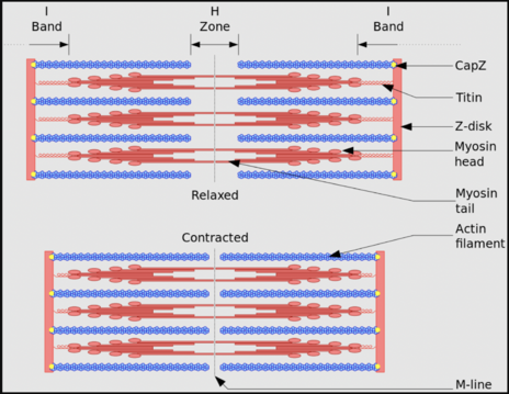
Myosin heads
Globular structures on thick filaments that bind to actin and pivot using ATP energy, enabling sarcomere shortening and muscle contraction.They are connected by a hinged region that allows them to pivot.
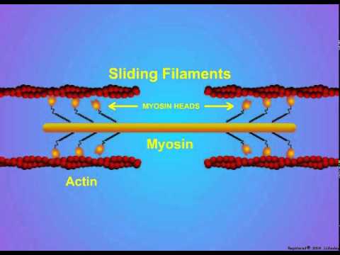
What allows myosin heads to change shape during muscle contraction?
Myosin heads change shape using energy from ATP hydrolysis.
During muscle contraction, myosin heads on thick filaments bind to actin on thin filaments. These myosin heads are attached to the thick filament by a hinged region that allows them to pivot. When ATP is hydrolyzed (broken down into ADP and Pi), the energy released changes the shape of the myosin head, causing it to pivot and pull the actin filament inward. This pulling action shortens the sarcomere, the basic unit of muscle contraction, and as many sarcomeres shorten together, the whole muscle contracts.
Sliding filament thoery steps

Cross-bridge cycle
A repeating series of events where myosin heads bind to actin, pivot, release, and reset using ATP, resulting in sarcomere shortening and muscle contraction.

What provides the energy for the cross-bridge cycle?
ATP hydrolysis provides the energy needed for myosin head movement.
Is ATP alone responsible for muscle contraction? No, muscle contraction is regulated by excitation-contraction coupling, not just by the presence of ATP.
Excitation-contraction coupling
The process that links muscle fiber stimulation (excitation) by a nerve to the contraction of the muscle, involving calcium signaling and regulatory proteins
What two things does the cross-bridge cycle depend on?
The presence of ATP and whether myosin can bind to actin.
What is the main control point for muscle contraction if ATP is available?
Whether myosin can bind to actin.
Tropomyosin
A filamentous protein that wraps around actin and blocks myosin binding sites in the resting state, preventing contraction.
Troponin
A globular protein attached to tropomyosin with a high-affinity binding site for calcium; it controls tropomyosin’s position.
How does calcium allow muscle contraction to begin?
Calcium binds to troponin, causing it to change shape and move tropomyosin, exposing binding sites on actin so myosin can attach.
Calcium ions control when contraction happens by regulating troponin and tropomyosin.
What is a synapse?
A synapse is a junction where a neuron communicates with another cell—either another neuron or an effector like a muscle or gland.
Synapse (CNS vs PNS)
CNS: The target cell is typically another neuron.
PNS: The target cell can be a neuron, muscle, or gland.
Neuromuscular junction
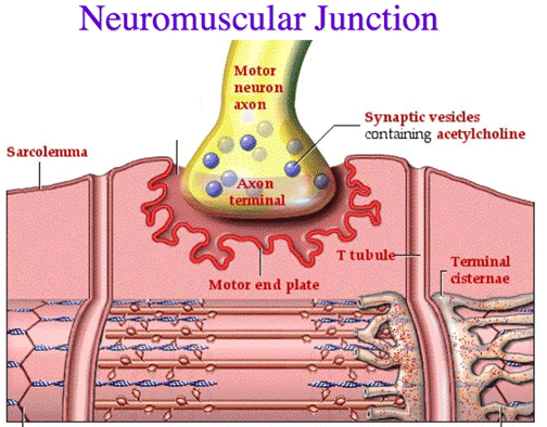
A specific type of synapse where a motor neuron communicates with a muscle fiber to initiate contraction.
How do most synapses transmit signals?
Most use chemical transmission, where neurotransmitters are released by the presynaptic neuron and affect the membrane potential of the postsynaptic cell.
Neurotransmitter
Chemical messenger released at a synapse that can excite or inhibit the postsynaptic cell depending on the type of receptor it binds to.
What is an electrical synapse and where is it found?
An electrical synapse directly passes current via gap junctions. It is found in:
Some brain neurons
Cardiac muscle
Smooth muscle
Glial cells
Gap junctions
Specialized protein channels made of connexins that allow ions to pass directly between adjacent cells, enabling fast electrical communication.
Gap junctions can be opened or closed by phosphorylation (adding a phosphate group) or dephosphorylation (removing a phosphate group) of connexin proteins.

What are the types of synapses based on signal transmission?
Chemical synapses: Use neurotransmitters to send signals.
Electrical synapses: Use direct electrical currents via gap junctions.
Chemical Synapse
A type of synapse where neurotransmitters are released from the presynaptic neuron’s terminal boutons to influence the postsynaptic cell.
Electrical Synapse
A type of synapse where ions flow directly between adjacent cells through gap junctions, allowing rapid signal transmission.
What molecules help hold presynaptic and postsynaptic cells together at chemical synapses?
Cell adhesion molecules (CAMs).
What are the three types of neuron-to-neuron synapses?
Axodendritic: Axon to dendrite
Axosomatic: Axon to cell body
Axoaxonic: Axon to another axon
Is signal transmission at neuron-neuron synapses usually bidirectional?
No, it is almost always one-way: from the presynaptic neuron's axon to the postsynaptic neuron.
Where are neurotransmitters stored before release?
In small, membrane-bound vesicles inside the presynaptic boutons (axon terminals).

What triggers neurotransmitter release into the synaptic cleft?
The vesicle fuses with the presynaptic membrane, releasing neurotransmitters via exocytosis.
Why can't neurotransmitters cross the membrane on their own?
Because they are hydrophilic and cannot pass through the lipid bilayer without vesicle fusion and exocytosis.
What is a miniature postsynaptic potential?
The small postsynaptic response caused by the release of a single vesicle of neurotransmitter.
How is synaptic strength related to firing rate?
More frequent action potentials cause more vesicles to release neurotransmitters, producing a stronger effect on the postsynaptic neuron.
What happens to vesicle membranes after neurotransmitter release?
They are recycled back into the presynaptic neuron for reuse.
What triggers neurotransmitter release at the axon terminal?
The arrival of an action potential at the axon terminal triggers the opening of voltage-gated calcium channels, allowing Ca²⁺ to enter and initiate neurotransmitter release.
Docked vesicles
Synaptic vesicles that are already positioned at the presynaptic membrane, ready for rapid fusion when an action potential arrives.
Why is neurotransmitter release so fast (under 100 μs)?
Because many synaptic vesicles are already docked near voltage-gated Ca²⁺ channels, allowing immediate fusion and release once calcium enters the terminal.
What role does calcium play in neurotransmitter release?
Ca²⁺ acts as a direct signal that triggers vesicle fusion with the axon terminal membrane, leading to neurotransmitter exocytosis.

Calmodulin
A calcium-binding protein that, when activated by Ca²⁺, stimulates protein kinases that phosphorylate other proteins involved in vesicle release.
What happens after Ca²⁺ binds to calmodulin?
The Ca²⁺-calmodulin complex activates protein kinases, which phosphorylate regulatory proteins, triggering a cascade that supports vesicle exocytosis.
Synaptotagmin-1
A calcium sensor protein on synaptic vesicles that, upon binding Ca²⁺, promotes vesicle fusion by interacting with the SNARE complex.
What are the roles of the C2A and C2B domains in synaptotagmin-1?
These domains bind calcium and insert into the presynaptic membrane, pulling the SNARE complex to drive vesicle fusion and neurotransmitter release.

What type of ion channels are found on the postsynaptic membrane and how are they activated?
hey are chemically regulated ion channels (neurotransmitter receptors) that open when a neurotransmitter binds to them, not in response to voltage changes
Chemically regulated ion channel
A type of ion channel on the postsynaptic membrane that opens in response to neurotransmitter binding, unlike voltage-gated channels.
What is an excitatory postsynaptic potential (EPSP)?
A depolarization caused by positive ions (like Na⁺) entering the cell, making it more likely to reach the threshold for an action potential.
What is an inhibitory postsynaptic potential (IPSP)?
A hyperpolarization caused by negative ions (like Cl⁻) entering or positive ions (like K⁺) leaving the cell, making it less likely to fire an action potential.
Where are neutotransmitter receptors found?
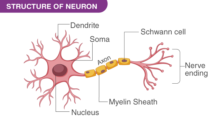
on the dendrites and cell body of a neuron, which allow these parts of the neuron to respond to neurotransmitters released from potentially thousands of different synapses onto the cell’s surface.
Decremental spread
The gradual weakening of a graded potential (like an EPSP) as it spreads from the dendrites/cell body toward the axon hillock.
What happens when neurotransmitters bind to receptors and cause an EPSP?
A depolarization occurs, which spreads toward the axon hillock with decreasing strength (decremental spread).
What must happen for an action potential to be generated at the axon hillock?
The EPSP must be strong enough to reach the threshold and open voltage-gated channels at the axon hillock.
Axon hillock
The part of the neuron where graded potentials are summed, and if threshold is reached, action potentials are initiated.
What kind of channels are found at the axon hillock, and what opens them?
Voltage-gated channels that open when the membrane reaches threshold depolarization.
4 major classes of neurotransmitters

What determines the influence of an ion on membrane potential?
The ion’s concentration gradient across the membrane
The membrane’s permeability to that ion
Since the membrane is more permeable to K⁺ than other ions, K⁺ largely determines the membrane potential.
What is the Nernst equation used for in neuroscience?
It calculates the equilibrium potential (Eₓ) for an ion—the membrane voltage at which there’s no net movement of that ion across the membrane.

Why does K⁺ reach equilibrium across the membrane?
Electrical attraction pulls K⁺ into the cell, but diffusion pushes it out. When these forces balance, K⁺ stops moving overall—this is the K⁺ equilibrium potential (Eₖ).
What is the value of Eₖ (K⁺ equilibrium potential), and what does the negative sign mean?
Eₖ ≈ -90 mV. The negative sign indicates the inside of the cell is more negative than the outside.
What happens if the membrane potential becomes more or less negative than -90 mV?
More negative: K⁺ is pulled into the cell
Less negative: K⁺ diffuses out of the cell
Thus, -90 mV keeps K⁺ concentrations stable inside and outside.
Why is the resting membrane potential not exactly -90 mV?
Because not all K⁺ channels are open (only ~30%) and other ions slowly leak across, the resting potential is usually around -70 mV, between -65 to -85 mV.
Action Potential
A rapid shift in a neuron’s or muscle cell’s membrane potential, crucial for transmitting nerve impulses along an axon.
What triggers the start of an action potential?
Mechanical or ligand-gated ion channels open in response to a stimulus, changing the resting potential; if the depolarization reaches the threshold (around -55 mV), voltage-gated Na⁺ channels open.
What happens when voltage-gated Na⁺ channels open?
Na⁺ rapidly enters the cell, making the membrane potential rise quickly and become less negative, approaching sodium’s equilibrium potential.
Sodium Equilibrium Potential (ENa)
Predicted by the Nernst equation to be around +66 mV, based on Na⁺ being more concentrated outside (145 mM) than inside (12 mM) the cell.
Steps of an action pitentual
Resting State: The neuron’s membrane is at -70 mV, maintained by the sodium-potassium pump and leaky potassium channels.
Threshold Reached (-55 mV): A stimulus depolarizes the membrane to -55 mV, triggering voltage-gated sodium channels to open.
Rising Phase: Rapid sodium influx causes the membrane potential to rise sharply toward +30 mV.
Peak: Sodium channels close and potassium channels open, stopping Na⁺ entry and starting K⁺ exit.
Repolarization: Potassium ions leave the cell, making the inside more negative and returning the membrane potential toward resting.
Hyperpolarization: Potassium channels close slowly, causing the membrane potential to briefly become more negative than resting.
Return to Resting: The sodium-potassium pump restores the original ion concentrations and membrane potential returns to -70 mV.
Refractory Periods: No new action potential can start during the absolute refractory period, and a stronger stimulus is needed during the relative refractory period.


What is the relative refractory period?
It's the phase after an action potential when the membrane is hyperpolarized and requires a stronger-than-normal stimulus to trigger another action potential.
What role does the Na⁺/K⁺ ATPase pump play after an action potential?
It restores original ion concentrations by pumping 3 Na⁺ out and 2 K⁺ in using ATP, re-establishing the resting membrane potential and ion gradients.
All-or-None Law
Once the threshold is reached, an action potential fires fully or not at all. The size (~+30mV peak) and duration of the action potential do not depend on stimulus strength.
Does a stronger stimulus produce a bigger or longer action potential?
No. The action potential’s size and duration are always the same once triggered, regardless of stimulus strength.

Propagation of the Action Potential
After initiation at the axon hillock, the action potential travels down the axon by sequentially depolarizing adjacent membrane segments, like a domino effect.

Why can action potentials only travel forward along the axon?
Because the recently activated section enters a refractory period, making it temporarily unresponsive to new stimuli, preventing backward propagation.
Action Potential Conduction in Muscle Fibers
Skeletal muscle action potentials resemble those in neurons (triggered by Na⁺ influx). However, Cl⁻ permeability plays a bigger role in resting potential and recovery, unlike neurons where K⁺ dominates.