BCHM ch 5, 6, 7 and 10
 Call Kai
 Learn
 Practice Test
 Spaced Repetition
 Match
 Flashcards
 Knowt Play
1/75

Earn XP
Description and Tags
1-20:ch5 21-40:ch6 41-61:ch7
76 Terms
Functions of globular proteins
• Storage of ions and molecules
– myoglobin, ferritin
• Transport of ions and molecules
– hemoglobin, serotonin transporter
• Defense against pathogens
– antibodies, cytokines
• Muscle contraction
– actin, myosin
• Biological catalysis
– chymotrypsin, lysozyme
Define ligand and binding site. What interactions are involved in protein-ligand binding, and is binding permanent?
A ligand is a molecule, typically small, that binds to a protein. The binding site is the region where it binds. Binding involves non covalent interactions and is usually reversible and transient.
Define prosthetic group, cofactor, and coenzyme. How do they differ?
Prosthetic group: permanently attached to a protein.
Cofactor: a helper that is not permanently attached; metal ions are examples.
Coenzyme: a non permanently attached organic molecule that assists a protein.
What are Ka and Kd, and how are they related to binding affinity?
Ka is the equilibrium association constant; Kd is the equilibrium dissociation constant. (Kd=1/Ka).
High Ka and low Kd indicate strong binding and high affinity. Low Ka and high Kd indicate weak binding and low affinity.
How do you determine Kd and compare affinity using a binding curve?
Kd is the free ligand concentration at 50% binding. A curve reaching 50% binding at a lower ligand concentration has a lower Kd and higher affinity.
Describe myoglobin’s structure, function, and oxygen-binding curve.
Myoglobin is a globular protein composed mainly of alpha helices. It stores oxygen in muscle and has a hyperbolic oxygen-binding curve.

Compare the lock-and-key and induced-fit models of protein-ligand binding.
Lock-and-key assumes a preformed binding site complementary to the ligand in size, shape, charge, and hydrophobic/hydrophilic character.
Induced fit involves conformational changes in the protein, and sometimes the ligand, to improve binding.
It allows for tighter binding of the ligand and high affinity for different ligands.
What is heme, why do globins use it, and what happens to its Soret band when oxygen binds?
The heme group is a strong chromophore that absorbs both in UV and visible range. Ferrous form (FE2+) without oxygen has an intense soret band at 429 nm oxygen binding alters the electronic properties of the heme and shifts the position of the soret band to 414 nm.
Why does CO bind heme more strongly than O₂, and why is CO toxic?
because the Carbon in CO has a filled lone electron pair that can be donated to vacant d-orbitals on the Fe2+.
Define positive and negative cooperativity. How are they reflected in binding curves?
Cooperativity: Protein with multiple binding sites and binding sites interact with each other.
Positive cooperativity: binding at one site increases affinity at remaining sites; associated with a sigmoidal curve.
Negative cooperativity: binding at one site decreases affinity at remaining sites. Hyperbolic
Describe hemoglobin’s T and R states, the T-to-R transition, and its oxygen-binding curve.
T (tense) has more interactions, greater stability, and lower O₂ affinity. R (relaxed) has fewer interactions, greater flexibility, and higher O₂ affinity. Oxygen binding triggers T → R by breaking ion pairs at the α₁–β₂ interface. Hemoglobin has a sigmoidal O₂-binding curve.
What is a Hill plot, and what do the different Hill coefficients indicate?
A Hill plot analyzes cooperativity. The Hill coefficient indicates:
(n=1): no co-operativity
(n>1): positive co-operativity
(n<1): negative co-operativity
Cooperativity = positive homotropic regulation
Define allosteric protein, homotropic regulation, and heterotropic regulation.
An allosteric protein allows ligand binding at one site to affect binding at another site. Homotropic regulation occurs when the normal ligand acts as the allosteric regulator. Heterotropic regulation occurs when a different ligand affects binding of the normal ligand. Positive homotropic regulation is cooperativity.
Compare myoglobin and hemoglobin in structure, function, and oxygen binding.
Myoglobin is a single polypeptide rich in alpha helices and stores oxygen in muscle. Hemoglobin is a tetramer (α₂β₂) that transports oxygen in blood. Their subunits have similar structures and contain heme. Myoglobin has a hyperbolic curve and no cooperativity; hemoglobin has a sigmoidal curve and positive cooperativity.
How do pH and CO₂ affect hemoglobin’s oxygen affinity, and what is the Bohr effect?
Lower pH and increased H⁺ decrease hemoglobin’s oxygen affinity and promote oxygen release. H⁺ stabilizes the T state; protonated His146 forms a salt bridge with Asp94. CO₂ contributes to H⁺ formation through CO₂ + H₂O ⇌ HCO₃⁻ + H⁺, catalyzed by carbonic anhydrase. The pH difference between lungs and metabolic tissues increases efficiency of the O2 transport, this is the Bohr effect.
What is 2,3-BPG, how does it regulate hemoglobin, and how does it help at high altitude?
BPG is a small negatively charged molecule produced from a glycolysis intermediate. It is a negative heterotropic regulator that binds hemoglobin’s positively charged central cavity and stabilizes the T state, lowering O₂ affinity. At high altitude, increased BPG helps hemoglobin release oxygen to tissues.
Compare fetal and adult hemoglobin, including structure, BPG binding, oxygen affinity, and the timing of HbF production.
HbF is α₂γ₂; HbA is α₂β₂. The gamma subunit has fewer positive charges than the adult beta subunit, so HbF binds BPG less strongly and has higher O₂ affinity. HbF production normally decreases after birth as its gene is switched off, around 2–4 months.
HbF is alkali-resistant
Why is fetal hemoglobin important in sickle cell disease, and how can its production be reactivated?
HbF can reduce sickle cell disease problems. Hydroxyurea can reactivate HbF production in adults.
What is HbS, how does it differ from normal hemoglobin, and how does it cause sickling?
HbS has a Glu → Val substitution at position 6 of the beta chain. The new hydrophobic valine side chain can bind to another hemoglobin molecule, forming strands that cause red blood cells to become sickle-shaped. Heterozygous individuals have increased resistance to malaria, according to the slides.
What are enzymes, and how are they different from inorganic catalysts?
Enzymes are biological catalysts, mostly proteins, that increase reaction rates without being used up. They are more specific, work under milder conditions (conductive to conditions in cells), and can be regulated.
How do enzymes speed up reactions?
Decrease activation energy
Bring reactive groups close together in proper orientation
Bind best to the transition state
Increase reaction rate
Are not consumed in reactions
Most are proteins
Do not change thermodynamic parameters (ΔG, Keq, ΔS, ΔH)
What are cofactors, coenzymes, prosthetic groups, apoenzymes, and holoenzymes?
Cofactor: Nonprotein component required by some enzymes.
Coenzyme: Organic cofactor.
Prosthetic group: Cofactor tightly or permanently attached to an enzyme.
Apoenzyme: Enzyme without its required prosthetic group.
Holoenzyme: Complete, functional enzyme with its required component(s).
What are ribozymes? Give two examples.
Ribozymes are RNA molecules that catalyze reactions. Examples include RNA involved in tRNA splicing is carried by tRNA itself, t does not require a spliceosome. and ribosomal RNA, which catalyzes peptide bond formation.
What are the three types of catalytic mechanisms?
Acid-base catalysis
Covalent catalysis
Metal ion catalysis
What factors affect the rate of an enzyme-catalyzed reaction?
Enzyme concentration, substrate concentration, effectors, temperature, and pH.
What is the Michaelis–Menten equation, and what does its graph look like?
The graph is a hyperbolic curve. At low substrate concentration, the reaction is first-order (rate increases with substrate concentration). At high substrate concentration, the enzyme becomes saturated and the reaction approaches zero-order kinetics (rate levels off).

What are Km and Vmax?
Km = substrate concentration when v₀ = ½ Vmax.
Vmax = maximum reaction velocity when the enzyme is saturated with substrate.
Michaelis-Menten equation:
v₀ = (Vmax × [S]) / (Km + [S])
How is Km different from Kd?
Km = reflects substrate binding and product formation.
Kd = measures how easily the enzyme-substrate complex dissociates.
Low Kd = strong binding (high affinity).
Km = Kd when substrate binding is at rapid equilibrium, typically when k₋₁ >> kcat.
What are kcat, turnover number, and enzyme efficiency?
kcat = number of substrate molecules converted to product per enzyme molecule per second when saturated.
Turnover number = kcat.
Enzyme efficiency = kcat / Km.
Higher kcat / Km = greater catalytic efficiency.
What is the steady-state assumption?
The concentration of the enzyme-substrate complex ([ES]) remains approximately constant because its rate of formation equals its rate of breakdown.
What graphs determine Km and Vmax?
Michaelis-Menten plot = plots initial velocity (v₀) versus substrate concentration ([S]).
Lineweaver-Burk plot = double-reciprocal plot of 1/v₀ versus 1/[S].
Y-intercept = 1/Vmax
X-intercept = -1/Km
Slope = Km/Vmax
It is useful for analyzing enzyme inhibition and two-substrate reactions.
What are irreversible and reversible inhibitors?
Irreversible inhibitor = One inhibitor molecule can permanently shut off one one enzyme molecule; often powerful toxins but also may be used as drugs.
Reversible inhibitor = structural analogs of substrates or products; often used as drugs to slow down a specific enzyme they can bind to
the free enzyme and prevent the binding of the substrate.
enzyme substrate complex and prevent the reaction.
What are the types of reversible inhibition?
Competitive:
Inhibitor binds free enzyme at the active site.- competes with substrate.
Km increases; Vmax stays the same.
Uncompetitive:
Inhibitor binds only to the enzyme-substrate complex (ES).
Km decreases; Vmax decreases.
Pure noncompetitive:
Inhibitor binds to free enzyme and ES equally at a regulatory (allosteric) site. equal affinity
Km stays the same; Vmax decreases.
Mixed:
Inhibitor binds free enzyme and ES at a regulatory site. different affinity
Km changes (inc or dec); Vmax decreases.
How do you identify inhibition on a Lineweaver-Burk plot?
Competitive = lines intersect on the y-axis.
Uncompetitive = lines are parallel.
Mixed = lines intersect left of the y-axis.
Pure noncompetitive = lines intersect on the x-axis.

How do Lineweaver-Burk plots identify two-substrate mechanisms?
Sequential mechanism = lines intersect.
Ping-pong mechanism = lines are parallel.
Random mechanism at equilibrium = lines intersect on the y-axis.
What is allosteric regulation?
Allosteric regulation = a molecule binds to a regulatory site and changes enzyme activity.
Properties of allosteric enzymes:
- Usually have multiple subunits.
- Have T (tense) and R (relaxed) states.
- Do not follow Michaelis-Menten kinetics.
- Each subunit has an active site.
What are allosteric activators, inhibitors, and cooperativity?
Allosteric activator = increases enzyme activity.
Allosteric inhibitor = decreases enzyme activity.
Positive cooperativity = binding at one site increases affinity at other sites.
Negative cooperativity = binding at one site decreases affinity at other sites.
How is ATCase regulated?
ATCase = an allosteric enzyme.
ATP = positive modulator (activator).
CTP = negative regulator (inhibitor).
Enzyme regulation by reversible covalent modification.(7)
What enzyme is regulated in multiple ways? and which ligands regulate it
Phosphorylation, Adenylylation, Acetylation, Myristoylation, Ubiquitination, ADP-ribosylation, Methylation
Enzyme regulated in multiple ways: glycogen phosphorylase
Important ligands that regulate it
AMP
ATP:
Glucose-6-phosphate:
Glucose:
What is the general formula for carbohydrates?
Cn(H2O)n
What are aldoses and ketoses? Give examples.
Aldose = carbohydrate with an aldehyde group.
Example: glucose.
Ketose = carbohydrate with a ketone group.
Example: fructose.
What are D and L isomers, and what molecule determines D/L configuration?
Glyceraldehyde is the standard molecule for D/L designation.
D sugar = OH on the right at the chiral carbon farthest from the carbonyl in a Fischer projection.
L sugar = OH on the left at that carbon.
D and L forms are enantiomers.
What are enantiomers, epimers, diastereomers, and anomers? Give examples.
Enantiomers = nonsuperimposable mirror images.
Example: D-glucose and L-glucose.
Epimers = stereoisomers that differ at only one chiral center.
Examples: D-mannose and D-glucose; D-galactose and D-glucose.
Diastereomers = stereoisomers that are not mirror images.
Example: D-threose and D-erythrose.
Anomers = sugars that differ at the anomeric carbon (α or β).
Example: α-D-glucose and β-D-glucose.
How do you identify sugar types from structures?
Which sugar has no chiral center?
Ketose = has a ketone group.
5-carbon sugar = pentose.
6-carbon sugar = hexose.
Dihydroxyacetone = has no chiral centers, so it has no D/L isomers.
Ribose = standard 5-carbon sugar.
Glucose = standard 6-carbon sugar.
Fructose = ketose form of glucose.
How do hemiacetals and hemiketals form during sugar cyclization?
Aldehyde + alcohol → hemiacetal.
Ketone + alcohol → hemiketal.
During cyclization, an internal alcohol attacks the sugar's carbonyl carbon, forming a ring.
The former carbonyl carbon becomes the anomeric carbon.
What are anomeric carbon, α and β forms, pyranoses, and furanoses?
Anomeric carbon = former carbonyl carbon that becomes a new chiral center during cyclization.
α form = anomeric OH is trans (opposite side) to CH2OH.
β form = anomeric OH is cis (same side) to CH2OH.
Pyranose = 6-membered ring containing oxygen.
Furanose = 5-membered ring containing oxygen.
What are reducing and non-reducing sugars?
Reducing sugar = has a free anomeric carbon.
Non-reducing sugar = has no free anomeric carbon available.
Example of a non-reducing sugar: sucrose.
What is a glycosidic bond, and how is it named?
A glycosidic bond joins sugars through the anomeric carbon of one sugar and a hydroxyl group of another.
The linkage is named by the anomeric configuration and carbon numbers.
Example: α(1→4) means the anomeric carbon 1 of one sugar links to carbon 4 of another.
What are homopolysaccharides and heteropolysaccharides?
Homopolysaccharide = made of one type of monomer.
Examples: starch, glycogen, cellulose, chitin.
Heteropolysaccharide = made of multiple types of monomers.
Examples: agar and glycosaminoglycans.
Compare starch, amylose, amylopectin, glycogen, cellulose, and chitin.
Amylose:
- Glucose monomers.
- Linear α(1→4) linkages.
- Part of starch; energy storage in plants.
Amylopectin:
- Glucose monomers.
- α(1→4) chains and α(1→6) branches every 24–30 residues.
- Part of starch; energy storage in plants.
Glycogen:
- Glucose monomers.
- α(1→4) chains and α(1→6) branches every 8–12 residues.
- Main energy-storage polysaccharide in animals.
Cellulose:
- Glucose monomers.
- Linear β(1→4) linkages.
- Hydrogen bonds within and between chains make it strong and insoluble.
- Structural component of plant cell walls.
Chitin:
- N-acetylglucosamine monomers.
- Linear β(1→4) linkages.
- Tough, flexible, insoluble; found in arthropod exoskeletons and mushroom cell walls.
What are the similarities and differences between starch, glycogen, cellulose, and chitin?
Similarities:
- All are polysaccharides.
- Each is a homopolysaccharide.
Differences:
- Starch and glycogen store energy.
- Cellulose and chitin provide structural support.
- Starch and glycogen use α linkages.
- Cellulose and chitin use β(1→4) linkages.
- Cellulose has extensive hydrogen bonding between chains.
- Glycogen has more frequent branching than amylopectin.
What are glycosaminoglycans (GAGs)? Name examples.
GAGs = linear polymers of repeating disaccharide units; negatively charged and highly hydrated.
Examples:
- Heparin
- Heparan sulfate
- Hyaluronan
- Chondroitin sulfate
- Keratan sulfate
What is a glycoconjugate, and what amino acids link to carbohydrates in glycoproteins?
Glycoconjugate = carbohydrate covalently attached to a protein or lipid.
Glycoprotein = protein with small oligosaccharides attached.
Common amino acids involved:
- Serine (Ser)
- Threonine (Thr)
- Asparagine (Asn)
About half of mammalian proteins are glycoproteins.
What are N-linked and O-linked glycoproteins?
N-linked = carbohydrate attaches to the nitrogen of asparagine (Asn).
O-linked = carbohydrate attaches to the oxygen of serine (Ser) or threonine (Thr).
What are glycolipids? Give examples and functions.
Glycolipids = lipids with covalently attached carbohydrates.
Examples:
Blood group antigens
Sphingolipids
Lipopolysaccharides (LPS):
What are proteoglycans? Give examples.
Proteoglycans = proteins with attached glycosaminoglycan (GAG) chains.
Examples:
- Syndecans
- Glypicans
What is the difference between syndecans and glypicans?
Syndecans = have a single transmembrane domain.
Glypicans = anchored to the cell membrane through a lipid anchor.
Both interact with neighboring-cell receptors and help regulate cell growth.
What are the functions of carbohydrates?
- Energy source.
- Energy storage.
- Structural support in cell walls and exoskeletons.
- Cell-cell recognition and signaling.
- Attachment to proteins and lipids to form glycoconjugates.
What is the difference between a reducing and non-reducing end in a polysaccharide?
Reducing end = has a free anomeric carbon.
Non-reducing end = does not have a free anomeric carbon.
Glycogen and amylopectin each have one reducing end but many non-reducing ends.
What are the main biological functions of lipids? (9)
Energy storage
Insulation and mechanical protection
Waterproofing
Buoyancy control
Cell membrane structure
Enzyme cofactors
Signaling molecules
Pigments
Antioxidants
SLIDE 12
Ribose is the standard five-carbon sugar.
• Glucose is the standard six-carbon sugar.
• Galactose is an epimer of glucose.
• Mannose is an epimer of glucose.
• Fructose is the ketose form of glucose.
How are lipids classified into two major categories?
Lipids containing fatty acids: complex lipids, including storage and membrane lipids.
Lipids that do not contain fatty acids: cholesterol, vitamins, and pigments.
What are fatty acids? What is the difference between saturated, monounsaturated, and polyunsaturated fatty acids?
Fatty acids are carboxylic acids with hydrocarbon chains containing 4–36 carbons.
Saturated: no double bonds.
Monounsaturated: one double bond.
Polyunsaturated: more than one double bond.
How do chain length and double bonds affect fatty acid solubility and melting point?
Solubility increases as chain length decreases.
Melting point decreases as chain length decreases.
Melting point decreases as the number of double bonds increases.
How do saturated, cis-unsaturated, and trans-unsaturated fatty acids differ in conformation?
Saturated fatty acids adopt extended conformations.
Cis double bonds create kinks in the chain.
Trans double bonds allow a more extended conformation and more regular packing.

What are triacylglycerols? What are their structure, linkage, and function?
Made of one glycerol molecule and three fatty acids.
Fatty acids attach to glycerol through ester linkages.
Primary storage form of lipids.
Solid triacylglycerols are fats; liquid ones are oils.
Used for long-term energy storage.
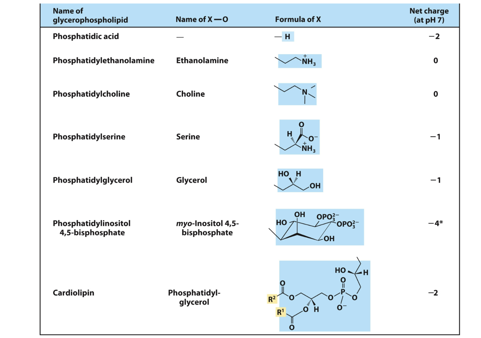
What are glycerophospholipids? Give examples and describe their structure.
Primary constituents of cell membranes.
Contain two fatty acids attached to glycerol-3-phosphate through ester linkages.
Have a charged phosphate group that may attach to an alcohol head group.
Examples: phosphatidylcholine, phosphatidylserine, and phosphatidylethanolamine.
Amphipathic: contain polar heads and nonpolar tails.
What are ether lipids? Give examples and describe their linkages.
Ether lipids contain an ether linkage instead of the typical ester linkage at one position.
Examples:
Plasmalogen: vinyl ether analog of phosphatidylethanolamine; common in vertebrate heart tissue.
Platelet-activating factor (PAF): aliphatic ether analog of phosphatidylcholine.
What is platelet-activating factor (PAF), and what are its functions?
First signaling lipid to be identified.
Stimulates aggregation of blood platelets.
Plays a role in mediation of inflammation.
Has an acetate group esterified at position C2.
What are sphingolipids? What is their backbone and what linkages do they contain?
Their backbone is sphingosine, a long-chain amino alcohol, NOT glycerol.
A fatty acid attaches to sphingosine through an amide linkage.
The polar head group attaches through a glycosidic or phosphodiester linkage.

What are examples of sphingolipids, and what is sphingomyelin made of?
Sphingomyelin
Gangliosides, such as GM2
Sphingomyelin consists of:
Ceramide (sphingosine + amide-linked fatty acid)
Phosphocholine attached to the alcohol group.
Sphingomyelin is abundant in the myelin sheath surrounding some nerve cells in animals
How do glycosphingolipids determine blood groups?
Blood groups are determined in part by the sugars on glycosphingolipid head groups.
O: no active glycosyltransferase; O antigen.
A: glycosyltransferase transfers N-acetylgalactosamine.
B: glycosyltransferase transfers galactose.
What is cholesterol, and what are its functions and products?
Cholesterol is a sterol with four fused rings, a hydroxyl group, and nonpolar side chains.
Found in most eukaryotic cell membranes.
Modulates membrane fluidity and permeability.
Thickens the plasma membrane.
Products derived from cholesterol:
Steroid hormones
Vitamin D
Bile acids
What are signaling lipids? Give examples and their functions.
Signaling lipids are biologically active lipids present in much smaller amounts than storage or structural lipids.
Arachidonic acid derivatives:
Prostaglandins: inflammation and fever.
Thromboxanes: blood clot formation.
Leukotrienes: smooth muscle contraction in lungs.
What is the precursor of vitamins A and D, and what is the function of retinoic acid?
Vitamin A (retinol) is derived from β-carotene.
Vitamin D is synthesized from cholesterol.
Retinol is involved in visual pigment and is a precursor for other hormones involved in signaling.
What are medicinal lipids (polyketides)?
Polyketides are a diverse family of biologically active compounds.
Synthesized similarly to fatty acid biosynthesis.
Commonly secondary metabolites with specialized functions.
Lovostatin, erythmacin, amphotericin B