Workshop- Leukaemia
 Call Kai
 Learn
 Practice Test
 Spaced Repetition
 Match
 Flashcards
 Knowt Play
1/32

There's no tags or description
Looks like no tags are added yet.
33 Terms
What is leukaemia?
A neoplastic condition of the blood and/or bone marrow in which neoplastic cells of lymphoid or non-lymphoid stem cells or their progeny undergo clonal expansion with or without cellular differentiation

What change in WBC numbers do we see in leukaemia?
§Leucocytosis = high numbers of leukaemic cells in circulation (much higher than we would see in infection/inflammation)
Can be an increase in any type of WBC but overall will see large increase in WBC count overall

Describe leukaemia masses
Leukaemia cells proliferate within the bone marrow and then spill into the bloodstream.
Because the cancerous cells circulate in blood, the disease is already systemic — so it doesn't need to "metastasize" in the traditional sense (like a solid tumour spreading through lymph or blood).
You don't typically see solid tumour masses in the marrow. Instead, you see diffuse infiltration of abnormal cells replacing normal marrow.
While not "metastases," leukaemic cells can infiltrate other organs, such as:
Liver and spleen → causing hepatosplenomegaly
Lymph nodes → mild enlargement (especially in lymphoid leukaemias)
Central nervous system → especially in acute lymphoblastic leukaemia (ALL)
Skin, gums, testes, kidneys → sometimes seen in acute myeloid leukaemia (AML)
So yes — organ infiltration is common, but it's due to circulating leukaemic cells migrating and accumulating, not due to "metastasis" from a primary tumour.
What causes clinical signs in leukaemia?
§Failure of normal marrow function
§Infiltrated organ dysfunction
§Hyperviscosity
§Paraneoplastic syndromes (IMHA, hypercalcaemia)
When should you suspect leukaemia?
Unspecific clinical signs
Lethargy, anorexia, and weight loss are common early signs due to anemia, infection, or organ infiltration.
Abnormal complete blood count
Severe or persistent leukocytosis, especially with immature or atypical cells (>100,000/μL in acute leukemia), should raise suspicion.
Moderate leukocytosis can be seen in chronic leukemia.
Atypical cells on blood smear review
Presence of blasts or dysplastic cells is highly suggestive of leukemia.
Persistent cytopenias
Anaemia, thrombocytopenia, or neutropenia caused by bone marrow replacement - weakness, bleeding, or recurrent infections.
Splenomegaly and/or hepatomegaly
Enlargement may occur due to infiltration by leukemic cells, especially in chronic myeloid leukemias.
Hyperglobulinemia
Persistent hyperglobulinemia, especially if confirmed on serum protein electrophoresis, may indicate chronic lymphoid leukemia.
Hypercalcemia
Occasional finding in some lymphoid leukemias due to paraneoplastic PTHrP production.
Incidental finding on routine bloodwork
Chronic leukemias (e.g., CLL) are often discovered before clinical signs when persistent leukocytosis is detected.
Describe the two different routes for haematopoiesis in the blood
Haematopoietic pleuripotent stem cell develops into either a CMP (which can become RBCs or platelets or some WBCs - monocytes, macrophages, eosinophils, basophils, neutrophils) or CLP (which become other WBCs- NK cell, b lymphocytes, plasma cells, t lymphocytes)

How do we categorise leukaemias?
Leukaemias are classified according to:
The rate of progression → acute or chronic
The cell lineage involved → lymphoid (B or T lymphocytes) or myeloid (myeloid precursors: granulocytes, monocytes, RBCs, platelets)

Describe ALL
Acute lymphoid leukaemia (ALL)
Rapid proliferation of immature lymphoblasts. Bone marrow heavily infiltrated with blasts. Poor prognosis.
Dogs (esp. young to middle-aged); rare in cats
Describe CLL
Chronic lymphoid leukaemia (CLL)
Slow proliferation of mature lymphocytes. Often incidental finding on blood tests. May respond to chemotherapy.
Older dogs, cats
Why do we sometimes see hyperglobulinemia with CLL?
Persistent hyperglobulinemia can be a clue for chronic lymphoid leukaemia because the malignant lymphocytes may overproduce antibodies, even before other clinical signs appear.
Describe AML
Acute myeloid leukaemia (AML)
Malignant proliferation of immature myeloid precursors (granulocytic, monocytic, erythroid, or megakaryocytic). Aggressive disease.
Dogs > cats
Describe CML
Chronic myeloid leukaemia (CML)
Overproduction of more mature myeloid cells; may resemble a severe leukocytosis. Rare in animals.
Dogs, rarely cats
what cell type disappears first in leukaemia
- neutrophils - shortest half life ***
- platelets - days
- RBCs - months
This is about as far as you can go in first opinion practice. Oncologists will do further tests to get to bottom line.
What are the different types of leukaemia?

What kind of leukaemia if polycythaemia vera
Polycythaemia vera is a type of chronic leukaemia
Describe the features of acute leukaemia
§Neoplastic transformation during stem cell proliferation
§Large numbers of immature (undifferentiated) cells (we call these blasts if they are from lymphoblasts or from myeloblasts)
§Aggressive, rapid disease
§Cytopaenia is very common
--Marrow disruption by neoplastic expansion (taken over by undifferentiated cell production and release happening so acutely)
--e.g. neutropaenia, thrombocytopaenia, anaemia
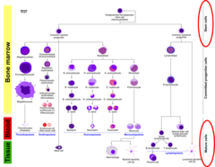
In acute leukaemia, when there is marrow disruption, lineage kinetics result in disappearance of ?
When there is marrow disruption, lineage kinetics result in disappearance of neutrophils first, then platelets then RBCs
Half-life of different cell types:
§Neutrophils (hours)
§Platelets (days)
§RBCs (months)

What is the consequence of cytopenia in leukaemia?
Red Blood Cells (Anaemia)
Low red blood cell count means less oxygen is delivered to tissues.
Signs include weakness, lethargy, pale gums, and rapid breathing.
Neutrophils (Neutropenia)
Low neutrophils reduce the body's ability to fight infections.
Signs include frequent infections, fever, slow-healing wounds, and oral or skin infections.
Platelets (Thrombocytopenia)
Low platelets impair blood clotting.
Signs include easy bruising, nosebleeds, bleeding gums, and small red spots on the skin.
Describe the features of chronic leukaemia
§Neoplastic transformation following differentiation
§Gives rise to large numbers of mature (differentiated) cells
§Slow progression and less aggressive in comparison to acute disease
§Cytopaenia is rare
§Lymphoid vs myeloid
§Chronic lymphoid leukaemia (CLL) most common type of leukaemia in dogs
§Chronic myeloid leukaemias are very rare
§Polycythaemia vera (Hct > 65%)
§Essential thrombocytosis (platelets >600,000/μL)
§Chronic neutrophilic leukaemia
§Reactive increases of the same cells (e.g. leukaemoid reaction - extreme neutrophilia, reactive thrombocytosis) are more common than chronic lymphoid leukaemias!
§Diagnosis of exclusion

If not chronic or acute leukaemia, then what could a leukaemia be?
Lymphoma in leukaemic phase
Leukaemia = starts in bone marrow and spreads outward.
Lymphoma = starts in lymphoid tissues and may spread into the marrow or blood later.
This refers to lymphoma that has spread into the blood and/or bone marrow, making it look similar to leukaemia.
The primary cancer originates as a solid tumour (usually in lymph nodes or spleen from lymphoid tissue).
Over time, neoplastic lymphocytes enter circulation, so you start to see them in blood smears.
When this happens, we say the lymphoma is in its "leukaemic phase."
In this stage, the disease can be hard to distinguish from leukaemia, since both have abnormal lymphoid cells in the blood.
What can I do in first opinion practice to diagnose leukaemia?
§Diagnose leukaemia (acute vs. chronic) via Blood smear examination
§Diagnose lymphoma via Lymph node cytology
§Exclude other causes of leucocytosis (e.g. infectious)
§Refer to oncology
What is a hallmark of acute leukaemia on doing a blood smear?
In acute leukaemias, cells of different lineages cannot be identified conclusively by morphologic features
Can just see blasts (immature cells):
§Large cells
§Round/oval nuclei
§Small amount of blue cytoplasm
§+/- prominent nucleoli

Describe the morphology seen in sells with chronic leukaemias
§In chronic leukaemias, the relevant WBCs look close to normal appearance (i.e. we can tell what cell line they come from) - there are just way too many of them
§Differentiated (mature cells)
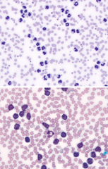
What is a leukaemic phase of lymphoma?
§Originates outside of the bone marrow (extramedullary)
§Document lymphoma in periphery (e.g. lymph nodes, spleen, liver)
§In the leukaemic phase (stage V), there are lymphoma cells in the blood and/or bone marrow
§No or mild cytopaenias
§Bone marrow infiltration usually

How do we diagnose leukaemia with a lab test using bone marrow and blood samples, (and sometimes lymph node tissue)
- immunophenotyping
- lymphocyte clonality PCR
How do we diagnose leukaemia with a lab test: immunophenotyping?
It looks at proteins (antigens) on the surface of blood or bone marrow cells.
This helps identify what type of cell is abnormal — for example, whether it's a B-cell, T-cell, or myeloid cell.
- rest of info is nice to know:
§Lab antibodies against cell surface markers (CD antigens)
§Immunocytochemistry/immunohistochemistry of marrow aspirates or biopsies
§Immuno-labelled flow cytometry of EDTA blood or marrow

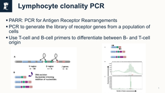
How do we diagnose leukaemia with a lab test: lymphocyte clonality PCR?
It examines the genetic rearrangements of immunoglobulin (Ig) or T-cell receptor (TCR) genes in lymphocytes.
Normal lymphocytes have diverse gene rearrangements (polyclonal).
In leukaemia or lymphoma, all malignant cells come from one clone, so they share an identical rearrangement (monoclonal).
Confirms whether an abnormal lymphocyte population is malignant (clonal) or reactive (polyclonal).
- rest of info is nice to know:
§PARR: PCR for Antigen Receptor Rearrangements
§PCR to generate the library of receptor genes from a population of cells
§Use T-cell and B-cell primers to differentiate between B- and T-cell origin
What are the prognoses of the different types of leukaemia in dogs?
Acute lymphoblastic leukemia (ALL)
Prognosis: 1-3 months (3-6 months with chemo)
Reason: Rapid proliferation of immature lymphocytes crowds the bone marrow, causing severe cytopenias and organ dysfunction.
Acute myeloid leukemia (AML)
Prognosis: 1-3 months
Reason: Immature myeloid cells replace normal marrow cells, leading to life-threatening cytopenias.
Chronic lymphocytic leukemia (CLL)
Prognosis: 1-3 years
Reason: Slowly accumulating mature lymphocytes usually cause mild or late-onset cytopenias, allowing longer survival.
Chronic myeloid leukemia (CML)
Prognosis: 3-12 months
Reason: Gradual marrow replacement by mature myeloid cells eventually leads to cytopenias and organ compromise.
What are the prognoses of the different types of leukaemia in cats?
Acute leukemia (usually FeLV-associated)
Prognosis:
SUMMARY: For acute myeloid leukaemia
- PE findings
- Haemogram findings
- Bone marrow changes
- Phenotyping tests
Mild or no enlargement of lymphoid organs (can be moderate to rarely severe). Rarely presents as a mass.
Large number of blasts; multiple cytopaenias (non-regenerative anaemia, neutropaenia, thrombocytopaenia) are typical but not always seen.
Large number of blasts which comprise >20% of marrow cells (per WHO definition), with reduced to minimal numbers of normal hematopoietic precursors. Tumour comprises >70% of marrow cells, with rare cases having a lower percentage of tumour cells (can be mistaken for stage V lymphoma).
Positive for monocyte, granulocyte, or megakaryocytic markers (erythroid markers are lacking in animals).
SUMMARY: For acute lymphoid leukaemia
- PE findings
- Haemogram findings
- Bone marrow changes
- Phenotyping tests
Mild or no enlargement of lymphoid organs (can be moderate to rarely severe).
Large number of blasts (or small to intermediate cells); multiple cytopaenias (non-regenerative anaemia, neutropaenia, thrombocytopaenia) are typical but not always seen.
Large number of blasts (these can be small, intermediate or large) with >25% of marrow cells (per WHO definition) found diffusely in the marrow (usually >50%), with reduced numbers of normal hematopoietic precursors.
Positive for B- or T-lymphoid markers.
SUMMARY: For leukemic phase of lymphoma (Stage V)
- PE findings
- Haemogram findings
- Bone marrow changes
- Phenotyping tests
Moderate to severe enlargement of lymphoid organs.
Variable number of lymphoma cells (usually resemble counterpart in tissues); typically no or mild cytopenias (mild non-regenerative anaemia most common; moderate thrombocytopaenia can be seen with variants of T-cell lymphoma).
Variable numbers of lymphoma cells (resemble counterpart in tissues); may still have some normal blood cell precursors. Lymphoma cells usually in foci or patches and usually
See cases on onenote